Prevalence of Celiac Disease in Latin America:
A Systematic Review and Meta-Regression
Rafael Parra-Medina
1, Nicolás Molano-Gonzalez
1, Adriana Rojas-Villarraga
1,
Nancy Agmon-Levin
2,3, Maria-Teresa Arango
1,2,5, Yehuda Shoenfeld
2,4,
Juan-Manuel Anaya
1*
1Center for Autoimmune Diseases Research (CREA), School of Medicine and Health Sciences, Universidad del Rosario, Carrera 24 #63-C-69, Bogotá, Colombia,2The Zabludowicz Center for Autoimmune Diseases, Sheba Medical Center, Tel-Hashomer, Israel,3Sackler Medical School, Tel Aviv University, Tel Aviv, Israel,4Incumbent of the Laura Schwarz-Kip Chair for Research of Autoimmune Diseases, Sackler Faculty of Medicine, Tel-Aviv University, Tel Aviv, Israel,5Doctoral Program in Biomedical Sciences Universidad del Rosario, Bogotá, Colombia
*anayajm@gmail.com
Abstract
Background
Celiac disease (CD) is an immune-mediated enteropathy triggered by the ingestion of
gluten in susceptible individuals, and its prevalence varies depending on the studied
population. Given that information on CD in Latin America is scarce, we aimed to
investi-gate the prevalence of CD in this region of the world through a systematic review and
meta-analysis.
Methods and Findings
This was a two-phase study. First, a cross-sectional analysis from 981 individuals of the
Co-lombian population was made. Second, a systematic review and meta-regression analysis
were performed following the Preferred Reporting Items for Systematic Meta- Analyses
(PRISMA) guidelines. Our results disclosed a lack of celiac autoimmunity in the studied
Co-lombian population (i.e., anti-tissue transglutaminase (tTG) and IgA anti-endomysium
(EMA)). In the systematic review, 72 studies were considered. The estimated prevalence of
CD in Latin Americans ranged between 0.46% and 0.64%. The prevalence of CD in
first-degree relatives of CD probands was 5.5%. The coexistence of CD and type 1 diabetes
mellitus varied from 4.6% to 8.7%, depending on the diagnosis methods (i.e.,
autoantibod-ies and/or biopsautoantibod-ies).
Conclusions
Although CD seems to be a rare condition in Colombians; the general prevalence of the
disease in Latin Americans seemingly corresponds to a similar scenario observed in
Europeans.
OPEN ACCESS
Citation:Parra-Medina R, Molano-Gonzalez N, Rojas-Villarraga A, Agmon-Levin N, Arango M-T, Shoenfeld Y, et al. (2015) Prevalence of Celiac Disease in Latin America: A Systematic Review and Meta-Regression. PLoS ONE 10(5): e0124040. doi:10.1371/journal.pone.0124040
Academic Editor:Domenico Coppola, H. Lee Moffitt Cancer Center & Research Institute, UNITED STATES
Received:November 27, 2014
Accepted:March 10, 2015
Published:May 5, 2015
Copyright:© 2015 Parra-Medina et al. This is an open access article distributed under the terms of the Creative Commons Attribution License, which permits unrestricted use, distribution, and reproduction in any medium, provided the original author and source are credited.
Data Availability Statement:All relevant data are within the paper and its Supporting Information files.
Funding:The authors have no support or funding to report.
Introduction
Celiac disease (CD) is an autoimmune intestinal disorder. This disease occurs due to an
im-mune-mediated enteropathy triggered by ingested prolamins present in wheat, barley, and rye
(generically called gluten). It occurs in susceptible individuals carrying the HLA-DQ2 and
HLA-DQ8 haplotype [
1
].
Recently the American College of Gastroenterology and the British Society of
Gastroenterol-ogy provided recommendations to perform an initial screen with IgA anti-tissue
transglutami-nase (tTG) [
2
,
3
]. Meanwhile, the ESPGHAN (European Society of Pediatric Gastroenterology
and Nutrition) proposed various criteria for diagnosis over time. Initially, the diagnosis required
a sequence of three small intestinal biopsies, but recently the guidelines indicated that
symptom-atic children with high levels of tTG and positive anti-endomysium (EMA) as well as HLA
DQ2/8 do not need the biopsy for disease diagnosis [
4
–
6
]. IgA tTG and IgA EMA
autoantibod-ies have a sensitivity and specificity of 98
–
100% [
7
]. They have been used for the CD diagnosis
since the 1990s [
8
]. Antigliadin (AGA) and anti-reticulin antibodies have also been previously
used, but they are currently considered obsolete for diagnosis because of their low sensitivity
and specificity [
9
]. In brief, individuals that have tested positive for celiac autoantibodies are
considered as potentially diagnosed patients, regardless of the biopsy results [
10
].
The distribution of CD has been associated with migratory patterns and changes in feeding
habits over time. In the early years, humans were not exposed to gluten contained within
cere-als. Approximately 10,000 years ago in a small region of the Middle East, farmers successfully
cultivated wild wheat and barley grains due to favorable environmental conditions. These
peo-ple then migrated to the Mediterranean area (Northern Africa and Southern Europe) and
Cen-tral Europe, searching for new lands to cultivate [
11
].
CD is a very frequent disorder in highly populated countries where inhabitants have a white
ancestry, primarily in Europe and North America. However, CD has also been reported among
people with Amerindian and African origins [
12
,
13
]. CD affects 0.6 to 1.0% of the population
worldwide and exhibits a female-to-male ratio of 2.8:1 [
14
]. Also the age of onset distribution
shows a first peak between nine months to two years and a second peak during the fourth
de-cade [
14
]. The frequency of CD is likely to increase in many developing countries due to an
in-creased prevalence of a
“
westernized
”
diet, involving greater wheat production. For instance,
over the past 30 years, the prevalence of CD in the United States has increased five-fold,
dou-bling approximately every 15 years [
15
].
The presence of CD in the Latin American (LA) population is uncertain. Latin America is
the geographical area defined by Mexico, Central America, islands of the Caribbean, and South
America. It is a rapidly growing region with almost 600 million inhabitants [
16
]. The LA
popu-lation is mixed with ancestries including Africans, Caucasians, and Amerindians [
17
]. In the
present study, we aimed to analyze the prevalence celiac autoimmunity (i.e., tTG and EMA) in
a Colombian population as a surrogate of CD [
18
]. We evaluated the presence of these
autoan-tibodies in healthy individuals and in patients with other autoimmune conditions, given that
CD may coexist with and share similar immunopathological mechanisms with other
autoim-mune diseases (ADs) [
19
]. In addition, we performed a systematic literature review and
meta-regression analysis to determine the estimated prevalence of CD in LA.
Methods
Study population
unaffected individuals from Northwest Colombia (Group 1), whilst Group 2 (Central
Colom-bia) was comprised of 180 AD patients and 120 non-AD controls, taken from an original
co-hort of 1667, and sampled according to a stratified sampling design where the strata were
different cohorts of patients with ADs.
All of the patients with ADs fulfilled the international classification criteria. These include:
the American College of Rheumatology (ACR) criteria for RA and SLE, the McDonald criteria
for MS and the American Diabetes Association (ADA) criteria for T1DM [
20
–
23
]. Patients
with SS met the American-European Consensus Group criteria; including a positive minor
sali-vary gland biopsy (MSGB) [
24
]. General and clinical characteristics of these patients have been
previously described [
25
–
46
].
The information regarding patient demographics as well as cumulative clinical and
labora-tory data was obtained by physical examination, interviewing or chart reviews as described
elsewhere [
25
–
46
]. All data were collected in an electronic and secure database. The review
board and the ethics committee of Universidad del Rosario approved the study according to
the ethical guidelines of the Helsinki Declaration and Resolution 008430 of 1993 of the
Minis-try of Health in Colombia. The study was classified as minimal risk research. All patients
com-pleted the written informed consent.
Detection of Autoantibodies
In Group 1, IgA and IgG antibodies against AGA and tTG were assessed by the Rad
Bio-Plex 2200 system (Bio-Rad Laboratories, Hercules, California, USA) as previously described
[
25
,
27
,
28
,
36
–
46
].
In Group 2, IgA h-tTG (native human tissue transglutaminase) antibodies were assessed
using the ELISA method (INOVA Diagnostic, USA. Cat. 708730) in the DYNEX DS2 ELISA
analyzer. According to the manufactured instructions, samples were classified as negative
(
<
20 units), weakly positive (20
–
30 units) or positive (
>
30 units). Weakly positive samples
were analyzed by indirect immunofluorescence (IFI) assay to evaluate the presence of IgA
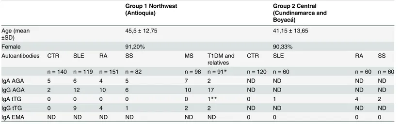
Table 1. Characteristics and results of the antibodies in two populations.
Group 1 Northwest (Antioquia)
Group 2 Central (Cundinamarca and Boyacá)
Age (mean ±SD)
45,5±12,75 41,15±13,65
Female 91,20% 90,33%
Autoantibodies CTR SLE RA SS MS T1DM and
relatives
CTR SLE RA SS
n = 140 n = 119 n = 151 n = 82 n = 98 n = 91* n = 120 n = 60 n = 60 n = 60
IgA AGA 5 6 4 5 7 2 ND ND ND ND
IgG AGA 2 12 10 6 10 17 ND ND ND ND
IgA tTG 0 0 0 0 0 1** 0 1 4 2
IgG tTG 0 9 4 1 2 2 ND ND ND ND
IgA EMA ND ND ND ND ND ND 0 0 0 0
*67 children with T1DM and 24first-degree relatives.
**T1DM Relative.
EMA, using two additional commercial kits (INOVA Diagnostics, USA. Cat. 508154, and
AESKU.Diagnostics, Germany. Cat. 512.050).
Search strategy
The search was done using the following databases: PubMed, Cochrane, Scopus, SciELO, and
Virtual Health Library, which includes BIREME, LILACS and many other LA sources. The
search was related to CD in LA and included articles published up to July 2013. The PRISMA
guidelines were followed during data extraction, analysis, and reporting [
47
].
No limits regarding language, publication type, or publication period were taken into
ac-count. The search was done with the following MeSH terms (Medical Subject Headings) and
key words in PubMed, Scopus and Chocrane:
celiac disease
,
transglutaminase antibody
,
anti-gliadin antibody
,
gliadin antibody
,
deamidated gliadin antibody
,
endomysial antibody
,
anti
endomysium antibody
,
HLA-DQ2
and
HLA-DQ8
. Each one was cross-referenced with the
fol-lowing MeSH terms:
Latin America
,
Hispanic Americans
,
Hispanics
,
South America
,
Argentina
,
Belize
,
Bolivia
,
Brazil
,
Chile
,
Colombia
,
Costa Rica
,
Cuba
,
Dominican Republic
,
Ecuador
,
El
Sal-vador
,
Guatemala
,
Haiti
,
Honduras
,
French Guiana
,
Mexico
,
Nicaragua
,
Panama
,
Paraguay
,
Peru
,
Puerto Rico
,
Surinam
,
Uruguay
, and
Venezuela
.
The same methodology and the term
celiac disease
(without other cross terms) were used to
explore sources of information in Spanish, Portuguese, and English through the SciELO and
Virtual Health Library databases. Each MeSH term and keyword was translated into DeCS
(Health Sciences Descriptors).
Study selection, data extraction, and quality assessment
Inclusion criteria for the systematic review were the following: (a) studies with screening tests
for autoantibodies of CD such as AGA, EMA, tTG, or deamidated gliadin peptide (DGP) in
healthy individuals or in patients without CD diagnosis; (b) studies with screening tests for
au-toantibodies of CD and a positive biopsy in healthy individuals or in patients without CD
diag-nosis; (c) studies that include the LA population. Studies were excluded if they were reviews or
case reports or if they discussed topics not related to CD. Unpublished data were also excluded.
A primary reviewer who screened all of the titles and abstracts from the publications performed
an eligibility assessment. Retrieved articles were rejected if the eligibility criteria were not met.
Also, a secondary reviewer was consulted when eligibility criteria were unclear. References
from the articles that seemed to be relevant for our review were hand-searched.
The extracted data from each article were: author name, country where the study took place,
year of publication, study design, number of patients, screening protocol and evaluated
out-comes. Several studies used different screening protocols to evaluate the presence of
autoanti-bodies. Therefore, we considered the results to be positives based on the cutoff of each protocol.
All articles were assessed according to the Oxford Center for Evidence based Medicine: 2011
Levels of Evidence [
48
].
Meta-regression
A population variable was created which addressed the nature of the cases in each article:
Pop-ulation A: Healthy individuals; PopPop-ulation B: First-degree relatives of CD patients; PopPop-ulation
C: T1DM (type 1 diabetes mellitus) patients; Population D: Patients with other ADs;
Popula-tion E: Patients with other condiPopula-tions. Studies published previously with the same populaPopula-tion
were excluded. The data obtained in the present study were involved in the meta-analyses.
diagnostic protocols separately; the first set were studies that evaluated the presence of IgA tTG
and EMA (sensitivity and specificity of 98
–
100%), and the second set were the studies that
eval-uated the positive autoantibodies and positive biopsy, following the statistical model-building
approach described below.
The meta-analysis was performed for each diagnostic protocol separately, by fitting a
meta-re-gression model with random effects and testing different combinations of predictor variables
available in all articles: a) year of study, b) country and c) population (nature of cases in each
study, as described previously) to explain the log-prevalence of the disease. The final predictors
included in the two meta-regression models (one for each diagnostic protocol) were selected
ac-cording to the likelihood ratio test and AIC criteria as described in [
49
]. Over the selected model,
routine diagnostic tests of meta-analysis were performed (test for funnel plot asymmetry,
I
2, H
2,
among others). The analysis was performed with the R2.15.2 package METAFOR [
49
].
Results
Colombian population
In Group 1, IgA tTG was positive only in one relative of a T1DM patient. In Group 2, seven
in-dividuals were positive or weakly positive for h-tTG using the reference values from the
com-mercial Kit. In all of these cases, the evaluation of IgA EMA was negative (confirmed by two
different kits). These results are shown in
Table 1
.
Systematic Literature Review
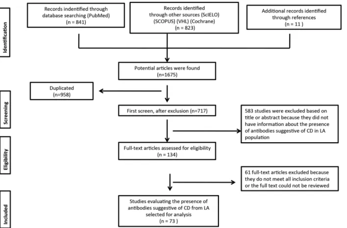
We identified 841 articles in the PubMed database search. Additional records identified through
other sources included 823 articles (Scopus, SciELO, Virtual Health Library and Cochrane).
Elev-en additional records were idElev-entified through hand searching. The database searches provided a
total of 1,675 publications. Of these, 958 were identified as duplicates. A total of 717 full text
arti-cles were assessed for eligibility. Finally, 73 artiarti-cles that contained interpretable data and fulfilled
the eligibility criteria were included [
50
–
122
]. In one paper, the data extraction was made from
its abstract [
50
]. Eight articles were from Argentina [
51
–
58
], 41 from Brazil [
50
,
59
–
98
], 3 from
Chile [
99
–
101
], 11 from Cuba [
102
–
112
], 5 from Mexico [
113
–
117
], 2 from Peru [
118
,
119
], 2
from Venezuela [
120
,
121
] and one paper was from the Hispanic residents in the United States
[
122
]. The flowchart for systematic literature review and articles included in the analysis is
shown in
Fig 1
. Detailed information is shown in
S1 Table
.
Meta-analysis
Meta-regression for tTG and EMA protocol.
A total of 27 studies comprising 28 articles
were included in the model [
50
,
52
,
54
,
55
,
66
,
69
,
71
–
74
,
78
,
80
,
82
,
83
,
85
–
87
,
89
,
90
,
94
,
97
,
100
,
114
,
116
–
118
,
121
,
122
]. The work of Remes-troche,
et al
. was analyzed as one study because they evaluate
the presence of CD in the same population, but in two different time periods [
114
,
117
]. In five
studies obtained from the systematic review, the population was divided into subpopulations
[
66
,
69
,
78
,
89
,
90
]. In addition, our two subpopulations from the central area of Colombia were
in-cluded in the analysis (i.e., healthy individuals and patients with ADs). In summary, 34
popula-tions were analyzed (
S1 Fig
).
Fig 1. Flow chart of the systemic literature review.VHL: virtual health library. CD: Celiac disease. LA: Latin America. doi:10.1371/journal.pone.0124040.g001
Table 2. Meta-regression model for tTG and EMA protocol.
estimate s.d. pval 95% C.I.
intrcpt (Brazil & Pop. A) -5.0462 0.2455 <.0001 -5.5275 -4.565
Argentina 0.3878 0.4508 0.3897 -0.4958 1.2714
Chile 0.5883 1.4914 0.6932 -2.3348 3.5114
Colombia -0.6819 1.1855 0.5652 -3.0053 1.6416
Mexico 0.1383 0.5408 0.7981 -0.9216 1.1983
Peru 0.0824 1.1771 0.9442 -2.2246 2.3895
USA -2.7854 1.1725 0.0175 -5.0835 -0.4872
Population B 2.1509 0.3873 <.0001 1.3919 2.91
Population C 2.6059 0.4567 <.0001 1.7109 3.501
Population D 0.0759 0.9788 0.9382 -1.8425 1.9943
Population E 1.5298 0.3882 <.0001 0.7689 2.2907
estimated tau: 0.5612 I^2: 66.14% H^2: 2.95
Test for Residual Heterogeneity: p-val<.0001 Test of Moderators: p-val<.0001
(Prevalence in log-scale). Population: A: Healthy individuals; B: First-degree relatives of CD patients; C: T1DM patients; D: Patients with other ADs; E: Patients with other conditions.
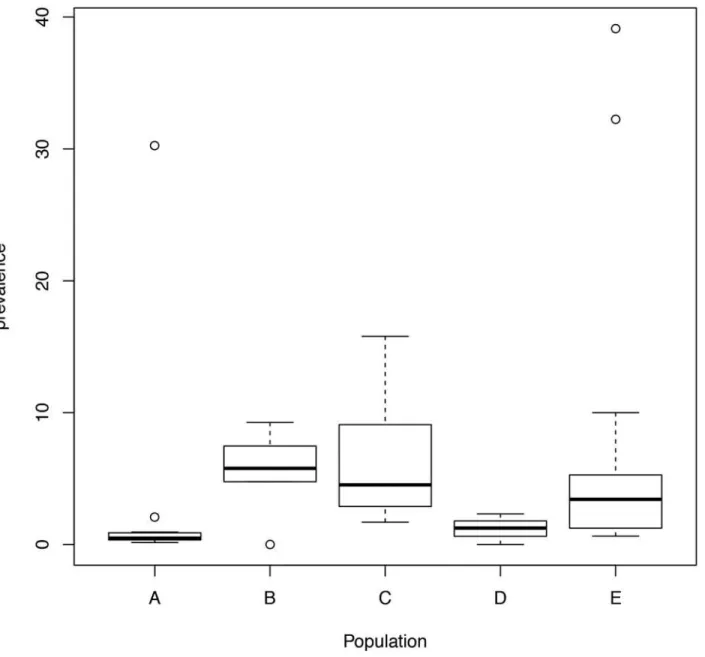
The parameters associated with countries and populations refer to the log-prevalence
be-tween various country and population combinations when compared against Brazil, regarding
population A (healthy individuals). The intercept parameter was the log-prevalence for
Brazil-ian individuals regarding controls. Note that this BrazilBrazil-ian population was chosen as a
refer-ence level due to its large number of studies. Taking these parameters into account, only
Hispanics from the USA had a log-prevalence less than that of the Brazilians, despite the
differ-ent population groups that were analyzed. However, populations B, C and E had a higher
prev-alence of the disease than population A. Furthermore, the observed parameters of population
D were not significant. This suggests that the prevalence of the disease is higher in individuals
with first-degree relatives diagnosed with CD and T1DM (
Fig 2
and
S1 Fig
).
Fig 2. Boxplot of the prevalence of the disease for each population using the model for tTG and EMA protocol.Population: A: Healthy individuals; B: First-degree relatives of CD patients; C: T1DM patients; D: Patients with other ADs; E: Patients with other conditions.
We did not find any evidence of publication bias (test for funnel plot asymmetry: z = -1.8174,
p = 0.0692). Additionally, two subpopulations in two different studies presented lack of fit to this
model [
64
,
66
] (
S2 Table
and
S3 Fig
). This discrepancy could be explained by the characteristic
of the studied population (i.e., pediatric inpatients and outpatients).
Meta-regression for autoantibodies positive and biopsy positive protocol.
A total of
49 studies were included in this model [
50
,
52
–
55
,
58
–
73
,
75
,
77
,
79
–
81
,
83
,
85
,
88
,
90
,
94
,
96
,
99
–
102
,
104
,
107
,
109
–
113
,
116
–
121
]. Within three studies obtained from the systematic review,
the population was divided into subpopulations. As a result, 51 populations were analyzed.
Again, the most parsimonious model (according to the AIC criteria and likelihood ratio
tests) was a meta-regression with random effects. As was the case with the previous diagnostic
protocol, the only moderators in this model were the population and country variables.
There-fore, interpretation of meta-regression parameters was the same as before.
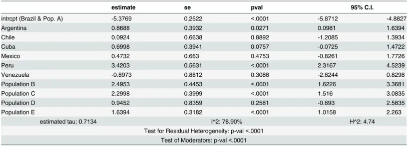
Table 3
shows the
estimated parameters of the selected model.
Populations from Peru and Argentina have a higher log-prevalence than Brazilian
individu-als, whilst populations B, C and E had a higher prevalence of the disease than population A.
Furthermore, the observed parameters of population D were not significant. As previously
found in the tTG and EMA protocol, the prevalence of the disease was higher in individuals
with first-degree relatives diagnosed with CD and T1DM (
Fig 3
and
S2 Fig
).
Note that we found evidence of publication bias (test for funnel plot asymmetry: z = -3.4489,
p = 0.0006). Additionally, two subpopulations in two different studies exhibited a lack of fit to
the model [
58
,
66
] (
S3 Table
and
S4 Fig
). This discrepancy could once again be explained by the
characteristic of the studied population (pediatric inpatients and patients with clinical
suspi-cion), as suggested in the meta-regression analysis for the tTG and EMA protocol.
Discussion
Although CD seems to be a rare condition in Colombians; the general prevalence of the disease
in Latin Americans seemingly corresponds to a similar scenario observed in Europeans (0.46%
Table 3. Meta-regression model for autoantibodies positive and biopsy positive.
estimate se pval 95% C.I.
intrcpt (Brazil & Pop. A) -5.3769 0.2522 <.0001 -5.8712 -4.8827
Argentina 0.8688 0.3932 0.0271 0.0981 1.6394
Chile 0.0924 0.6638 0.8892 -1.2085 1.3934
Cuba 0.6998 0.3941 0.0757 -0.0725 1.4722
Mexico 0.4732 0.663 0.4753 -0.8261 1.7726
Peru 3.4203 0.5631 <.0001 2.3167 4.5239
Venezuela -0.8973 0.8812 0.3086 -2.6244 0.8298
Population B 2.4953 0.4453 <.0001 1.6226 3.3681
Population C 2.2998 0.3999 <.0001 1.516 3.0835
Population D 0.9452 0.8359 0.2581 -0.693 2.5835
Population E 1.6394 0.3182 <.0001 1.0158 2.263
estimated tau: 0.7134 I^2: 78.90% H^2: 4.74
Test for Residual Heterogeneity: p-val<.0001 Test of Moderators: p-val<.0001
(Prevalence in log-scale). Population: A: Healthy individuals; B: First-degree relatives of CD patients; C: T1DM patients; D: Patients with other ADs; E: Patients with other conditions.
to 0.64%) [
123
,
124
]. Our study is the first to summarize and analyze all published studies
about CD in the LA population. The presence of the disease was evaluated in a population with
different characteristics, and we found 801 patients positive for IgA tTG and IgA EMA and 454
patients positive for autoantibodies and biopsy (
S1 Table
).
The LA population presents with a notable racial, genetic, and cultural diversity [
17
].
There-fore in LA, some countries such as Argentina, Brazil, Chile, Cuba, Uruguay and Venezuela,
have
>
50% of Caucasian component, and certain regions within these nations are more
Cau-casian than others [
111
,
125
]. The highest prevalence of CD has been reported in those
coun-tries with a high Caucasian admixture (
S1 Table
). However, in nations such as Chile, Uruguay
and Venezuela, the prevalence of CD is uncertain. Brazil is the only country where the presence
of CD has been widely studied. This country is an important migratory destination for
Fig 3. Boxplot of the prevalence of the disease for each population using the model for autoantibodies positive and biopsy positive.Population: A: Healthy individuals; B: First-degree relatives of CD patients; C: T1DM patients; D: Patients with other ADs; E: Patients with other conditions.
European Caucasians. These immigrants are located primarily in the Southern region of Brazil,
and most cases of CD have been reported in this region. In contrast, the North and the
North-east region of Brazil have large Amerindian and African ancestral influences respectively. In
those regions the presence of the disease is low [
126
]. In addition, the presence of the disease in
native Indian and African-derived communities from the Northeast Brazilian region is null
[
84
,
95
] (
S1 Table
).
Our meta-regressions also show that Hispanics in the United States (i.e., a person of
Mexi-can, Puerto RiMexi-can, Cuban, South or Central American (except for Brazil), or other Spanish
cul-ture or origin, regardless of race) have a different behavior than individuals of other countries.
The prevalence of CD in Hispanic was estimated in 2.519 individuals (1.686 Mexican
Ameri-cans and 833 as other Hispanic groups). Only one patient was positively detected by
autoanti-bodies (IgA tTG and IgA EMA), whilst the other two patients were detected by interview data.
However, none of these patients were Mexican-American [
122
]. Nevertheless, these results are
very different from those observed in the Mexican population (Tables
1
and
2
,
S1 Table
, Figs
2
and
3
, and
S1
and
S2
Figs). Thus, this discrepancy in the prevalence on native Mexicans vs.
Mexican Americans highlights the major effect of environmental factors over genetic factors in
the risk of developing CD.
Polyautoimmunity (i.e., the presence of two or more ADs in a single patient) [
19
] was
ob-served in our study. The estimated prevalence of CD in the T1DM patients was 4.6 to 8.7%.
This number is similar to the prevalence reported in other populations [
127
]. The association
among CD and T1DM can be explained by the presence of common risk alleles including
HLA-DQA1
0501 and DQB1
0201 [
128
]. However, the estimated prevalence of CD in the
group of patients with ADs was 0.7% to 1%. This surprisingly low prevalence may be explained
by the small number of studies available and the lack of awareness about polyautoimmunity
[
98
]. In fact, most of the studies evaluating this association came from case reports or from
studies with a small sample size (
S1 Table
).
Both familial CD [
129
] and familial autoimmunity (i.e., the presence of diverse ADs in
rela-tives of CD probands) are frequent conditions [
130
,
131
], indicating aggregation of the
autoim-mune trait. The prevalence of CD in first-degree and second-degree relatives is 10% and 5%
respectively [
132
]. In our results, the estimated prevalence of CD in first-degree relatives was
about 5.6%, whereas the estimated prevalence in populations with other conditions, such as
at-risk populations, was 0.24% to 0.3% (
S1 Table
). This result could be explained by the
heteroge-neity of the study population and the small sample size.
Although the two Colombian population analyzed in this study may disclose a different
ge-netic background [
133
], no celiac autoimmunity was observed in neither. Colombians in
gen-eral still eat cultivated food, such as potatoes, tapioca and corn [
134
]. The seven individuals
with positive or weakly positive results from Central Colombia were interpreted as false
posi-tives, because the IgA EMA test was negative. In fact, false positive results for the IgA tTG have
been also reported in other ADs [
6
,
135
].
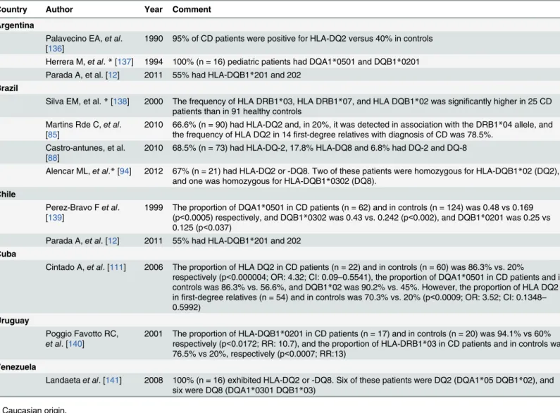
During the search, we found that some studies have reported the presence of HLA-DQ2
and HLA-DQ8 haplotype in the LA population with high titers of CD autoantibodies and
diag-nosis of CD (
Table 4
) [
12
,
85
,
88
,
111
,
136
–
141
]. HLA DQ2.5 is common in the LA population,
whilst approximately 90% of studied Amerindian individuals carry the DQ8 haplotype
[
14
,
142
,
143
].
Study Limitations
previously evaluated are now considered obsolete. For this reason, some older studies were
excluded from the statistical model. Second, in some studies, the biopsy was not performed
in all antibody-positive individuals. Also, in some studies, the biopsy was not obtained from
the duodenum or the interpretation was not performed with the respective histological
classi-fication. Thus, in order to obtain accurate results, data from those studies were not included
in the statistical model. Third, different ELISA protocols for testing the presence of tTG were
used across the studies, so the dilution of positive IgA EMA was different among our
analy-ses. Fourth, most of the data about CD came from populations at high risk. Therefore the
prevalence might be overestimated.
Data from Colombia were not considered for estimation of disease prevalence. In addition,
the meta-regression has some limitations, because the country effect is a confounding factor in
the analysis of populations. All the included studies did not represent the whole country
’
s
pop-ulation where they were performed.
Table 4. Presence of HLA in celiac disease patients.
Country Author Year Comment
Argentina
Palavecino EA,et al.
[136] 1990 95% of CD patients were positive for HLA-DQ2 versus 40% in controls Herrera M,et al.*[137] 1994 100% (n = 16) pediatric patients had DQA1*0501 and DQB1*0201
Parada A, et al. [12] 2011 55% had HLA-DQB1*201 and 202 Brazil
Silva EM, et al.*[138] 2000 The frequency of HLA DRB1*03, HLA DRB1*07, and HLA DQB1*02 was significantly higher in 25 CD patients than in 91 healthy controls
Martins Rde C,et al.
[85] 2010 66.6% (n = 90) had HLA-DQ2 and, in 20%, it was detected in association with the DRB1the frequency of HLA DQ2 in 14first-degree relatives with diagnosis of CD was 78.5%. *04 allele, and Castro-antunes, et al.
[88] 2010 68.5% (n = 73) had HLA-DQ-2, 17.8% HLA-DQ8 and 6.8% had DQ-2 and DQ-8
Alencar ML,et al.*[94] 2012 67% (n = 21) had HLA-DQ2 or -DQ8. Two of these patients were homozygous for HLA-DQB1*02 (DQ2),
and one was homozygous for HLA-DQB1*0302 (DQ8). Chile
Perez-Bravo Fet al.
[139] 1999 The proportion of DQA1(p<0.0005) respectively, and DQB1*0501 in CD patients (n = 62) and in controls (n = 124) was 0.48 vs 0.169*0302 was 0.43 vs. 0.242 (p<0.002), and DQB1*0201 was 0.25 vs 0.125 (p<0.037)
Parada A,et al. [12] 2011 55% had HLA-DQB1*201 and 202
Cuba
Cintado A,et al. [111] 2006 The proportion of HLA DQ2 in CD patients (n = 22) and in controls (n = 60) was 86.3% vs. 20%
respectively (p<0.000004; OR: 4.32; CI: 0.09–0.5541), the proportion of DQA1*0501 in CD patients and in controls was 86.3% vs. 56.6%, and DQB1*02 was 90.2% vs. 45%. However, the proportion of HLA DQ2 infirst-degree relatives (n = 54) and in controls was 70.3% vs. 20% (p<0.0009; OR: 3.52; CI: 0.1348– 0.5992)
Uruguay
Poggio Favotto RC,
et al. [140]
2001 The proportion of HLA-DQB1*0201 in CD patients (n = 17) and in controls (n = 20) was 94.1% vs 60% respectively (p<0.0172; RR: 10.7), and the proportion of HLA-DRB1*03 in CD patients and in controls was 76.5% vs 20%, respectively (p<0.0007; RR:13)
Venezuela
Landaetaet al. [141] 2008 100% (n = 16) exhibited HLA-DQ2 or -DQ8. Six of these patients were DQ2 (DQA1*05 DQB1*02), and
six were DQ8 (DQA1*0301 DQB1*03)
*Caucasian origin.
Conclusions
In general the prevalence of CD in LA is similar to that reported in Europeans. CD in the LA
population is frequent and is primarily reported in populations and regions with Caucasian
an-cestry. Nevertheless, in certain countries with substantial Caucasian ancestry such as Uruguay,
the prevalence is unknown.
The low prevalence reported in some regions could be explained mainly by: the lack of
knowledge of the disease, low gluten consumption (which does not entail an autoimmune
re-sponse), changes in quantity and quality of cereal processing, microbiota or early childhood
ex-posure to infectious agents that could impair the natural development of the immune system
(i.e., hygiene hypothesis) [
144
].
Supporting Information
S1 Fig. Forest plot. Prevalence of the disease for each population using the model for tTG
and EMA protocol.
Population: A: Healthy individuals; B: First-degree relatives of CD
pa-tients; C: T1DM papa-tients; D: Patients with other ADs; E: Patients with other conditions.
(TIF)
S2 Fig. Forest plot. Prevalence of the disease for each population using the model for
auto-antibodies positive and biopsy positive.
Population: A: Healthy individuals; B: First-degree
relatives of CD patients; C: T1DM patients; D: Patients with other ADs; E: Patients with
other conditions.
(TIF)
S3 Fig. Funnel plot of the model for tTG and EMA protocol.
(TIF)
S4 Fig. Funnel plot for autoantibodies positive and biopsy positive.
(TIF)
S1 PRISMA Checklist. PRISMA checklist.
(DOC)
S1 Table. Presence of autoantibodies positive and biopsy positive in Latin American
pa-tients.
Abbreviations: AGA: Antigliadin antibodies; CD: Celiac disease; CMV:
Cytomegalovi-rus; DGP: deamidated gliadin peptide; EBV: Epstein barr viCytomegalovi-rus; EMA: anti-endomysium
antibody; GFD: Gluten free diet; HCV: Hepatitis C virus; HEV: Hepatitis E virus; JRA: juvenile
rheumatoid arthritis; MS: multiple sclerosis; N/A: Not available; T1DM: Type 1 Diabetes
Melli-tus; T2DM: Type 2 Diabetes MelliMelli-tus; tTG: anti-tissue transglutaminase antibody; RA:
Rheu-matoid arthritis; SLE: Systemic Lupus Erythematosus; SS: Sjögren syndrome.
€
: presumptive
celiac disease patients.
(DOC)
S2 Table. Studies with lack of fit to the final model (tTG and EMA protocol).
(DOC)
S3 Table. Studies with lack of fit to the final model (positive and biopsy positive).
(DOC)
Acknowledgments
Colombiana de Celiacos
”
and its president Ximena Guerrero for her continued support and
as-sistance in the searching of patients with possible CD in Colombians.
Author Contributions
Conceived and designed the experiments: JMA. Performed the experiments: RPM JMA NAL
MTA ARV NMG YS. Analyzed the data: NMG RPM MTA ARV JMA. Contributed reagents/
materials/analysis tools: JMA YS NAL ARV NMG RPM. Wrote the paper: RPM NMG ARV
MTA NAL YS JMA.
References
1. Schuppan D, Dennis MD, Kelly CP. Celiac disease: epidemiology, pathogenesis, diagnosis, and nutri-tional management. Nutr Clin care. 2005; 8: 54–69. PMID:16013224
2. Rubio-Tapia A, Hill ID, Kelly CP, Calderwood AH, Murray JA, American College of Gastroenterology. ACG clinical guidelines: diagnosis and management of celiac disease. Am J Gastroenterol. 2013; 108: 656–676. doi:10.1038/ajg.2013.79PMID:23609613
3. Ludvigsson JF, Bai JC, Biagi F, Card TR, Ciacci C, Ciclitira PJ, et al. Diagnosis and management of adult coeliac disease: guidelines from the British Society of Gastroenterology. Gut. 2014; 63: 1210–
1228. doi:10.1136/gutjnl-2013-306578PMID:24917550
4. Meeuwisse GW. Diagnostic criteria in coeliac disease. Acta Paediatr Scand. 1970; 59: 461–463.
5. McNeish AS Harms HK Rey J Shmerling DH Visakorpi JK Walker-Smith J. The diagnosis of coeliac disease. Arch Dis Child. 1979; 54: 783–786. PMID:507902
6. Husby S, Koletzko S, Korponay-Szabó IR, Mearin ML, Phillips a, Shamir R, et al. European Society for Pediatric Gastroenterology, Hepatology, and Nutrition guidelines for the diagnosis of coeliac dis-ease. J Pediatr Gastroenterol Nutr. 2012; 54: 136–160. doi:10.1097/MPG.0b013e31821a23d0
PMID:22197856
7. Admou B, Essaadouni L, Krati K, Zaher K, Sbihi M, Chabaa L, et al. Atypical Celiac Disease: From Recognizing to Managing. Gastroenterol Res Pract. 2012; 2012: 637187. doi:10.1155/2012/637187
PMID:22811701
8. Petaros P, Martelossi S, Tommasini a, Torre G, Caradonna M, Ventura A. Prevalence of autoimmune disorders in relatives of patients with celiac disease. Dig Dis Sci. 2002; 47: 1427–1431. PMID:
12141796
9. Rostom A, Dubé C, Cranney A, Saloojee N, Sy R, Garritty C, et al. The diagnostic accuracy of serolog-ic tests for celiac disease: A systematserolog-ic review. Gastroenterology. 2005; 128: S38–S46. PMID:
15825125
10. Ludvigsson JF, Leffler DA, Bai JC, Biagi F, Fasano A, Green PHR, et al. The Oslo definitions for coeli-ac disease and related terms. Gut. 2013; 62: 43–52. doi:10.1136/gutjnl-2011-301346PMID:
22345659
11. Greco L. Epidemiology of coeliac disease. Proceedings of the Seventh International Symposium on Coeliac Disease Tampere: Finland; 1996.
12. Parada A, Araya M, Pérez-Bravo F, Méndez M, Mimbacas A, Motta P, et al. Amerindian mtDNA hap-logroups and celiac disease risk HLA haplotypes in mixed-blood Latin American patients. J Pediatr Gastroenterol Nutr. 2011; 53: 429–434. doi:10.1097/MPG.0b013e31821de3fcPMID:21505366
13. Brar P, Lee AR, Lewis SK, Bhagat G, Green PHR. Celiac disease in African-Americans. Dig Dis Sci. 2006 May; 51: 1012–5. PMID:16642428
14. Gujral N, Freeman HJ, Thomson ABR. Celiac disease: prevalence, diagnosis, pathogenesis and treatment. World J Gastroenterol. 2012; 18: 6036–6059. doi:10.3748/wjg.v18.i42.6036PMID:
23155333
15. Lionetti E, Catassi C. New clues in celiac disease epidemiology, pathogenesis, clinical manifesta-tions, and treatment. Int Rev Immunol. 2011; 30: 219–231. doi:10.3109/08830185.2011.602443
PMID:21787227
16. González Burchard E, Borrell LN, Choudhry S, Naqvi M, Tsai H-J, Rodriguez-Santana JR, et al. Latino populations: a unique opportunity for the study of race, genetics, and social environment in epidemio-logical research. Am J Public Health. 2005; 95: 2161–2168. PMID:16257940
18. Dahlbom I, Korponay-Szabó IR, Kovács JB, Szalai Z, Mäki M, Hansson T. Prediction of clinical and mucosal severity of coeliac disease and dermatitis herpetiformis by quantification of IgA/IgG serum antibodies to tissue transglutaminase. J Pediatr Gastroenterol Nutr. 2010; 50: 140–146. doi:10.1097/ MPG.0b013e3181a81384PMID:19841593
19. Anaya JM. The diagnosis and clinical significance of polyautoimmunity. Autoimmun Rev. 2014; 13:423–426. doi:10.1016/j.autrev.2014.01.049PMID:24424171
20. Arnett FC, Edworthy SM, Bloch DA, McShane DJ, Fries JF, Cooper NS, et al. The American Rheuma-tism Association 1987 revised criteria for the classification of rheumatoid arthritis. Arthritis Rheum. 1988; 31: 315–324. PMID:3358796
21. Tan EM, Cohen AS, Fries JF, Masi AT, McShane DJ, Rothfield NF, et al. The 1982 revised criteria for the classification of systemic lupus erythematosus. Arthritis Rheum. 1982; 25: 1271–1277. PMID: 7138600
22. McDonald WI, Compston A, Edan G, Goodkin D, Hartung HP, Lublin FD, et al. Recommended diag-nostic criteria for multiple sclerosis: guidelines from the International Panel on the diagnosis of multiple sclerosis. Ann Neurol. 2001; 50: 121–127. PMID:11456302
23. Report of the Expert Committee on the Diagnosis and Classification of Diabetes Mellitus. Diabetes Care. 1997; 20: 1183–1197. PMID:9203460
24. Vitali C, Bombardieri S, Jonsson R, Moutsopoulos HM, Alexander EL, Carsons SE, et al. Classifica-tion criteria for Sjögren’s syndrome: a revised version of the European criteria proposed by the Ameri-can-European Consensus Group. Ann Rheum Dis. 2002; 61: 554–558. PMID:12006334
25. Anaya J-M, Castiblanco J, Tobón GJ, García J, Abad V, Cuervo H, et al. Familial clustering of autoim-mune diseases in patients with type 1 diabetes mellitus. J Autoimmun. 2006; 26: 208–214. PMID:
16503115
26. Tobon GJ, Arango a, Abad V, García J, Cuervo H, Velásquez a, et al. Clinical and immunological characteristics of type 1 diabetes mellitus in a northwestern Colombian population. Diabetes Res Clin Pract. 2006; 72: 170–175. PMID:16325957
27. Krause I, Anaya JM, Fraser A, Barzilai O, Ram M, Abad V, et al. Anti-infectious antibodies and autoim-mune-associated autoantibodies in patients with type I diabetes mellitus and their close family mem-bers. Ann N Y Acad Sci. 2009; 1173: 633–639. doi:10.1111/j.1749-6632.2009.04619.xPMID: 19758209
28. Shor DB-A, Barzilai O, Ram M, Izhaky D, Porat-Katz BS, Chapman J, et al. Gluten sensitivity in multi-ple sclerosis: experimental myth or clinical truth? Ann N Y Acad Sci. 2009; 1173: 343–9. doi:10.1111/ j.1749-6632.2009.04620.xPMID:19758171
29. Amaya-Amaya J, Sarmiento-Monroy JC, Caro-Moreno J, Molano-González N, Mantilla RD, Rojas-Villarraga A, et al. Cardiovascular disease in latin american patients with systemic lupus erythemato-sus: a cross-sectional study and a systematic review. Autoimmune Dis. 2013; 2013: 794383. doi:10. 1155/2013/794383PMID:24294522
30. Barragán-Martínez C, Amaya-Amaya J, Pineda-Tamayo R, Mantilla RD, Castellanos-de la Hoz J, Bernal-Macías S, et al. Gender differences in Latin-American patients with rheumatoid arthritis. Gend Med. 2012; 9: 490–510. doi:10.1016/j.genm.2012.10.005PMID:23217568
31. Cárdenas Roldán J, Amaya-Amaya J, Castellanos-de la Hoz J, Giraldo-Villamil J, Montoya-Ortiz G, Cruz-Tapias P, et al. Autoimmune thyroid disease in rheumatoid arthritis: a global perspective. Arthri-tis. 2012; 2012: 864907. doi:10.1155/2012/864907PMID:23209899
32. Amador-Patarroyo MJ, Arbelaez JG, Mantilla RD, Rodriguez-Rodriguez A, Cárdenas-Roldán J, Pineda-Tamayo R, et al. Sjögren’s syndrome at the crossroad of polyautoimmunity. J Autoimmun. 2012; 39: 199–205. doi:10.1016/j.jaut.2012.05.008PMID:22749530
33. Pérez-Fernández OM, Mantilla RD, Cruz-Tapias P, Rodriguez-Rodriguez A, Rojas-Villarraga A, Anaya J-M. Spondyloarthropathies in autoimmune diseases and vice versa. Autoimmune Dis. 2012; 2012: 736384. doi:10.1155/2012/736384PMID:22400103
34. Anaya J-M, Cañas C, Mantilla RD, Pineda-Tamayo R, Tobón GJ, Herrera-Diaz C, et al. Lupus nephri-tis in Colombians: contrasts and comparisons with other populations. Clin Rev Allergy Immunol. 2011; 40: 199–207. doi:10.1007/s12016-010-8249-4PMID:21287296
35. Franco J-S, Molano-González N, Rodríguez-Jiménez M, Acosta-Ampudia Y, Mantilla RD, Amaya-Amaya J, et al. The coexistence of antiphospholipid syndrome and systemic lupus erythematosus in colombians. PLoS One. 2014; 9: e110242. doi:10.1371/journal.pone.0110242PMID:25343509
36. Shapira Y, Agmon-Levin N, Selmi C, Petríková J, Barzilai O, Ram M, et al. Prevalence of anti-Toxoplasma antibodies in patients with autoimmune diseases. J Autoimmun. 2012; 39: 112–116. doi:
37. Shapira Y, Poratkatz B-S, Gilburd B, Barzilai O, Ram M, Blank M, et al. Geographical differences in autoantibodies and anti-infectious agents antibodies among healthy adults. Clin Rev Allergy Immunol. 2012; 42: 154–163. doi:10.1007/s12016-010-8241-zPMID:21229335
38. Orbach H, Amitai N, Barzilai O, Boaz M, Ram M, Zandman-Goddard G, et al. Autoantibody screen in inflammatory myopathies high prevalence of antibodies to gliadin. Ann N Y Acad Sci. 2009; 1173: 174–179. doi:10.1111/j.1749-6632.2009.04810.xPMID:19758147
39. Berkun Y, Zandman-Goddard G, Barzilai O, Boaz M, Sherer Y, Larida B, et al. Infectious antibodies in systemic lupus erythematosus patients. Lupus. 2009; 18: 1129–1135. doi:10.1177/
0961203309345729PMID:19880558
40. Zandman-Goddard G, Berkun Y, Barzilai O, Boaz M, Ram M, Anaya JM, et al. Neuropsychiatric lupus and infectious triggers. Lupus. 2008; 17: 380–384. doi:10.1177/0961203308090017PMID:18490412
41. Pordeus V, Barzilai O, Sherer Y, Luiz RR, Blank M, Bizzaro N, et al. A latitudinal gradient study of common anti-infectious agent antibody prevalence in Italy and Colombia. Isr Med Assoc J. 2008; 10: 65–68. PMID:18300578
42. Ram M, Anaya J-M, Barzilai O, Izhaky D, Porat Katz B-S, Blank M, et al. The putative protective role of hepatitis B virus (HBV) infection from autoimmune disorders. Autoimmun Rev. 2008; 7: 621–625. doi:10.1016/j.autrev.2008.06.008PMID:18603025
43. Barzilai O, Sherer Y, Ram M, Izhaky D, Anaya JM, Shoenfeld Y. Epstein-Barr virus and cytomegalovi-rus in autoimmune diseases: are they truly notorious? A preliminary report. Ann N Y Acad Sci. 2007; 1108: 567–577. PMID:17894021
44. Meron MK, Amital H, Shepshelovich D, Barzilai O, Ram M, Anaya J-M, et al. Infectious aspects and the etiopathogenesis of rheumatoid arthritis. Clin Rev Allergy Immunol. 2010; 38: 287–291. doi:10. 1007/s12016-009-8158-6PMID:19575154
45. Altman A, Szyper-Kravitz M, Agmon-Levin N, Gilburd B, Anaja J-M, Barzilai O, et al. Prevalence of rubel-la serum antibody in autoimmune diseases. Rev Bras Reumatol. 2012; 52: 307–318. PMID:22641586
46. Zandman-Goddard G, Berkun Y, Barzilai O, Boaz M, Blank M, Ram M, et al. Exposure to Epstein-Barr virus infection is associated with mild systemic lupus erythematosus disease. Ann N Y Acad Sci. 2009; 1173: 658–663. doi:10.1111/j.1749-6632.2009.04754.xPMID:19758212
47. Liberati A, Altman DG, Tetzlaff J, Mulrow C, Gøtzsche PC, Ioannidis JPA, et al. The PRISMA state-ment for reporting systematic reviews and meta-analyses of studies that evaluate health care inter-ventions: explanation and elaboration. J Clin Epidemiol. 2009; 62: e1–34. doi:10.1016/j.jclinepi.2009. 06.006PMID:19631507
48. OCEBM Levels of Evidence Working Group. The Oxford 2011 levels of evidence. Oxford Cent Evi-dence-Based Med. 2011.
49. Viechtbauer W. Conducting Meta-Analyses in R with the metafor Package. J Stat Softw. 2010; 36:1–48. 50. Machado AP de SL, Silva LR, Zausner B, Oliveira J de A, Diniz DR, de Oliveira J. Undiagnosed celiac
disease in women with infertility. J Reprod Med. 2013; 58: 61–66. PMID:23447921
51. Gomez JC, Selvaggio GS, Viola M, Pizarro B, la Motta G, de Barrio S, et al. Prevalence of celiac dis-ease in Argentina: screening of an adult population in the La Plata area. Am J Gastroenterol. 2001; 96: 2700–2704. PMID:11569698
52. Gomez JC, Selvaggio G, Pizarro B, Viola MJ, La Motta G, Smecuol E, et al. Value of a screening algo-rithm for celiac disease using tissue transglutaminase antibodies as first level in a population-based study. Am J Gastroenterol. 2002; 97: 2785–2790. PMID:12425549
53. González D, Sugai E, Gomez JC, Oliveri MB, Gomez Acotto C, Vega E, et al. Is it necessary to screen for celiac disease in postmenopausal osteoporotic women? Calcif Tissue Int. 2002; 71: 141–144. PMID:12200648
54. Rumbo M, Chirdo FG, Ben R, Saldungaray I, Villalobos R. Evaluation of coeliac disease serological markers in Down syndrome patients. Dig Liver Dis. 2002; 34: 116–121. PMID:11926554
55. Mora M, Litwin N, Toca M del C, Azcona MI, Solís Neffa R, Ortiz G, et al. Prevalence of celiac disease: multicentric trial among pediatric population in five urban districts of Argentina. 2010; 1: 26–31. doi:
10.1590/S0325-00752012000600006PMID:23224306
56. Bustos D, Moret A, Tambutti M, Gogorza S, Testa R, Ascione A, et al. Autoantibodies in Argentine women with recurrent pregnancy loss. Am J Reprod Immunol. 2006; 55: 201–207. PMID:16451354
57. Begué C, Beratarrechea AG, Varela E, Piccioni HL, Rodota L, Castro ME, et al. Enfermedad celíaca: prevalencia del diagnóstico en un hospital de comunidad. Acta Gastroenterol Latinoam. 2010; 40: 317–322. PMID:21381406
59. Gandolfi L, Pratesi R, Cordoba JC, Tauil PL, Gasparin M, Catassi C. Prevalence of celiac disease among blood donors in Brazil. Am J Gastroenterol. 2000; 95: 689–692. PMID:10710058
60. Gandolfi L, Catassi C, Garcia S, Modelli IC, Campos D Jr, Pratesi R. Antiendomysial antibody test reli-ability in children with frequent diarrhea and malnutrition: is it celiac disease? J Pediatr Gastroenterol Nutr. 2001; 33: 483–487. PMID:11698768
61. Kotze LM, Utiyama SR, Nisihara RM, Zeni MP, de Sena MG, Amarante HM. Antiendomysium antibod-ies in Brazilian patients with celiac disease and their first-degree relatives. Arq Gastroenterol. 2001; 38: 94–103. PMID:11793949
62. Pratesi R, Gandolfi L, Garcia SG, Modelli IC, Lopes de Almeida P, Bocca AL, et al. Prevalence of coe-liac disease: unexplained age-related variation in the same population. Scand J Gastroenterol. 2003; 38: 747–750. PMID:12889561
63. Pratesi R, Gandolfi L, Martins RC, Tauil PL, Nobrega YK, Teixeira WA. Is the prevalence of celiac dis-ease incrdis-eased among epileptic patients? Arq Neuropsiquiatr. 2003; 61: 330–334. PMID:12894262
64. Brandt KG, Silva GAP, Antunes MMC. Celiac disease in a group of children and adolescents with type 1 diabetes mellitus. Arq Bras Endocrinol Metab. 2004; 48: 823–827. PMID:15761555
65. Queiroz MS, Nery M, Cancado EL, Gianella-Neto D, Liberman B. Prevalence of celiac disease in Bra-zilian children of short stature. Braz J Med Biol Res. 2004; 37: 55–60. PMID:14689044
66. Trevisiol C, Brandt KG, Silva GA, Crovella S, Ventura A. High prevalence of unrecognized celiac dis-ease in an unselected hospital population in north-eastern Brasil (Recife, Pernambuco). J Pediatr Gastroenterol Nutr. 2004; 39: 214–215. PMID:15269636
67. Baptista ML, Koda YK, Mitsunori R, Nisihara, Ioshii SO. Prevalence of celiac disease in Brazilian chil-dren and adolescents with type 1 diabetes mellitus. J Pediatr Gastroenterol Nutr. 2005; 41: 621–624.
PMID:16254520
68. Lima VM, Gandolfi L, Pires JA, Pratesi R. Prevalence of celiac disease in dyspeptic patients. Arq Gas-troenterol. 2005; 42: 153–156. PMID:16200250
69. Nisihara RM, Kotze LM, Utiyama SR, Oliveira NP, Fiedler PT, Messias-Reason IT. Celiac disease in children and adolescents with Down syndrome. J Pediatr (Rio J). 2005; 81: 373–376. PMID:16247538
70. Tanure MG, Silva IN, Bahia M, Penna FJ. Prevalence of celiac disease in Brazilian children with type 1 diabetes mellitus. J Pediatr Gastroenterol Nutr. 2006; 42: 155–159. PMID:16456407
71. Pereira MA, Ortiz-Agostinho CL, Nishitokukado I, Sato MN, Damiao AO, Alencar ML, et al. Prevalence of celiac disease in an urban area of Brazil with predominantly European ancestry. World J Gastroen-terol. 2006; 12: 6546–6550. PMID:17072989
72. Melo SB, Fernandes MI, Peres LC, Troncon LE, Galvao LC. Prevalence and demographic character-istics of celiac disease among blood donors in Ribeirao Preto, State of Sao Paulo, Brazil. Dig Dis Sci. 2006; 51: 1020–1025. PMID:16758312
73. De Bem RS, Da Ro Sa Utiyama SR, Nisihara RM, Fortunato JA, Tondo JA, Carmes ER, et al. Celiac disease prevalence in Brazilian dilated cardiomyopathy patients. Dig Dis Sci. 2006; 51: 1016–1019. PMID:16758314
74. Araújo J, Pontes Da Silva GA, De Melo FM. Serum prevalence of celiac disease in children and ado-lescents with type 1 diabetes mellitus. J Pediatr (Rio J). 2006; 82: 210–214. PMID:16729151
75. Crovella S, Brandao L, Guimaraes R, Filho JL, Arraes LC, Ventura A, et al. Speeding up coeliac dis-ease diagnosis in the developing countries. Dig Liver Dis. 2007; 39:900–902. PMID:17706474
76. Nisihara RM, Skare TL, Silva MB. Rheumatoid Arthritis and anti-endomysial antibodies. Acta Reum Port. 2007; 32: 163–167.
77. Oliveira RP, Sdepanian VL, Barreto J a, Cortez AJP, Carvalho FO, Bordin JO, et al. High prevalence of celiac disease in Brazilian blood donor volunteers based on screening by IgA antitissue transgluta-minase antibody. Eur J Gastroenterol Hepatol. 2007; 19: 43–49. PMID:17206076
78. Utiyama SR da R, Nass FR, Kotze LM da S, Nisihara RM, Ambrosio AR, Messias-Reason IT. [Sero-logical screening of relatives of celiac disease patients: antiendomysium antibodies, anti-tissue trans-glutaminase or both?]. Arq Gastroenterol. 2007; 44: 156–161. PMID:17962863
79. Whitacker FCF, Hessel G, Lemos-Marini SH V, Paulino MFVM, Minicucci WJ, Guerra-Júnior G. [Prev-alence and clinical aspects when it comes to the association between type 1 diabetes mellitus (DM1) and celiac disease]. Arq Bras Endocrinol Metab. 2008; 52: 635–641. PMID:18604376
81. Mont-Serrat C, Hoineff C, Meirelles RMR, Kupfer R. [Diabetes and autoimmune diseases: prevalence of celiac disease in children and adolescents with type 1 diabetes] Arq Bras Endocrinol Metabol. 2008; 52: 1461–1465. PMID:19197454
82. Brandt KG, Silva GAP da. [Soroprevalência da doença celíaca em ambulatório pediátrico, no nor-deste do Brasil] Arq Gastroenterol. 2008; 45: 239–242. PMID:18852954
83. Koehne Vde B, Bahia M, Lanna CC, Pinto MR, Bambirra EA, Cunha AS. Prevalence of serological markers for celiac disease (IgA and IgG class antigliadin antibodies and IgA class antiendomysium antibodies) in patients with autoimmune rheumatologic diseases in Belo Horizonte, MG, Brazil. Arq Gastroenterol. 2010; 47: 250–256. PMID:21140085
84. Utiyama SR, Ribas JL, Nisihara RM, Kotze LM, de Messias-Reason IJ. Celiac disease in native Indi-ans from Brazil: A clinical and epidemiological survey. N Am J Med Sci. 2010; 2: 138–142. doi:10. 4297/najms.2010.3138PMID:22624128
85. Martins Rde C, Gandolfi L, Modelli IC, Almeida RC, Castro LC, Pratesi R. Serologic screening and ge-netic testing among brazilian patients with celiac disease and their first degree relatives. Arq Gastro-enterol. 2010; 47: 257–262. PMID:21140086
86. Dias Mdo C, De Castro LCG, Gandolfi L, De Almeida RC, Córdoba MS, Pratesi R. Screening for celiac disease among patients with Turner syndrome in Brasília, DF, Midwest region of Brazil. Arq Gastroen-terol. 2010; 47: 246–249. PMID:21140084
87. Modelli IC, Gandolfi L, Almeida RC de, Araújo GMAC, Picanço M de A, Pratesi R. Serological screen-ing for celiac disease in symptomatic 12 to 36 month-old children. Arq Gastroenterol. 2010; 47: 61–65.
PMID:20520977
88. Castro-Antunes MM, Magalhaes R, Nobre JM, Duarte BP, Silva GA. Celiac disease in first-degree rel-atives of patients. J Pediatr (Rio J). 2010; 86: 331–336. doi:doi:10.2223/JPED.2013PMID:20711550
89. Nass FR, Kotze LMDS, Nisihara RM, De Messias-Reason IJ, Ramos Da Rosa Utiyama S. Serological and clinical follow-up of relatives of celiac disease patients from Southern Brazil. Digestion. 2010; 83: 89–95. doi:10.1159/000320451PMID:21042020
90. Ribeiro-Cabral VL, da-Silva-Patrício FR, Ambrogini-Junior O, Jankiel-Miszputen S. Anti-tissue trans-glutaminase antibodies (IgA and IgG) in both Crohn’s disease and autoimmune diabetes. Rev Esp Enferm Dig. 2011; 103: 453–457. PMID:21951113
91. Goeldner I, Skare TL, de Messias Reason IT, Nisihara RM, Silva MB, da Rosa Utiyama SR. Autoanti-bodies for gastrointestinal organ-specific autoimmune diseases in rheumatoid arthritis patients and their relatives. Clin Rheumatol. 2011; 30: 99–102. doi:10.1007/s10067-010-1540-1PMID:20683740
92. Nisihara R, Utiyama SR, Azevedo PM, Skare TL. Celiac disease screening in patients with scleroder-ma. Arq Gastroenterol. 2011; 48: 163–164. PMID:21709960
93. Andretta MA, Vieira TD, Nishiara R, Skare TL. Anti-Saccharomyces cerevisiae (ASCA) and anti-endomysial antibodies in spondyloarthritis. Rheumatol Int. 2012; 32:551–554. doi: 10.1007/s00296-010-1722-9PMID:21305298
94. Alencar ML, Ortiz-Agostinho CL, Nishitokukado L, Damião AOMC, Abrantes-Lemos CP, Leite AZ de A, et al. Prevalence of celiac disease among blood donors in São Paulo: the most populated city in Brazil. Clinics (Sao Paulo). 2012; 67: 1013–1018. PMID:23018296
95. Almeida RC, Gandolfi L, De Nazare Klautau-Guimaraes M, Ferrari I, Sousa SM, Abe-Sandes K, et al. Does celiac disease occur in Afro-derived Brazilian populations? Am J Hum Biol. 2012; 24:710–712. doi:10.1002/ajhb.22271PMID:22508149
96. Menezes TM, Motta ME. Celiac disease prevalence in children and adolescents with myocarditis and dilated cardiomiopathy. J Pediatr (Rio J). 2012; 88: 439–442. doi:doi:10.2223/JPED.2219PMID:
23093320
97. Almeida LM, Castro LC, Uenishi RH, de Almeida FC, Fritsch PM, Gandolfi L, et al. Decreased preva-lence of celiac disease among Brazilian elderly. World J Gastroenterol. 2013; 19: 1930–1935. doi:10. 3748/wjg.v19.i12.1930PMID:23569338
98. Skare T, Nisihara RM, Utiyama SRR. Is it worth investigating coeliac disease in patients with rheumat-ic disorders? Rheumatology (Oxford). 2013; 52: 217–218. doi:10.1093/rheumatology/kes229PMID:
22956552
99. Araya M, Mondragón a, Pérez-Bravo F, Roessler JL, Alarcón T, Rios G, et al. Celiac disease in a Chil-ean population carrying Amerindian traits. J Pediatr Gastroenterol Nutr. 2000; 31: 381–386. PMID:
11045834
100. Calderon HP, Valdes AP, Zemelman D V, Poniachik TJ, Hurtado HC, Garmendia MM, et al. [Frequen-cy of celiac disease among patients with psoriasis]. Rev Med Chil. 2007; 135: 1296–1303. doi:
101. Madrid SA, Diaz SM, Hurtado HC, Aguilera OL, Mena UB. [Silent celiac disease among 21 patients with cryptogenic epilepsy]. Rev Med Chil. 2011; 139: 587–591. doi:/S0034-98872011000500004
PMID:22051708
102. Sorell L, Garrote JA, Galvan JA, Velazco C, Edrosa CR, Arranz E. Celiac disease diagnosis in pa-tients with giardiasis: high value of antitransglutaminase antibodies. Am J Gastroenterol. 2004; 99: 1330–1332. PMID:15233673
103. Castañeda C, Alvarez Fumero R, Sorell L, Galván JA CF. Screening for celiac disease in risk groups in Cuba. J Pediatr Gastroenterol Nutr. 2004; 39: S211–S212.
104. Sorell L, Galván JA AB. Screening of celiac disease in Cuba. The Global Village of Coeliac Disease Perspectives on Coeliac Disease. 2005. pp. 131–5.
105. Galván JA, Castañeda C, Rodríguez EA, Alvarez R, Turcaz N, Novoa LI, et al. Screening for celiac disease in a healthy Cubans children cohort from Pinar del Río province. 2005; II: 5–7.
106. Sanchez JC, Cabrera-Rode E, Sorell L, Galvan JA, Hernandez A, Molina G, et al. Celiac disease as-sociated antibodies in persons with latent autoimmune diabetes of adult and type 2 diabetes. Autoim-munity. 2007; 40: 103–107. PMID:17364501
107. Galván JA, Cabrera-Rode E, Molina G, Díaz-Horta O, Palenzuela DO, Novoa LI, et al. Celiac dis-ease-associated antibodies in type 1 diabetes patients in Cuba. Biotecnol Apl. 2008; 25: 47–50. 108. Santana-Porbén S, Castellanos-Fernández M. [Malnutrition in adults with gastrointestinal disorders:
A new reservoir of celiac disease?] Rev Gastroenterol. 2009; 74: 202–211. PMID:19858008
109. Galvan JA, Lemos G, Fernandez de Cossio ME, Ruenes C, Martinez Y, Tejeda Y, et al. Silent celiac disease in a cohort of healthy adults. Autoimmunity. 2009; 42: 705–708. doi:10.3109/
08916930903214009PMID:19886741
110. Guerreiro Hernández AM, Villaescusa Blanco R, Morera Barrios LM, Alonso Valle M, Martínez Cardet L, Junco González Y. Detection of anti-gliadin and anti-transglutaminase antibodies in patients with possible celiac disease. Rev Cubana Hematol Inmunol Hemoter. 2010; 26: 28–32.
111. Cintado A, Sorell L, Galván JA, Martínez L, Castañeda C, Fragoso T, et al. HLA DQA1*0501 and DQB1*02 in Cuban celiac patients. Hum Immunol. 2006; 67: 639–642. PMID:16916661
112. Sarmiento L, Galvan JA, Cabrera-Rode E, Aira L, Correa C, Sariego S, et al. Type 1 diabetes associ-ated and tissue transglutaminase autoantibodies in patients without type 1 diabetes and coeliac dis-ease with confirmed viral infections. J Med Virol. 2012; 84: 1049–1053. doi:10.1002/jmv.23305
PMID:22585721
113. Madrazo de la Garza JA, Santiago-Lomelí M M-AJ et al. Prevalence of serum IgA anti-transglutami-nase antibodies(anti-tTG) in an open population in Mexico. Rev Gastroenterol Mex. 2006; 71 suppl: 118–9. PMID:17037795
114. Remes-Troche JM, Ramírez-Iglesias MT, Rubio-Tapia A, Alonso-Ramos A, Velazquez A, Uscanga LF. Celiac disease could be a frequent disease in Mexico: prevalence of tissue transglutaminase anti-body in healthy blood donors. J Clin Gastroenterol. 2006; 40: 697–700. PMID:16940881
115. Remes-Troche JM, Rios-Vaca A, Ramírez-Iglesias MT, Rubio-Tapia A, Andrade-Zarate V, Rodrí-guez-Vallejo F, et al. High prevalence of celiac disease in Mexican Mestizo adults with type 1 diabetes mellitus. J Clin Gastroenterol. 2008; 42: 460–465. doi:10.1097/MCG.0b013e318046ea86PMID: 18344893
116. Worona L, Coyote N VP. Prevalencia de la enfermedad celiaca en un grupo de pacientes con diabe-tes mellitus tipo I del Hospital Infantil de México. Rev Mex Gastroenterol. 2009; 74 (Supl 2): 67. 117. Remes-Troche JM, Nuñez-Alvares C, Uscanga-Dominguez LF. Celiac disease in Mexican
popula-tion: An update. Am J Gastroenterol. 2013; 108: 283–284. doi:10.1038/ajg.2012.408PMID:
23381082
118. Arevalo F, Roe E, Arias-Stella-Castillo J, Cardenas J, Montes P, Monge E. Low serological positivity in patients with histology compatible with celiac disease in Peru. Rev Esp Enferm Dig. 2010; 102: 372–375. PMID:20575597
119. Llanos O, Matzumura M, Tagle M, Huerta-Mercado J, Cedron H, Scavino J, et al. [Celiac disease: de-scriptive study at the Anglo American clinic]. Rev Gastroenterol Peru. 2012; 32: 134–140. PMID:
23023175
120. Landaeta N Fernandez L. Enfermedad Celiaca en pacientes pediátricos con Diábetes Mellitus Tipo 1. Gen. 2008; 62: 96–99.
121. Landaeta N, Rodríguez M, Fernandez A, Padrón D, Arredondo C. Screening para enfermedad celiaca en familiares de primer grado de niños celiacos. 2009; 108–10.
122. Rubio-Tapia C A, Ludvigsson JF, Brantner TL, Murray J a, Everhart JE. The Prevalence of Celiac Dis-ease in the United States. Am J Gastroenterol. 2012; 107: 1538–1544. doi:10.1038/ajg.2012.219
123. Kang JY, Kang AHY, Green A, Gwee KA, Ho KY. Systematic review: worldwide variation in the fre-quency of coeliac disease and changes over time. Aliment Pharmacol Ther. 2013; 38: 226–245. doi: 10.1111/apt.12373PMID:23782240
124. Kratzer W, Kibele M, Akinli A, Porzner M, Boehm BO, Koenig W, et al. Prevalence of celiac disease in Germany: A prospective follow-up study. World J Gastroenterol. 2013; 19:2612–20. doi:10.3748/wjg. v19.i17.2612PMID:23674868
125. Sans M. Admixture studies in Latin America: from the 20th to the 21st century. Hum Biol. 2000; 72: 155–177. PMID:10721616
126. Pena SDJ, Di Pietro G, Fuchshuber-Moraes M, Genro JP, Hutz MH, Kehdy F de SG, et al. The geno-mic ancestry of individuals from different geographical regions of Brazil is more uniform than ex-pected. PLoS One. 2011; 6: e17063. doi:10.1371/journal.pone.0017063PMID:21359226
127. Bybrant MC, Örtqvist E, Lantz S, Grahnquist L. High prevalence of celiac disease in Swedish children and adolescents with type 1 diabetes and the relation to the Swedish epidemic of celiac disease: a co-hort study. Scand J Gastroenterol. 2014; 49: 52–8. doi:10.3109/00365521.2013.846403PMID: 24164443
128. Concannon P, Rich SS, Nepom GT. Genetics of type 1A diabetes. N Engl J Med. 2009; 360: 1646–1654. doi:10.1056/NEJMra0808284PMID:19369670
129. Greco L, Romino R, Coto I, Di Cosmo N, Percopo S, Maglio M, et al. The first large population based twin study of coeliac disease. Gut. 2002; 50: 624–628. PMID:11950806
130. Cárdenas-Roldán J, Rojas-Villarraga A, Anaya J-M. How do autoimmune diseases cluster in families? A systematic review and meta-analysis. BMC Med. 2013; 18: 11:73.
131. Neuhausen SL, Steele L, Ryan S, Mousavi M, Pinto M, Osann KE, et al. Co-occurrence of celiac dis-ease and other autoimmune disdis-eases in celiacs and their first-degree relatives. J Autoimmun. 2008; 31: 160–165. doi:10.1016/j.jaut.2008.06.001PMID:18692362
132. Bai JC, Fried M, Corazza GR, Schuppan D, Farthing M, Catassi C, et al. World gastroenterology orga-nisation global guidelines on celiac disease. J Clin Gastroenterol. 2013; 47: 121–126. doi:10.1097/ MCG.0b013e31827a6f83PMID:23314668
133. Wang S, Ray N, Rojas W, Parra M V, Bedoya G, Gallo C, et al. Geographic patterns of genome admix-ture in Latin American Mestizos. PLoS Genet. 2008; 4: e1000037. doi:10.1371/journal.pgen.1000037
PMID:18369456
134. Bourges H, Bengoa J, O0Donnell A. Historias de la Nutrición en América Latina. Soc Latinoam Nutr; 2009.
135. Fasano A, Catassi C. Clinical practice. Celiac disease. N Engl J Med. 2012 20; 367: 2419–2426. doi:
10.1056/NEJMcp1113994PMID:23252527
136. Palavecino EA, Mota AH, Awad J, Derosa S, Herrera M, Chertkoff L, et al. HLA and celiac disease in Argentina: involvement of the DQ subregion. Dis Markers. 1990; 8: 5–10. PMID:2311351
137. Herrera M, Theiler G, Augustovski F, Chertkoff L, Fainboim L, DeRosa S, et al. Molecular characteri-zation of HLA class II genes in celiac disease patients of Latin American Caucasian origin. Tissue An-tigens. 1994; 43: 83–87. PMID:8016846
138. Silva EM, Fernandes MI, Galvao LC, Sawamura R, Donadi EA. Human leukocyte antigen class II al-leles in white Brazilian patients with celiac disease. J Pediatr Gastroenterol Nutr. 2000; 31: 391–394. PMID:11045836
139. Pérez-Bravo F, Araya M, Mondragón a, Ríos G, Alarcón T, Roessler JL, et al. Genetic differences in HLA-DQA1*and DQB1*allelic distributions between celiac and control children in Santiago, Chile. Hum Immunol. 1999; 60: 262–267. PMID:10321965
140. Poggio Favotto RC, Mimbacas AB, Crispino B, Jasinski C, Cardoso H. [Association between HLA-DQB1 and HLA-DRB1 alleles and coeliac disease in hospitalized patients] Rev med Urug. 2001; 17: 107–113.
141. Landaeta N, Fernández-Mestre M, Rodríguez M, Padrón D, Medina M, Layrisse Z. Polimorfismo HLA-DQ en pacientes pediátricos con enfermedad Celíaca. Gen. 2008; 62: 92–95.
142. Layrisse Z, Guedez Y, Domínguez E, Paz N, Montagnani S, Matos M, et al. Extended HLA haplotypes in a Carib Amerindian population: the Yucpa of the Perija Range. Hum Immunol. 2001; 62: 992–1000. PMID:11543901
143. Gonzalez-Galarza FF, Christmas S, Middleton D, Jones AR. Allele frequency net: a database and on-line repository for immune gene frequencies in worldwide populations. Nucleic Acids Res. 2011; 39: D913–919. doi:10.1093/nar/gkq1128PMID:21062830