The relation between orthodontic fixed appliances use and enamel demineralization is well established. Different preventive approaches have been suggested to this problem, but controversy remains about which is the best. The aim of this study was to perform a systematic review of clinical trials that investigated the effectiveness of materials containing fluorides to lute brackets or cover the bonding interface in order to inhibit the development and progression of white spot lesions. The null hypothesis was that fluoride materials do not affect the incidence of white spot lesions around brackets. A MEDLINE search was conducted for randomized clinical trials evaluating the development of white spot lesions in patients using fixed orthodontic appliances, followed by meta-analysis comparing the results for patients for whom dental materials containing fluorides were used (experimental group) to those for whom these materials were not used (control group). The pooled relative risk of developing white spot lesions for the experimental group was 0.42 (95% confidence interval: 0.25 to 0.72); hence, when fluoride-releasing materials are used, the patient has 58% less risk of white spot lesion development. Regarding white spot lesion extent, the pooled mean difference between the experimental and control groups was not statistically significant (-0.12; 95% confidence interval: –0.29 to 0.04). In conclusion, the results of the present systematic review suggest that fluoride-releasing materials can reduce the risk of white spot lesions around brackets. However, when white spot lesions had already occurred, there is no evidence that fluoride-releasing materials reduce the extent of these lesions.
Fluoride-Releasing Materials to Prevent
White Spot Lesions around Orthodontic
Brackets: A Systematic Review
Patrícia Layane de Menezes Macêdo Nascimento1, Micaelle Tenório Guedes Fernandes1, Fabricio Eneas Diniz de Figueiredo2, André Luis Faria-e-Silva1,2
1Graduate Program in Dentistry,
UFSE - Universidade Federal de Sergipe, Aracaju, SE, Brazil 2Graduate Program in Health
Sciences, UFSE - Universidade Federal de Sergipe, Aracaju, SE, Brazil
Correspondence: Prof. Dr. André Luis Faria-e-Silva, Rua Cláudio Batista, s/n. Bairro Sanatório, 49060-100 Aracaju, Sergipe, Brasil. Tel: +55-79-2105-1821. Email: [email protected]
Key Words: orthodontic brackets, dental enamel solubility, cariostatic agents, meta-analyses.
Introduction
There is a well-established relationship between orthodontic fixed appliance use and enamel demineralization. A cross-sectional study showed a 3-fold increase in the prevalence of white spot lesions (WSLs), which is the first clinical sign of enamel demineralization, in patients wearing such appliances compared to patients not wearing them (1). Many prior studies have reported that patients who use brackets have an up to 85% risk of developing WSLs (2-5). Orthodontic brackets increase the risks of developing WSL by hampering dental hygiene, which results in increased plaque retention including acidogenic bacteria such as Streptococcus mutans and various Lactobacilli (6). In addition to compromising the aesthetics, presence of untreated WSLs may lead to tooth cavitation, requiring further restorative procedures. Therefore, both orthodontists and patients must act to prevent their development. The main strategies involve mechanical plaque control methods and enamel resistance enhancement using topical fluoride and/or materials releasing fluorides.
The use of fluorides to prevent carious lesions has been largely described, as the presence of F interferes with de- and remineralization events (7). During an acid challenge, hydroxyapatite is dissolved in a dental tissue process of
demineralization, whereas when pH is reestablished (higher than 5.5) this mineral is formed on enamel surface (remineralization). In presence of F in the biofilm fluid, fluorapatite is formed at the same time that hydroxyapatite is dissolved when pH is between 4.5 and 5.5, decreasing the demineralization of enamel. (8). Apart from the daily use of toothpastes and/or mouth rinses containing fluorides effective to prevent WSL development, these approaches have the disadvantage of depending on patient compliance and their results can be disappointing when solely these strategies of caries prevention are used in high-risk patients, such as those using orthodontic appliances (9).
An attempt to improve the availability of fluorides to reduce the occurrence of carious lesions is to add fluoride to dental materials used close to orthodontics bracket. Fluoride can be added to the cement itself, to the adhesive, and to the sealants or varnishes used to protect the bonding interface. Besides the advantage of not depending on patient collaboration, these treatments are usually implemented in a single dental office visit (6,9). Prior reviews have sought to assess the preventive effectiveness of this strategy, however, little evidence is available to support it (10-13).
Thus, the aim of this study was to perform a systematic
ISSN 0103-6440
Brazilian Dental Journal (2016) 27(1): 101-107
P
.L.M.M. Nascimento et al.
review of clinical trials that have investigated the effects of fluoride materials used to lute brackets or applied close to the bonding interface on the development and progression of WSLs. The null hypothesis was that the occurrence of WSL is not affected when fluoride materials are used.
Material and Methods
This systematic review and meta-analysis was conducted following the PRISMA (Preferred Reporting Items for Systematic Reviews and Meta-analyses) statement (14). The eligibility criteria and search strategy were based on PICO elements (population, intervention, comparison and outcome).
Eligibility Criteria
Studies were eligible for inclusion in this systematic review if they were clinical trials evaluating the development of WSLs in patients using fixed orthodontic appliances and if they compared the use of dental materials containing fluorides (experimental group) to those not using these materials (control group). Only studies evaluating the risk of WSL in terms of a binary outcome (presence or absence of WSL) were included. Studies were excluded if data could not be extracted for the main outcome of interest; in vitro studies and studies in which the main outcome of interest was enamel hardness rather than WSL development were also excluded.
Search Strategy
A MEDLINE search for controlled trials was conducted from inception to June 2015 using the following key terms: (“Orthodontic Brackets”[Mesh] OR “Bracket, Orthodontic” OR “Brackets, Orthodontic” OR “Orthodontic Bracket”) AND (“Dental Caries”[Mesh] OR “Dental Decay” OR “Caries, Dental” OR “Decay, Dental” OR “Carious Dentin” OR “Carious Dentins” OR “Dentin, Carious” OR “Dentins, Carious” OR “Dental White Spot” OR “White Spots, Dental” OR “White Spots” OR “Spot, White” OR “Spots, White” OR “White Spot” OR “Dental White Spots” OR “White Spot, Dental”) AND (clinical[Title/Abstract] AND trial[Title/ Abstract]) OR clinical trials as topic[MeSH Terms] OR clinical trial[Publication Type] OR random*[Title/Abstract] OR random allocation[MeSH Terms] OR therapeutic use[MeSH Subheading])
The search also included a manual search of cross-references from original articles and reviews to identify additional studies that could not be located in the MEDLINE database. No language or publication year criteria were established.
Data Extraction and Outcomes
Two independent reviewers screened the search results
and identified studies that were potentially relevant, based on the papers’ titles and abstracts. Relevant studies were read in full and selected according to the eligibility criteria. To record the study characteristics, methodological quality and results, reviewers used a data extraction form according to the CONSORT 2010 statement (15) to collect the following characteristics: title, authors, materials used prior to or during bracket cementation (fluoride-releasing or not), and incidence of WSL. Disagreement between the 2 reviewers was solved either by consensus or by a third reviewer.
The risk of patients developing WSLs was defined as the primary outcome. For surfaces already presenting WSLs, the means and standard deviations of the lesions’ extent (secondary outcome) were extracted following the scale proposed by Gorelik et al. (16).
Bias Risk Assessment
The Cochrane Risk of Bias Tool was used to assess the study methodology quality for the eligible randomized controlled trials. The study quality was assessed independently by 2 reviewers using the following 7 criteria: random sequence generation, allocation concealment (selection bias), blinding of participants and personnel (performance bias), blinding of outcome assessment (detection bias), incomplete outcome data (attrition bias), selective reporting (reporting bias), and other biases (mainly related to absence of control over the use of other fluoride sources by the patient). The response for each criterion was reported as low, high or unclear risk of bias (17).
Data Synthesis and Analysis
The Review Manager 5.3 (Cochrane IMS, Copenhagen, Denmark) was used for meta-analysis. The heterogeneity of studies was evaluated using the I2 test. Pooled estimates were calculated using random effects models in order to take potential inter-study heterogeneity into account and to adopt a more conservative approach. Data from similar fluoride-releasing materials were grouped in subgroup analyzes seeking to identify sources if heterogeneity. Mantel-Haenszel’s statistical method was used for dichotomous variable WSL risk, and the inverse of variance was used as a continuous variable measuring WSL extent. Publication bias was not assessed as there were not enough studies for inclusion in a funnel plot (18)
Results
Search Results and Study Characteristics
Braz Dent J 27(1) 2016
103
White spot lesions around brack
ets
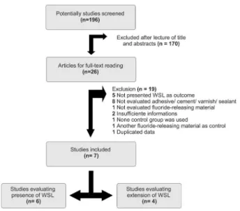
in Figure 1, resulting in a total of 7 (19-25) studies that met the eligibility criteria and were included in the meta-analysis. Four of them evaluated the addition of fluoride to cementation materials, one evaluated its addition to sealant and two evaluated its addition to varnish. The studies were published between 1989 and 2009. The pooled population comprised 1867 teeth. Among them, 247 developed WSL (Table 1).
Risk of Bias
It was found that all included studies showed a low risk of reporting bias. However, this was the only criterion for which low risk was observed. Randomization and allocation concealment procedures were inadequate or unclear in all trials except the one performed by Behan et al. (25), which had a low risk of selection bias. Failures in randomization and allocation concealment allow the staff to predict the upcoming treatment allocation, thereby leading to selection bias. In addition, neither the patients nor the investigators were blinded to the treatment performed in the majority of trials; hence, they were classified as having a high risk of performance and detection bias. Only 2 studies (21,24) showed a risk of attrition bias. The quality assessment of the included trials is shown in detail in Figure 2.
Risk of White Spot Lesions
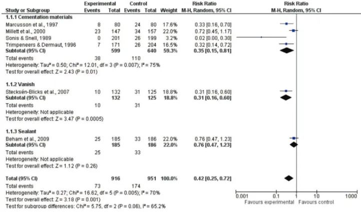
Data on the risk of developing WSLs were available from 6 studies (Table 1). The pooled relative risk of developing WSLs for the experimental group, compared to control group, was 0.42 (95% confidence interval: 0.25 to 0.72); hence, when fluoride-releasing materials are used, the patient has 58% less risk of WSL development (Fig. 3).
However, substantial heterogeneity (p<0.005; I²=70%) across studies was observed.
As the sealant and varnish subgroups were evaluated by two or fewer studies each, a subgroup analysis was unable to show the reasons for heterogeneity. In addition, the risk reduction was statistically significant for the cementation materials and varnish subgroups but not for the sealant subgroup.
Extent of White Spot Lesions
Data on WSL extent were extracted from 4 studies (Table 1). The pooled mean difference between the experimental and control groups was not statistically significant (mean reduction: 0.12; 95% confidence interval: –0.29 to 0.04). Moderate heterogeneity was observed across studies (p<0.16; I²=51%). Subgroup analyses showed a significant reduction only for the cementation materials subgroup (Fig. 4).
Discussion
The results of this systematic review showed a statistically significant reduction in the risk of WSL development when fluoride-releasing agents were used to cover or cement orthodontic brackets. Thus, the hypothesis of the study was rejected. Despite results favoring the use of fluoride around brackets, substantial heterogeneity was observed between the studies included in this review. Subgroup analyses showed that the type of material used to deliver fluorides near the brackets affected the risk. Using fluoride in the luting material or varnish reduced the risk of WSL development (65% and 69% of risk reduction,
Figure 1. Flowchart showing the article selection for systematic review.
16
wher
eas fluoride
in the sealant
did not
result
eduction in WSL risk. This differ
ence on risk
elated to materials’ pr
operties and fr
equency of
ackets,
esented incr
eased solubility in or
al
onment. It is thus expected loss of sealant/varnish
esulting in r
educed action of fluoride
on caries
inhibition. Thus, the periodic r
eplacement of varnish in the
included studies incr eased its action, wher eas the sealant
was not r
eplaced during the follow-up.
Regar
ding the experimental design of the studies
included in this r
eview
, only the study performed by
Stecksén-Blicks et al. (24), which evaluated varnish, used
a par
allel design; the other studies used a cr
ossover design
Study Treatments Methodology No. of teeth with WSL/
No. of teeth evaluated Mean (SD) of WSL scores
Cementation Materials
Sonis and Snell, 1989
Control: Regular resin Experimental: FR resin
-Brackets luted with a resin; - Appointments at each 3-4 weeks;
- Follow-up: NA; - Measurement by photography.
Control: 26/199
Experimental: 0/201 NA
1
Trimpeneers and Dermaut, 1996
Control: Regular resin Experimental: FR resin
-Brackets luted with a resin; - Appointments every 4-5 weeks; - Follow-up: average of 21 (9-33) months;
- Measurement by photography.
Control: 26/204
Experimental: 7/171 NA
Marcusson et al., 1997
Control: Regular resin Experimental: FRGIC
-Brackets luted with a material; - Appointments every 4-6 weeks;
- Follow-up: until 24 months; - Measurement by photography.
Control: 24/80
Experimental: 8/80 NA
Millett et al., 2000
Control: Regular adhesive resin Experimental: FR compomer
-Brackets luted with a material; - Appointments: NA. - Follow-up: until 24 months; - Measurement by photography.
Control: 34/157 Experimental: 23/147
Control: 1.35 (0.60) Experimental:1.09 (0.29)
Vanish
Vivaldi-Rodrigues et al.,2006
Control: No varnish Experimental:
FR varnish
- Applications every 3 months during experimental period of 1 year;
- Follow-up: 12 months; - Measurement by photography.
NA Control
2: 0.61 (1.15)
Experimental2: 0.34 (0.64)
Stecksén-Blicks et al., 2007
Control: Placebo varnish Experimental:
FR varnish
- Application of varnish at all scheduled checkups. - Appointments every 6 weeks; - Follow-up: average of 21 (ranging
from 9 to 33) months; - Measurement by photography.
Control: 31/125 Experimental: 10/132
Control2: 1.08 (0.28)
Experimental2: 1.09 (0.30)
Sealant
Beham et al., 2009
Control: Regular resin Experimental: Prior
use of sealant
- Application of sealant to entire enamel surface before bracket cementation;
- Appointments: NA. - Follow-up: NA
- Measurement by clinical evaluation.
Control: 33/186 Experimental: 25/185
Control: 1.17 (0.47) Experimental: 1.20 (0.48)
Braz Dent J 27(1) 2016
105
White spot lesions around brack
ets
(split-mouth). Because oral hygiene quality and patients’ use of other fluoride sources affect caries development, the split-mouth design provides superior bias control. However, in a similar systematic review, Lessafre et al. (26) pointed out that when a crossover design is used to evaluate fluoride effectiveness, there is a possibility of cross-contamination between experimental and control teeth in the same
mouth, either between maxilla and mandible or between the sides of the mouth; this might lead to underestimate the effectiveness of any fluoride material.
Regarding WSL extent, lesions were reduced by an average of 0.12 (for scores ranging from 0 to 3) when fluoride-releasing materials were used, but this reduction was not statistically significant. A moderate heterogeneity
Figure 3. Forest plot showing estimated effect for outcome risk to white spot lesions.
P
.L.M.M. Nascimento et al.
was observed among studies included in the WSL extent analysis, and the subgroup partially explained the heterogeneity. Only cementation materials demonstrated a statistical reduction in WSL extent, but just a single study evaluated this outcome. The protective effect of fluoride agents against enamel demineralization is well known. Therefore, a significant reduction in both the risk of development and extent was expected. However, this review did not find any significant reduction of WSL extent. Despite the large number of enamel surfaces evaluated in the included studies, only those already presenting WSLs were evaluated, reducing the sample size. In addition, only three studies evaluated the extent of WSLs. As a result, the sample size for the summary effect of WSL extent was around 20% of the sample used to summarize the risk WSL development. This reduced sample size may help to explain the absence of statistical differences between the experimental and control conditions. Among the three studies that evaluated WSL extent, only the one performed by Steksen-Blicks et al. (17) reported a sample-size calculation, and it was done to detect a difference in WSL incidence, not in WSL extent.
One limitation of meta-analysis involves studies with high heterogeneity. Despite the difficulty of establishing the limit of heterogeneity to indicate a meta-analysis of data, several studies have proposed limit values between 50 and 75%. The analysis of WSL risk in the present review showed an I2 value of 70%, which is within the limits mentioned above. The subgroup analysis showed a 65% heterogeneity between materials, indicating that the type of material partially explained the heterogeneity. Thus, the different approaches for delivering fluorides near brackets could be related to the observed heterogeneity. Eliminating the study that evaluated sealants (25) reduces the I2 to 46%, which is similar to the value for the WSL extent analysis. However, this study places great importance on summary effects due the high number of analyzed events, and maintaining this study increased the values of the power test.
Another important limitation of the present systematic review was related to possible publication bias, since a single database was used to search the studies. Despite the MEDLINE being the main database for studies in dentistry, studies indexed to other databases could improve the quality of evidence. Furthermore, all included studies presented a high risk of bias, indicating that the evidence is weak. Only one study (25) described the generation of random sequence for allocation of participants in the study; while no study included described allocation concealment. Regarding that blinding of operator is not possible for some studies, only one study (24) blinded the operator/patient and less than 50% of studies showed blinded evaluation of outcomes. Thus, this review also showed the need for further
well-designed clinical studies evaluating the effectiveness of fluoride-releasing materials on caries prevention around orthodontic brackets.
Despite the low quality of the evidence, delivering fluoride-releasing materials near brackets seems to be an effective approach to reduce the risk of WSL development in patients wearing fixed orthodontic appliances. This reduction could be even more meaningful for patients who have difficulties following oral hygiene instructions and who thus are under higher risk. However, when white spot lesions are present, fluoride-releasing materials seem to have no effect upon lesions’ extent. Similar findings were described by other systematic reviews, which also concluded that the evidence to indicate fluoride-releasing material on caries prevention around brackets requires further well-conducted studies. Therefore, orthodontists should bear in mind that there is limited evidence to support the use of fluoride-releasing materials in order to prevent WSL development.
Resumo
A relação entre o uso de aparelhos ortodônticos fixos e desmineralização do esmalte é bem estabelecida. Diferentes abordagens preventivas têm sido sugeridas para este problema, mas ainda permanece controverso qual é o melhor. O objetivo deste estudo foi realizar uma revisão sistemática de ensaios clínicos que investigaram a efetividade de materiais contendo fluoretos para cimentação de bráquetes ou cobrindo a interface de união buscando inibir o desenvolvimento e progressão de lesões de mancha branca. A hipótese nula foi que materiais fluoretados não afetam a incidência de lesões de mancha branca em volta de bráquetes. Uma busca no MEDLINE foi conduzida para ensaios clínicos controlados avaliando o desenvolvimento de lesões de mancha branca em pacientes usando aparelhos ortodônticos fixos, seguido por meta-análise comparando os resultados de pacientes em que materiais usando fluoretos foram utilizados (grupo experimental) com aqueles em que tais materiais não foram usados. O risco relativo agrupado de desenvolvimento de lesões de mancha branca para o grupo experimental foi 0,42 (95% de intervalo de confiança: 0,25 a 0,72); enquanto que, quando materiais liberando fluoretos foram utilizados, o paciente teve 58% menos risco de desenvolver lesões de mancha branca. Em relação à extensão das lesões de mancha branca, a diferença média agrupada entre os grupos experimental e controle não foi estatisticamente significante (-0,12; 95% de intervalo de confiança: -0,29 a 0,04). Em conclusão, os resultados da presente revisão sistemática sugerem que materiais que liberam fluoretos podem reduzir o risco de lesões de mancha branca em volta de bráquetes. Entretanto, quando lesões de mancha branca já ocorreram, não há evidência que materiais que liberam fluoretos reduzem a extensão da lesão.
References
1. Lucchese A, Gherlone E. Prevalence of white-spot lesions before and during orthodontic treatment with fixed appliances. Eur J Orthod 2013;35:664-668.
2. Trimpeneers LM, Dermaut LR. A clinical evaluation of the effectiveness of a fluoride-releasing visible light-activated bonding system to reduce demineralization around orthodontic brackets. Am J Orthod Dentofac Orthod 1996;110:218-222.
3. Julien KC, Buschang PH, Campbell PM. Prevalence of white spot lesion formation during orthodontic treatment. Angle Orthod 2013;83:641-647.
2014;12:458-Braz Dent J 27(1) 2016
107
White spot lesions around brack
ets
466.
5. Akin M, Tezcan M, Ileri Z, Ayhan F. Incidence of white spot lesions among patients treated with self- and conventional ligation systems. Clin Oral Investig 2015;19:1501-1506.
6. Srivastava K, Tikku T, Khanna R, Sachan K. Risk factors and management of white spot lesions in orthodontics. J Orthod Sci 2013;2:43-49. 7. Lynch RJ, Navada R, Walia R. Low-levels of fluoride in plaque and
saliva and their effects on the demineralization and remineralization of enamel; role of fluoride toothpastes. Int Dent J 2004;54:304-309. 8. Cury JA, Tenuta LMA. Enamel remineralization: controlling the caries
disease or treating the early caries lesions? Braz Oral Res 2009;23:23-30.
9. Zabokova-Bilbilova E, Popovska L, Kapusevska B, Stefanoska E. White spot lesions: prevention and management during orthodontic treatment. Prilozi 2014;35:161-168.
10. Derks A, Katsaros C, Frencken JE, van't Hof MA, Kuijpers-Jagtman AM. Caries-inhibiting effect of preventive measures during orthodontic treatment with fixed appliances. A systematic review. Caries Res 2004;38:413-420.
11. Rogers S, Chadwick B, Treasure E. Fluoride-containing orthodontic adhesives and decalcification in patients with fixed appliances: a systematic review. Am J Orthod Dentofacial Orthop 2010;138:390. e1-e8.
12. Benson PE, Parkin N, Dyer F, Millett DT, Furness S, Germain P. Fluorides for the prevention of early tooth decay (demineralised white lesions) during fixed brace treatment. Cochrane Database Syst Rev 2013; 12:CD003809
13. Cheng LL. Limited evidence indicates fluoride may prevent demineralized white lesions during orthodontic treatment. J Am Dent Assoc 2015;146: 699-701.
14. Moher D, Liberati A, Tetzlaff J, Altman DG. Preferred reporting items for systematic reviews and meta-analyses: the PRISMA statement. Int J Surg 2010;8:336-341.
15. Schulz KF, Altman DG, Moher D, CONSORT Group. CONSORT 2010 statement: updated guidelines for reporting parallel group randomized trials. Ann Intern Med 2010;52:726-732.
16. Gorelick L, Geiger AM, Gwinnett AJ. Incidence of white spot formation
after bonding and banding. Am J Orthod 1982;81:93-98.
17. Higgins JPT, Altman DG, Gotzsche PC, Juni P, Moher D, Oxman AD, et al.. The Cochrane Collaboration's tool for assessing risk of bias in randomized clinical trials. BMJ 2011;343:d5928.
18. Biljana M, Jelena M, Branislav J, Milorad R. Bias in meta-analysis and funnel plot asymmetry. Stud Health Technol Inform 1999;68:323–328. 19. Sonis AL, Snell W. An evaluation of a fluoride-releasing, visible light-activated bonding system for orthodontic bracket placement. Am J Orthod Dentofac Orthop 1989;95:306-311.
20. Trimpeneers LM, Dermaut LR. A clinical evaluation of the effectiveness of a fluoride-releasing visible light-activated bonding system to reduce demineralization around orthodontic brackets. Am J Orthod Dentofac Orthop 1996;110:218-222.
21. Marcusson A, Norevall LI, Persson M. White spot reduction when using glass ionomer cement for bonding in orthodontics: a longitudinal and comparative study. Eur J Orthod 1997;19:233-242.
22. Millet DT, McCluskey L-A, McAuley F, Creanor SL, Newell J, Love J. A comparative clinical trial of a compomer and a resin adhesive for orthodontic bonding. Angle Orthod 2000;70:233-240.
23. Vivaldi-Rodrigues G, Demito CF, Bowman J, Ramos AL. The effectiveness of a fluoride varnish in preventing the development of white spot lesions. World J Orthod 2006;7:1-7.
24. Stecksén-Blicks C, Renfors G, Oscarson ND, Bergstrand F, Twetman S. Caries-preventive effectiveness of a fluoride varnish: a randomized controlled trial in adolescents with fixed orthodontic appliances. Caries Res 2007;41:455-459.
25. Benham AW, Campbell PM, Buschang PH. Effectiveness of pit and fissure sealants in reducing white spot lesions during orthodontic treatement: a pilot study. Angle Orthod 2009;79:338-345.
26. Lessafre E, Philstrom B, Needleman I, Worthington H. The design and analyses of split-mouth studies: what statisticians and clinicians should know. Stat Med 2009;28:3470-3482.