TaqMan MGB Probe Fluorescence Real-Time Quantitative
PCR for Rapid Detection of Chinese Sacbrood Virus
Ma Mingxiao1*, Liu Jinhua2, Song Yingjin3, Li Li1, Li Yongfei1
1Department of Laboratory Animal Center, Liaoning Medical University, Jinzhou, China,2Jilin Entry-Exit Inspection and Quarantine Bureau, Changchun, China, 3Agriculture and Biology Engineering College, Tianjin University, Tianjin, China
Abstract
Sacbrood virus (SBV) is a picorna-like virus that affects honey bees (Apis mellifera) and results in the death of the larvae. Several procedures are available to detect Chinese SBV (CSBV) in clinical samples, but not to estimate the level of CSBV infection. The aim of this study was develop an assay for rapid detection and quantification of this virus. Primers and probes were designed that were specific for CSBV structural protein genes. ATaqMan minor groove binder (MGB) probe-based, fluorescence real-time quantitative PCR was established. The specificity, sensitivity and stability of the assay were assessed; specificity was high and there were no cross-reactivity with healthy larvae or other bee viruses. The assay was applied to detect CSBV in 37 clinical samples and its efficiency was compared with clinical diagnosis, electron microscopy observation, and conventional RT-PCR. The TaqMan MGB-based probe fluorescence real-time quantitative PCR for CSBV was more sensitive than other methods tested. This assay was a reliable, fast, and sensitive method that was used successfully to detect CSBV in clinical samples. The technology can provide a useful tool for rapid detection of CSBV. This study has established a useful protocol for CSBV testing, epidemiological investigation, and development of animal models.
Citation:Mingxiao M, Jinhua L, Yingjin S, Li L, Yongfei L (2013) TaqMan MGB Probe Fluorescence Real-Time Quantitative PCR for Rapid Detection of Chinese Sacbrood Virus. PLoS ONE 8(2): e52670. doi:10.1371/journal.pone.0052670
Editor:Wenzhe Ho, Temple University School of Medicine, United States of America
ReceivedAugust 20, 2012;AcceptedNovember 19, 2012;PublishedFebruary 8, 2013
Copyright:ß2013 Mingxiao et al. This is an open-access article distributed under the terms of the Creative Commons Attribution License, which permits unrestricted use, distribution, and reproduction in any medium, provided the original author and source are credited.
Funding:This work was supported by grants from the National Natural Science Foundation of China (No. 30972200) and the Science and Technology Department of Liaoning Province of China (No. 2011408004). The funders had no role in study design, data collection and analysis, decision to publish.
Competing Interests:The authors have declared that no competing interests exist.
* E-mail: [email protected]
Introduction
Sacbrood virus (SBV) is a picorna-like virus that affects the honey bee (Apis mellifera) and results in the death of the larvae [1]. Larvae that have been infected with SBV fail to pupate and ecdysial fluid, rich in SBV, accumulates beneath their unshed skin. Infected larvae change in color from white to pale yellow and then die. Shortly after death they dry out and form a distinctive dark brown gondola-shaped scale [2]. SBV causes a fatal infection in bee larvae, but may also infect the adult bee and infected workers may have decreased life spans [3,4]. When this infection occurs in adults, however, obvious physical signs of disease are lacking [3,5]SBV infection occurs most frequently in the spring and this timing is believed to reflect the availability of susceptible larvae and young adults that are at their height in this season when the colony is growing most rapidly [3].
The SBV that infects the Chinese honeybee was named Chinese SBV (CSBV). CSBV was first described in Guangdong China in 1972, and re-emerged in Liaoning China in 2008 [6], when it caused lethal disease in individual bees or the collapse of entire colonies.
Several procedures have been utilized previously to detect CSBV. These methods include reverse transcriptase polymerase chain reaction (RT-PCR) [6,7] and loop-mediated isothermal amplification (LAMP) assay [8]. Although these techniques can be used to characterize CSBV in clinical samples, they cannot estimate the level of CSBV infection. The quantitation of CSBV would permit a better understanding of virus infection, both in individual bees and in the hive.
Real-time RT-PCR detection methods have been developed recently for the detection and quantitation of bee viruses [9–13]. This paper describes a TaqMan MGB (minor groove binder) probe fluorescence real-time quantitative PCR assay to quantify CSBV. Standard curves from a plasmid that contained a partial sequence of the CSBV genome were used to obtain an absolute quantitation of CSBV. The specificity, sensitivity and stability of the method were validated from a standard DNA curve. The established TaqMan MGB probe fluorescence real-time quantitative PCR assay was applied to detect CSBV in 37 clinic samples, and its efficiency was compared with that of clinic diagnosis, electron microscopy observation, and conventional RT-PCR assay.
Materials and Methods
a volume of 5 ml (final density 1.38 g/cm3) with CsCl solution and centrifuged at 270 000g overnight. Two light-scattering bands were formed that were collected separately and diluted with NT buffer; virus was collected by sedimentation at 270 000gfor 1 h. Pellets were resuspended in NT buffer. Complementary DNA (cDNA) was synthesized using 12.5ml of eluted RNA, oligo(dT)18 primer [14] and avian myeloblastosis virus (AMV) reverse transcriptase XL (TaKaRa, Dalian, China) according to the manufacturer’s instructions.
Real-time quantitative PCR protocol by TaqMan MGB probe assay
Primer pairs and probes specific for CSBV were designed based on the nucleotide sequences of CSBV published previously in the GenBank database (accession no. AF469603 and HM237361.1). The primers and probes used for TaqMan MGB probe fluorescence real-time quantitative PCR were designed using the Primer5 software. The sequence of the forward primer is 59 -CCTGGGAAGTTTGCTAGTATTTACG-39and of the reverse primer is 59-CCTATCACATCCATCTGGGTCAG-39. The TaqMan MGB probe sequence was 59-
CGACATACCCG-CAAATTCAGCACGC-39, which was labeled with the fluores-cent reporter dye FAM (6-carboxyfluorescein) at the 59 end and with the fluorescent NFQ-MGB at the 39end. A 161-bp product was expected when these primers were used. The primers and the TaqMan MGB probes were synthesized and labeled by TaKaRa, Dalian, China.
TransStartTM Probe qPCR SuperMix (Beijing TransGen Biotech Co., Ltd.) was used for amplification by TaqMan PCR. The PCR reaction mixture contained 500 nM of each primer, 250 nM of the probe, 26PCR buffer 12.5ml, Reference 0.5ml and 2ml of standard template plasmid or cDNA in a 25ml total reaction volume. The thermal cycling conditions were 30 sec at 95uC, followed by 40 cycles that consisted of a denaturation step at 95uC for 5 sec, annealing and extension step at 60uC for 60 sec. The ABI Prism 7000 system (Applied Biosystems ABI) was used for amplification and detection.
Preparation of the plasmid DNA standard for calibration of the CSBV TaqMan MGB probe PCR assay
A standard DNA curve was generated with a 3.01 kb plasmid that was obtained by cloning a 320-bp PCR fragment located in
Figure 1. Standard curve ofTaqMan MGB probe real-time quantitative polymerase chain reaction (PCR) for Chinese sacbrood virus (CSBV).DNA standard curves of CSBVTaqMan PCR assay using a FAM-MGB-labeledTaqMan probe obtained with a 10-fold serial dilution (56108to 56103DNA copies) of a 3.1-kb plasmid that included a 320-bp fragment located in the CSBV VP1 gene. The standard curve was obtained by linear regression analysis of the CTmeasured for each amplification (y-axis) vs. the log copy number for each standard dilution (x-axis). The slope of the standard curve (–3.47) and the correlation coefficient are indicatedr2= 0.99 (Y = –3.47X
+34.60). The red box represents Ct.
the VP1 gene of CSBV into the pMDH 18-T Vector (TaKaRa, Dalian, China.). The plasmid was quantified based on the DNA concentration determined by UV (ultraviolet) light spectrometry; stock solutions were prepared in Tris–EDTA (TE) buffer that ranged in concentration from 56108 to 5 DNA copies per microliter. A standard DNA curve for the range of 56108 to 56103DNA copies per reaction was generated by analysing 2ml of each dilution in triplicate byTaqMan PCR.
Evaluation of the method
The limit of detection of the CSBV TaqMan PCR was determined by serial 10-fold dilution of the DNA standard prepared in triplicate.
The specificity of the CSBV TaqMan PCR was assessed by testing cDNAs generated from deformed wing virus (DWV), black queen cell virus (BQCV), acute bee paralysis virus (ABPV), and chronic bee paralysis virus (CBPV) isolates, which were provided by Professor Laurent Gauthier, healthy larvae and two positive samples were included as controls for the specific RT-PCR tests.
The reproducibility of the CSBV TaqMan PCR assay was demonstrated by evaluation of the variability of the CT values obtained after amplification of 10-fold serial dilutions of the plasmid DNA standard ranging from 102 to 106 copies per reaction in triplicate during the same experiment.
Validation of the CSBV TaqMan PCR method on clinical samples
Thirty-seven clinical samples collected from Qingyuan, Jinzhou, Tieling, Yingkou, Dalian and Liaozhong areas of Liaoning Province and Huinan Ji’an areas of Jilin Province, China from 2008 to 2011 were tested to determine the feasibility of the TaqMan PCR assay. At the same time, all mentioned samples were subjected to conventional RT-PCR and electron microscopy.
Results
Standardisation of the CSBV TaqMan PCR assay
The assay was calibrated using a plasmid DNA control as a standard. The standard curve generated from the amplification plot of the 10-fold serial dilution experiment (Fig. 1) showed a linear correlation between CTvalues and the DNA load over a 6-log range (r2= 0.99). The slope of the DNA standard curve was 3.47, which indicated that the efficiency of the CSBVTaqMan PCR was 95%.
The limit of detection of the CSBVTaqMan PCR was 50 CSBV genome equivalent copies (Fig. 2); this result compared favourably with conventional PCR for which the limit of detection of CSBV genome from plasmid DNA control was 56103copies (data not shown).
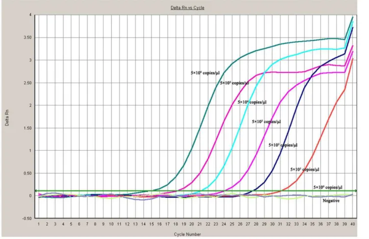
Figure 2. Sensitivity of theTaqMan MGB probe real-time quantitative polymerase chain reaction (PCR) for Chinese sacbrood virus (CSBV).To evaluate the sensitivity of theTaqMan MGB probe real-time RT-PCR assay. Amplification of seven 10-fold dilutions of DNA plasmid gave a titer that ranged from 56106to 56100DNA copies per reaction mixture. The reactions were performed in triplicate. The limit of detection of the TaqMan MGB probe real-time PCR was 50 CSBV genome equivalent copies.
doi:10.1371/journal.pone.0052670.g002
Specificity and reproducibility of the CSBV TaqMan PCR CBPV specificity was confirmed by a BLAST search of the amplicon (116 bp) generated by TaqMan PCR. No significant similarity was found. Furthermore, no amplification was detected when theseTaqMan PCR conditions were performed on cDNAs obtained from CBPV, ABPV, BQCV or DWV samples (Fig. 3). The coefficient of variation (CV) of the mean CTvalues obtained
from the DNA standard curve ranged from 0.27 to 1.4% (Table 1).
Validation of the CSBV TaqMan PCR method on clinical samples
The 37 collected clinical samples were tested to determine the feasibility of the TaqMan PCR assay. At the same time, all
Figure 3. Comparison of TaqMan PCR assay and reverse transcriptase polymerase chain reaction (RT-PCR) methods by testing collected clinical samples.The 16 collected clinical samples were tested using the TaqMan RT-PCR and RT-PCR, respectively. (A) RT-PCR test results. (B) TaqMan RT-PCR test results. The LNQY-1, LNJZ-3, LNYK-4, JLHN, LNQY-5 and LNSZ-2 samples tested positive using conventional RT-PCR and the TaqMan RT-PCR method, but the LNYK-6 samples tested positive using the TaqMan RT-PCR method, however the samples were negative by electron microscopy and conventional RT-PCR. lane 1. DNA marker DL2000, lane 2. LNQY-1, lane 3. LNQY-5, lane 4. LNQY-2, lane 5. LNYK-3, lane 6. LNYK-4, lane 7. LNYK-6, lane 8. LNLZ-1, lane 9. LNLZ-3, lane 10 JLJA, lane 11. JLHN, lane 12. LNJZ-1, lane 13. LNJZ-3, lane 14 LNJZ-5, lane 15 LNSZ-2 lane 16. JLBC.
mentioned samples were subjected to conventional RT-PCR and electron microscopy. As shown in Table 2 (Fig. 3), the LNQY-1, LNJZ-3, LNYK-4 and JLHN samples presented severe signs of sacbrood disease and all three methods yielded positive results. The LNQY-5 and LNSZ-2 samples tested positive using conventional RT-PCR and the TaqMan RT-PCR method, however the samples were negative by electron microscopy and for clinical symptoms, The LNYK-6 samples tested positive using
the TaqMan RT-PCR method, however the samples were negative by electron microscopy and conventional RT-PCR (Table 2). Furthermore, DWV, BQCV, ABPV and CBPV were also detected by RT-PCR, but these viruses were not detected by other means (Fig. 4).
Table 1.Reproducibility ofTaqMan minor groove binder (MGB) probe fluorescence real-time quantitative polymerase chain reaction (PCR).
No. of CSBV copies (amount of cDNA per mix) MGB-labelled CSBV probe
CTvalue CV (%)
56106copies/ml 14.9060.03383 0.39
56105copies/ml 17.9560.04509 0.44
56104copies/
ml 21.2660.1715 1.4
56103copies/ml 24.9660.08083 0.57
56102copies/ml 27.6360.04055 0.27
56101 copies/ml 31.1560.09045 0.73
The reproducibility of the Chinese sacbrood virus (CSBV)TaqMan PCR assay was demonstrated by evaluating the variability of the CTvalues obtained after amplification of 10-fold serial dilutions of the plasmid DNA standard ranging from 101to 106copies per reaction in triplicate during the same experiment. The coefficient of variation (CV) of the mean CTvalues obtained for the DNA standard curve ranged from 0.27 to 1.4%.
doi:10.1371/journal.pone.0052670.t001
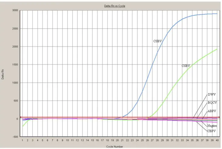
Figure 4. Comparison of the specificity of Chinese sacbrood virus (CSBV)TaqMan PCR assay for CSBV detection.Chinese sacbrood virus strains gave a positive reaction using theTaqMan PCR assay method, no cross-reactivity with other clinically related viruses, including deformed wing virus (DWV), black queen cell virus (BQCV), acute been paralysis virus (ABPV), and chronic bee paralysis virus (CBPV), was detected. doi:10.1371/journal.pone.0052670.g004
Discussion
Quantitative real-time polymerase chain reaction (qRT-PCR), is a laboratory technique based on PCR that is used to amplify and simultaneously quantify a targeted DNA molecule. The standard DNA probes are labeled at the 59 end with a fluorochrome reporter (usually 6-carboxyfluorescein [6-FAM]) and a fluoro-chrome quencher (6-carboxy-tetramethyl-rhodamine [TAMRA]) at the 39 end. A new technology of qRT-PCR that uses MGB probes [15] for fluorescence quantitative PCR, has been developed recently for the detection and quantitation DNA molecules. The
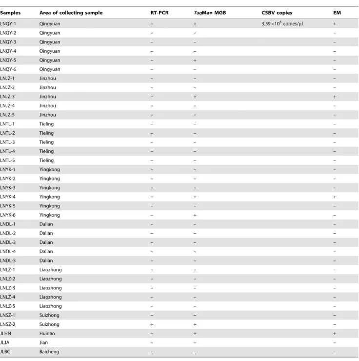
DNA probes with the MGB groups form extremely stable duplexes with single-stranded DNA targets, and therefore allow shorter probes to be used for hybridization-based assays. MGB probes have higher melting temperature (Tm) and increased specificity when compared with unmodified DNA, especially when a mis-match occurs in the MGB region of the duplex. The fluorogenic MGB probes were more specific for single-base mismatches and fluorescence quenching was more efficient, and therefore gave increased sensitivity. In summary, MGB probes were more sequence specific than standard DNA probes, especially for single-base mismatches at elevated hybridization temperatures. Table 2.Detection of 37 Chinese sacbrood virus (CSBV) clinical samples: ofTaqMan minor groove binder (MGB) probe fluorescence real-time quantitative PCR, reverse transcriptase polymerase chain reaction (RT-PCR) and electron microscopy methods.
Samples Area of collecting sample RT-PCR TaqMan MGB CSBV copies EM
LNQY-1 Qingyuan + + 3.596105copies/
ml +
LNQY-2 Qingyuan – – –
LNQY-3 Qingyuan – – –
LNQY-4 Qingyuan – – –
LNQY-5 Qingyuan + + –
LNQY-6 Qingyuan – – –
LNJZ-1 Jinzhou – – –
LNJZ-2 Jinzhou – – –
LNJZ-3 Jinzhou + + +
LNJZ-4 Jinzhou – – –
LNJZ-5 Jinzhou – – –
LNTL-1 Tieling – – –
LNTL-2 Tieling – – –
LNTL-3 Tieling – – –
LNTL-4 Tieling – – –
LNTL-5 Tieling – – –
LNYK-1 Yingkong – – –
LNYK-2 Yingkong – – –
LNYK-3 Yingkong – – –
LNYK-4 Yingkong + + +
LNYK-5 Yingkong – – –
LNYK-6 Yingkong – + –
LNDL-1 Dalian – – –
LNDL-2 Dalian – – –
LNDL-3 Dalian – – –
LNDL-4 Dalian – – –
LNDL-5 Dalian – – –
LNLZ-1 Liaozhong – – –
LNLZ-2 Liaozhong – – –
LNLZ-3 Liaozhong – – –
LNLZ-4 Liaozhong – – –
LNLZ-5 Liaozhong – – –
LNSZ-1 Suizhong – – –
LNSZ-2 Suizhong + + –
JLHN Huinan + + +
JLJA Jian – – –
JLBC Baicheng – – –
In this paper, a TaqMan MGB probe fluorescence real-time quantitative PCR assay was developed for the detection and quantitation of CSBV. Kerstin Wernike [16] describe aTaqMan MGB probe fluorescence real-time quantitative PCR assay for the detection of SBV, Howere the TaqMan MGB hasn’t been evaluated. This paper is the first to describe a TaqMan MGB probe fluorescence real-time quantitative PCR assay for the especially detection and quantitation of CSBV in honey bees.
TheTaqMan MGB probe with primers specific for the CSBV VP1 gene was used. The standard curve generated with the plasmid that contained a partial sequence of the CSBV genome showed that quantitation of this genome was linear over six orders of magnitude. The efficiency of the standard curve and its good correlation was confirmed. Quantitation of the positive control from CSBV-infected larvae gave similar results for both methods compared to the values obtained by UV spectrometry. These results validated the use of the DNA standard curve to quantify CSBV in bee samples. The limit of detection of thisTaqMan PCR method was 50 CSBV genome equivalent copy numbers, which represented an improvement in the limit of detection by thr
conventional the PCR method developed previously in our laboratory [5], for which the limit of detection was 56103CSBV copies. The reproducibility of several experiments showed the high reproducibility of the method for the standard curve (0.27 to 1.4%), and for the efficiency of cDNA synthesis from the positive control (0.95%). The feasibility of the TaqMan PCR assay was validated by detection of 37 clinical samples using conventional PCR and theTaqMan PCR. TheTaqMan PCR assay was more sensitivity than conventional PCR.
In summary, in this paper we provide a quantitative description of CSBV infection in larvae in China. Our data demonstrated that real-time quantitative RT-PCR is a specific, sensitive, robust, and reproducible assay with practical applications in the diagnosis of honey bee virus diseases and analysis of virus infection.
Author Contributions
Conceived and designed the experiments: MM SY. Performed the experiments: LJ LY LL. Analyzed the data: MM SY. Wrote the paper: MM SY.
References
1. Ritter W (1996) Diagnostik und Bekampfung von Bienenkrankheiten. Gustav Fischer Verlag Jena. Stuttgart. Germany, 104–114.
2. Bailey L (1975) Recent research on honeybee viruses. Bee World.56: 55–64. 3. Bailey L (1976) Viruses attacking the honeybee. Adv. Virus Res 20: 271–304. 4. Wang DI, Moller FE (1970) The division of labor and queen attendance
behaviour of nosema-infected worker honey bees. J. Econ. Entomol 63: 1539– 1541.
5. Anderson DL, Gibbs AJ (1989) Transpuparial transmission of Kashmir bee virus and sacbrood virus in the honeybee (Apis mellifera). Ann. Appl. Biol 114: 1–7. 6. Mingxiao M, Ming L, Chunying Y, Pengfei L, Yibo Z, et al. (2010) Development of a RT-PCR method for determination of Chinese sacbrood virus. Chin. J. Biol 23: 425–427(in chinese).
7. Yan X, Chen J, Han R (2009) Detection of Chinese sacbrood virus (CSBV) in Apis cerana by RT-PCR method. Sociobiology, 53: 687–694.
8. Mingxiao M, Chen M, Mng L, Wang S, Song Y, et al. (2011) Loop-mediated isothermal amplification for rapid detection of Chinese sacbrood virus. J. Virol. Methods176(1–2): 115–119.
9. Chen YP, Higgins JA, Feldlaufer MF (2005) Real-time quantitative reverse-time transcription-PCR analysis of deformed wing virus infection in the honeybee virus (DWV) (Apis mellifera L.). Appl. Environ. Microbio 71: 436–441. 10. Chantawannakul P, Ward L, Boonham N, Brown M (2006) A scientific note on
the detection of honey bee viruses using real-time PCR (TaqMan) in Varroa
mites collected from a Thai honeybee (Apis mellifera) apiary. J. Invertebr. Pathol 91: 69–73.
11. Blanchard P, Ribiere M, Celle O, Lallemand P, Schurr F, et al. (2007) Evaluation of a real-time two-step RT-PCR assay for quantitation of chronic bee paralysis virus (CBPV) genome in experimentally-infected bee tissues and in life stages of a symptomatic colony. J. Virol. Methods141: 7–13.
12. Kukielka D, Espero´n F, Higes M, Sa´nchez-Vizcaı´no JM (2008) A sensitive one-step real-time RT-PCR method for detection of deformed wing virus and black queen cell virus in honeybee Apis mellifera. J. Virol. Methods 147: 275–278. 13. Kukielka D, Sa´nchez-Vizcaı´no JM (2009) One-step real-time quantitative PCR
assays for the detection and field study of sacbrood honeybee and acute bee paralysis viruses. J. Virol. Methods161: 240–246.
14. Sambrook J, Russell DW (2001) Molecular Cloning: A Laboratory Manual. third ed. Cold Spring Harbor Laboratory Press. Cold Spring Harbor. N.Y. 15. Kutyavin IV, Afonina IA, Mills A, Gorn VV, Lukhtanov EA, et al. (2001)
39-Minor groove binder-DNA probes increase sequence specificity at PCR extension temperatures Nucleic Acids Res 28(2): 655–661.
16. Wernike K, Hoffmann B, Dauber M, Lange E, Schirrmeier H, et al. (2010) Detection and Typing of Highly Pathogenic Porcine Reproductive and Respiratory Syndrome.