Different methods of real-time PCR for detection of pseudorabies virus
Diferentes métodos de PCR em tempo real para detecção do vírus da pseudorraiva
Carolina Kymie Vasques Nonaka1 Antônio Augusto Fonseca Junior2 Estefânia Oliveira Guedes1 Régia Maria D´Ambros3 Graciela Kunrath Lima1 Marcelo Fernandes Camargos1 Marcos Bryan Heinemann4
ISSNe 1678-4596
INTRODUCTION
Pseudorabies (PR), also known as Aujeszky’s disease, is a highly contagious viral disease of great sanitary and economic importance in pigs (OIE, 2015). Pigs are considered the main reservoir of the virus and its most important disseminator. The disease is caused by Suid herpesvirus 1 (SuHV-1, family Herpesviridae) (DAVISON et al., 2009).
Cases of SuHV-1 infection can be found in parts of Europe, Southeast Asia, Central America and South America (OIE, 2015). Besides the infection of domestics pigs, common problem is the high number of feral pigs and wild boar infected by SuHV-1. In the United States, different publications
presented a percentage of positive samples varying from 0% to 60.91% in the states were the samples were collected (MÜLLER et al., 2011). Two
researches were done in Brazil. The first one used 358
samples from 1998-2001 collected from different farms and demonstrated prevalence varying from 0% to 55.9% positive samples in ELISA (CUNHA et al., 2006). The second research was done with 186 samples collected from feral pigs throughout the Brazilian wetland plain, obtaining 31.8% positive in serum neutralization (PAES et al., 2013). Due to
the health risk, the fact of being a notifiable disease
that imposes severe sanitary barriers, PR is still constantly studied to evaluate methods of rapid and accurate diagnosis (OIE, 2015).
1Escola de Veterinária, Departamento de Medicina Veterinária Preventiva, Universidade Federal de Minas Gerais (UFMG), Belo Horizonte, MG, Brasil. 2Laboratório Nacional Agropecuário Lanagro/MG, Ministério da Agricultura, Pecuária e Abastecimento, Pedro Leopoldo, MG, Brasil. 3Centro de Diagnóstico em Sanidade Animal (CEDISA), Concórdia, SC, Brasil.
4Departamento de Medicina Veterinária Preventiva, Faculdade de Medicina Veterinária e Zootecnia, Av. Prof. Dr. Orlando Marques de Paiva, 87, 5508-270, São Paulo, SP, Brasil. E-mail: [email protected]. Corresponding author.
ABSTRACT: Pseudorabies (PR) is a highly contagious viral disease of great animal health and economic importance in swine industry. The aim of this study was to evaluate different genomic regions, real-time PCR chemistries and equipment for the molecular diagnosis of PR. Eight primer pairs targeting four genes (gB, gC, gE, gD), three different qPCR chemistries (SybrGreen, hydrolysis probes and plexor) and two equip-ment (ABI7500, Rotorgene 3000) were evaluated. Oligonucleotides targeting gB using hydrolysis probes showed the best performance after
evaluating efficiency (99%), the detection limit (10-1.5 TCID50 mL-1) and diagnostic sensitivity and; therefore, those primers were selected for
performance verification factors such as repeatability, reproducibility and robustness (1.39% variance between days, 24% variance between analysts and 4.07% variance in analysis error). The qPCR standardized and validated in this research proved to be reliable for the diagnosis of PR and may be used in diagnostic laboratories that follow ISO 17025 and ISO 16140.
Key words: real time PCR, pseudorabies, diagnose.
RESUMO: Pseudorraiva (PR) é uma doença viral altamente contagiosa de grande importância sanitária e econômica na indústria suína. O objetivo deste estudo foi avaliar diferentes regiões genômicas, químicas de PCR em tempo real e equipamentos para o diagnóstico molecular de PR. Foram avaliados quatro genes (GB, GC, GE, gD), três diferentes produtos químicos (SybrGreen, sondas de hidrólise e plexor) e dois equipamentos (ABI7500, Rotorgene 3000). Oligonucleotídeos com alvo para gB baseados na química de sondas de hidrólise apresentaram o
melhor desempenho nos testes de eficiência (99%), de limite de detecção (10-1.5 TCID50 mL-1) e sensibilidade diagnóstica. Portanto, estes foram
selecionados para fatores de verificação de desempenho, tais como a repetibilidade, reprodutibilidade e robustez (1,39% de variância entre dias, 24% variância entre analistas e 4,07% de variância por erro de análise). A qPCR, padronizada e validada neste trabalho, mostrou-se confiável para o diagnóstico de PR e pode ser utilizada em laboratórios de diagnóstico que se seguem normas internacionais como ISO 17025 e ISO 16140.
Palavras-chave: PCR em tempo real, pseudorraiva, diagnóstico.
PR diagnosis is performed by detection
of the agent as virus isolation and identification
or polymerase chain reaction as well as by detecting a serological response against SuHV-1. Viral isolation has the disadvantage of relying on the presence of viable viruses and the possibility of contamination of cell cultures (JULIAN & SCHWAB, 2012). The tests based on the antibody detection have the disadvantage of depending on the seroconversion of the animal. Use of these tests during the acute phase of the disease can lead to false-negative results, because it takes at least 10 days after infection, on average, for the detection of antibodies. Another disadvantage is that younger animals may die before seroconversion, hindering serological diagnosis (KINKER et al., 1997).
A growing number of real time PCR (qPCR) chemistries are available to diagnostic laboratories. There are differences in performance of these methods concerning costs, detection limits,
specificity and practicability (BUH GASPARIC et
al., 2008). It is important to evaluate each chemistry, because its optimization and costs may have a impact in laboratory analysis. Detection limit, for example, occasionally changes according to qPCR chemistry used, which affects diagnostics sensitivity and indicates these molecular methodologies are not easily interchangeable (GHALMI et al., 2008).
International guides such as the Manual of Diagnostic Tests and Vaccines for Terrestrial (OIE, 2015) and laboratory quality systems (ISO 17025, 2010) highlight the importance of checking the performance of analytical methods and documentation of the validation research to obtain reliable and appropriate results for the intended use. The aim of this study was to develop and verify the performance of molecular methods for detection of SuHV-1 targeting different genes, real time PCR chemistries and equipment.
MATERIALS AND METHODS
Samples were allocated into three groups: 1 - sixteen isolated viruses (including standard sample Shope and vaccine strain Bartha), 2 - spiked samples for use in detection limit and performance
verification and 3-25 positive and 85 negative
clinical specimens (swine brain) with results in other techniques diagnosis (virus isolation for the tissue and virus neutralization from serum collected from each animal). Each group is described below.
Virus isolates used in this research were the standard strain Shope, vaccine strain Bartha and
fourteen Brazilian SuHV-1 isolates (four genotype I and ten genotype II) previously characterized by FONSECA JR. et al. (2010a) and FONSECA JR et al. (2012). Viruses were grown in PK15 cells and DNA extracted as described by FONSECA JR et al. (2010b) (Group 1). All cells were previously tested for contamination (PPV, PCV-2, Mycoplasma, BVDV) as described by PINHEIRO de OLIVEIRA et al. (2013).
The spiked samples were brains of pigs from farms located in the free zone for PR and without history of PR. Each tissue was spiked with the standard strain Shope multiplied in PK-15 cell (104.5 TCID
50 mL
-1). Six of these samples were used
to evaluate the detection limit. Virus suspension was diluted in base ten in TE (10mM Tris HCl 1mM EDTA, pH 8) from 10-1 to 10-6. Twenty five mg of
porcine brain tissue were spiked with approximately 600µL of the virus dilutions. DNA was extracted from the porcine brain tissues spiked and from the virus dilution in parallel so that the detection limit could be compared in the presence and absence of pig tissue (Group 2).
Clinical samples used were brains of domestic swine positive for PR and were described in a previous experiment (FONSECA JR et al., 2010b). DNA was extracted using phenol/chloroform method according to SAMBROOK et al. (2001). The evaluation of the extracted DNA and the presence of inhibitors were performed based on the detection of GAPDH gene using primers GAPDH.230.F ( 5 ´ G G G C G T G A A C C AT G A G A A G T 3 ´ ) a n d GAPDH.230.R (5´AAGCAGGGATGATGTTCTGG3´) in a 20µL reaction in the following conditions: 0,4µM of each primer, 10µL of SybrGreen PCR Master Mix (Applied Biosystems, USA) and thermocyling conditions according to the manufacturer.
SuHV-1 conserved sequences were se-lected as target for PCRs after alignment using the software BioEdit Sequence Alignment Edi-tor (HALL, 1999). Oligonucleotides (primers and probes) for SYBR Green and hydrolysis probe chemistries were designed using the software Primer3Plus. Primers for Plexor technology were designed using the software Plexor Primer Design (Promega, USA) (Table 1). Oligonucleotide
speci-ficity was tested in silico using PrimerBlast pro-gram (NCBI).
All qPCR tests were performed using trial and error adjustments in oligonucleotide concentration and annealing temperature. Each technique was assessed according to reaction
of the best oligonucleotides for each chemistry. All tests were performed with appropriate positive and negative controls, DNA extraction controls typically used for molecular methods. The following kits were used in each reaction: TaqMan® Fast Universal PCR Master Mix (Applied
Biosystems, USA); SybrGreen PCR Master Mix (Applied Biosystems, USA) and GoTaq® qPCR
Master Mix Plexor System (Promega, USA). The efficiency of the qPCR reactions were tested using dilutions of DNA samples spiked with the standard strain Shope. Each dilution was tested in triplicate with different DNA concentrations in order to determine the optimal amount of nucleic acid to be added to reaction mixes (50ng µL-1 or 100ng µL-1). DNA concentrations were
estimated by spectrophotometry (NanoVue®, GE
Healthcare, USA). The efficiency of each PCR technique was assessed using standard curves
and analyzing the values of R2, slope and limit
of detection (LD).
PCRs with the highest efficiencies in each
qPCR chemistries were selected to the following
tests: selectivity tests, diagnostic specificity and
diagnostic sensitivity. Only the qPCR with the best results in these tests was selected for repeatability and reproducibility.
Following the OIE criteria (OIE, 2015), selectivity tests were performed (exclusion and inclusion). The inclusion tests were conducted using fourteen SuHV-1 isolates from two different genetic groups and vaccine strain Bartha (FONSECA JR et al., 2012). Exclusion tests were performed with classical swine fever virus, African swine fever virus, bovine herpesvirus 1, porcine parvovirus, porcine circovirus 2 and Streptococcus suis.
Diagnostic specificity was tested using
seventy samples (swine brain) collected from three
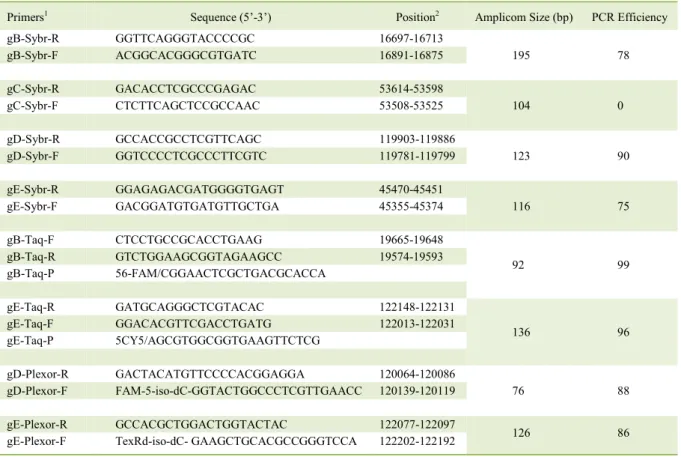
Table 1 - Primers used in this research for molecular detection of SuHV-1 targeting different genomic regions and using different qPCR chemistries.
Primers1 Sequence (5’-3’) Position2 Amplicom Size (bp) PCR Efficiency
gB-Sybr-R GGTTCAGGGTACCCCGC 16697-16713
195 78
gB-Sybr-F ACGGCACGGGCGTGATC 16891-16875
gC-Sybr-R GACACCTCGCCCGAGAC 53614-53598
104 0
gC-Sybr-F CTCTTCAGCTCCGCCAAC 53508-53525
gD-Sybr-R GCCACCGCCTCGTTCAGC 119903-119886
123 90
gD-Sybr-F GGTCCCCTCGCCCTTCGTC 119781-119799
gE-Sybr-R GGAGAGACGATGGGGTGAGT 45470-45451
116 75
gE-Sybr-F GACGGATGTGATGTTGCTGA 45355-45374
gB-Taq-F CTCCTGCCGCACCTGAAG 19665-19648
92 99
gB-Taq-R GTCTGGAAGCGGTAGAAGCC 19574-19593
gB-Taq-P 56-FAM/CGGAACTCGCTGACGCACCA
gE-Taq-R GATGCAGGGCTCGTACAC 122148-122131
136 96
gE-Taq-F GGACACGTTCGACCTGATG 122013-122031
gE-Taq-P 5CY5/AGCGTGGCGGTGAAGTTCTCG
gD-Plexor-R GACTACATGTTCCCCACGGAGGA 120064-120086
76 88
gD-Plexor-F FAM-5-iso-dC-GGTACTGGCCCTCGTTGAACC 120139-120119
gE-Plexor-R GCCACGCTGGACTGGTACTAC 122077-122097
126 86
gE-Plexor-F TexRd-iso-dC-GAAGCTGCACGCCGGGTCCA 122202-122192
1The names of each oligonucleotide are formed by the name of the protein encoded by the gene followed by the qPCR system. Sybr:
SybrGreen, Taq: hydrolysis probe, Plexor: Plexor (Promega, USA). 2
farms with no history of PR. Samples were collected from adult animals in a slaughterhouse. Animals were previously tested by virus neutralization test and the tissues submitted to a nested PCR (FONSECA JR et al., 2010b). Diagnostic sensitivity was evaluated
using twenty-five samples (swine brain collected
from animals form farms in the state of Santa Catarina in 2003; DNA extracted three years before this study) positive in viral isolation and in a nested PCR (FONSECA JR et al., 2010b). Viruses presented in clinical sample were characterized as genotype II in the cited publication.
Performance verification was done only with the methodology with better efficiency and
LD as proposed by FONSECA JR et al. (2013). Testing repeatability occurred on three consecutive days, using seven different samples in triplicate (a sample in LD concentration, three samples a log above LD and three samples ½ log above LD in the respective concentrations from, 10-1.5 TCID50
mL-1, 10-1.0 TCID50 mL-1 and 10-0.5 TCID50 mL-1).
The same procedures were repeated for a second person. Statistical analysis for reproducibility and repeatability was done by calculation of variances and as described by ISO 16140 (2003).
The robustness was evaluated with the same samples. It was considered relevant to the methodology: pipetting error, reagent change and thermocycler. Thus, the variables tested were: the variation of +1µL and
-1µL in the final volume of reaction; comparison of
the equipment Rotor Gene 3000 (Qiagen, USA) with results obtained in 7500 machine (Applied Biosystem, USA) and we used UNG-dUTP qPCR Master Mix Kit (6mM MgCl2) (Ampliqon, Germany) for comparison with the TaqMan® Universal Fast PCR Master Mix.
For interlaboratory reproducibility, thirty negative and thirty positive spiked samples were used. Negative samples were composed of approximately 25mg of tissue (swine brain) and 600µL of TE. Samples were divided in two parts, one was tested in the laboratory and another part sent to the Animal Virology Laboratory in Veterinary school at the Universidade Federal de Minas Gerais. Thermocycler used in the interlaboratory test was the Real-time 7500 (Applied Biosystems, USA). DNA extraction was performed with extraction kit AccuPrep®
Genomic DNA Extraction (Bioneer Corporation, USA). The PCR reaction used the TaqMan® Fast Universal PCR
Master Mix (Applied Biosystems, USA).
RESULTS
Oligonucleotides were analyzed for dimer formation and dG due to self anneling or
loop formation. The gB-Sybr had 15 primer dimers and a maximum dG of 16.30 kcal mol-1. gE-Sybr
had no primer dimer. Reaction efficiencies varied from 0% to 99% (Table 1). All qPCRs amplified
DNA from strain Shope, but reactions targeting gE did not amplify DNA from vaccine strain Bartha.
Only reactions with best efficiency for each qPCR
chemistrie were included in the following tests. Oligonucleotides targeted to gB (hydrolysis probe) and gD (system SYBRGreen and Plexor) were
selected due to higher efficiency.
In relation to the amount of DNA, concentrations of 50ng µL-1 and 100ng µL-1 were
tested. As there was no significant difference between the amounts of amplification of DNA
tested, it was decided that the lower amount of DNA would be used in all tests in order to obtain a test with less interfering substances.
The final volume of all qPCRs was
20µL (18µL of mix and 2µL of DNA). Reagents concentrations for the qPCR with the best
performances were: 0.375pmol µL-1 of each
primer, 10µL of 2X Master Mix and 0.8% DMSO (Amresco®). Probe concentration was 0.2pmol uL-1 in
the hydrolysis probe technique.
The first tests were performed only with
the dilutions of the standard strain Shope, followed by phenol/chloroform extraction. The following LD were found for the different detection systems:
10-0.5 TCID50 mL-1 for gD-SYBR, 10-1.5 TCID50
mL-1 for gD-Plexor, 10-2.5 TCID50 mL-1 for gB-Taq.
In the following test, a new standard curve was constructed with approximately 25mg of porcine brain tissue (per tube) spiked with virus suspension to determine the matrix interference in the
SuHV-1 DNA amplification. For all systems tested, a new
LOD was reported: 100.5 TCID50 mL-1 (gD-SYBR),
from 10-0.5 TCID50 mL-1 (gD-plexor), from 10-1.5
TCID50 mL-1 (gB-Taq).
Analytical specificity was tested to verify
the interference of other infectious agents. The three best qPCR for each chemistry were tested and
proven to be high specific (100%). The qPCR did
not amplify DNA from any organisms save from SuHV-1 isolates.
Twenty-five positive samples were
tested for diagnostic sensitivity. The qPCR-gB-Taq presented the highest diagnostic sensitivity,
amplificating all positive samples (100% diagnostic
sensitivity). qPCR-gD-Sybr and PCR-gD-plexor had 72% and 96% diagnostic sensitivity, respectively.
The three qPCRs did not present any amplification
We tested only qPCR-gB-Taq for
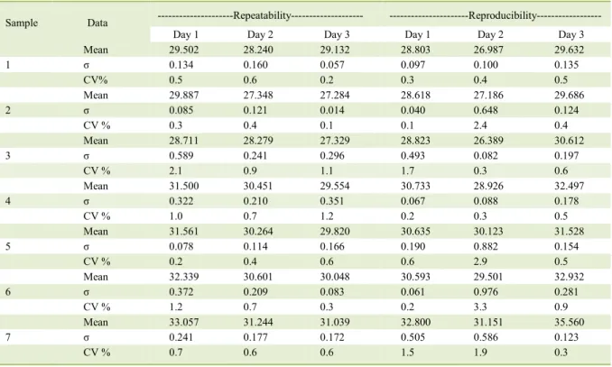
performance verification due to its high diagnostic sensitivity. Data analysis by the coefficient of
variance (CV), using seven samples in triplicates within three days of repeatability and reproducibility yielded the following results (Table 2). The CV was 5% for qPCR using 50ng µL-1 of DNA and 8% for
qPCR using 100ng µL-1 of DNA. These results show
a low dispersion of the data, demonstrating that the developed method was stable and homogenous for both DNA concentrations. Statistical analysis as described by ISO 16140 (2003) indicated 1.39% variance between days, 24% variance between analysts and 4.07% variance in analysis error. The sum of those errors is below 30%, which indicated that the tests are adequate.
The next step was the robustness evaluation.
Thus, variations considered significant for the method
were: thermal cycler (CV 5.5%), Master Mix (CV 1%) and pipetting error. The change of equipment and Master Mix did not affect the performance of the method. Reducing the reaction volume by 1µL did not interfere with the results and adding 1µL to the reaction mix suggested that an increase in the
final volume of the reaction could lead to increased
analytical sensitivity. There was 100% concordance in diagnostic sensitivity (positive samples) and 100%
concordance in specificity (negative samples) in the
interlaboratory tests.
DISCUSSION
Real-time PCR is an important tool in diagnosis. The viral detection by PCR in a latent or replicative state is a key tool in disease control (YOON et al., 2006). qPCR offer advantages like reduced risk of cross-contamination, shorter time required to perform the technique and excellent
sensitivity and specificity.
There are other PCRs for detection of SuHV-1 described in the literature. A nested-PCR (FONSECA JR et al., 2010a) tested using the same samples to test analytical sensitivity had the same re-sults for gD-plexor, was one log more sensible than gB-SYBR and one log less sensible than gB-Taq. Two hydrolysis probe qPCR for gE and gB described by MA et al. (2008) presented similar results, with gE less sensible than gB, but no PCR was able to amplify samples stored for more than 10 years as done in this research These PCRs, as well as others (YOON et al.,
Table 2 - Statistics generated from the Cts of qPCR-gB-Taq using samples at a concentration of 200 ng/uL.
Sample Data ---Repeatability--- ---Reproducibility---
Day 1 Day 2 Day 3 Day 1 Day 2 Day 3
Mean 29.502 28.240 29.132 28.803 26.987 29.632
1 σ 0.134 0.160 0.057 0.097 0.100 0.135
CV% 0.5 0.6 0.2 0.3 0.4 0.5
Mean 29.887 27.348 27.284 28.618 27.186 29.686
2 σ 0.085 0.121 0.014 0.040 0.648 0.124
CV % 0.3 0.4 0.1 0.1 2.4 0.4
Mean 28.711 28.279 27.329 28.823 26.389 30.612
3 σ 0.589 0.241 0.296 0.493 0.082 0.197
CV % 2.1 0.9 1.1 1.7 0.3 0.6
Mean 31.500 30.451 29.554 30.733 28.926 32.497
4 σ 0.322 0.210 0.351 0.067 0.088 0.178
CV % 1.0 0.7 1.2 0.2 0.3 0.5
Mean 31.561 30.264 29.820 30.635 30.123 31.528
5 σ 0.078 0.114 0.166 0.190 0.882 0.154
CV % 0.2 0.4 0.6 0.6 2.9 0.5
Mean 32.339 30.601 30.048 30.593 29.501 32.932
6 σ 0.372 0.209 0.083 0.061 0.976 0.281
CV % 1.2 0.7 0.3 0.2 3.3 0.9
Mean 33.057 31.244 31.039 32.800 31.151 35.560
7 σ 0.241 0.177 0.172 0.505 0.586 0.123
2005; YOON et al., 2006; PEREZ & ARCE, 2009), were not subjected to all steps in a validation such as described here (repeatability and reproducibility, interlaboratorial tests).
Repeatability and reproducibility tests indicated that qPCR-gB-Taq performs well under requirements described by ISO 1640 (2003). Robustness tests indicated that the qPCR-gB-Taq is not subject to the interferences analyzed. The CV in the use of other equipment was 5.5% and replacement of the master mix obtained a CV of 1%, demonstrating that exchanging equipment and replacement of Master Mix did not affect the performance of the
method. A significant CV must be greater than 15%.
The increased amount of mix suggested an increase in sensitivity, but further tests are needed to verify if this
would be beneficial to the assay, since increasing the final volume of the reaction also increased the cost of the reaction and, perhaps, decreased specificity.
This study demonstrated the importance of
performance verification and the use of clinical sam -ples in development of qPCR for diagnoses. The qPCR standardized and validated in this research proved to be reliable for the diagnosis of PR and may be used in diagnostic laboratories that follow Quality Assurance described by International Standard Guidelines like ISO 17025 (2010) and ISO 16140 (2003).
ACKNOWLEDGEMENTS
The authors are thankful for the financial support
from Fundação de Amparo à Pesquisa do Estado de Minas Gerais (Fapemig), Coordenação de Aperfeiçoamento de Pessoal de Nível Superior (Capes/Proex) and Conselho Nacional de
Desenvolvimento Científico e Tecnológico (CNPq). M. B. H. is research fellows of Conselho Nacional de Desenvolvimento
Científico e Tecnológico (CNPq).
REFERENCES
BUH GASPARIC, M. et al. Comparison of different real-time PCR
chemistries and their suitability for detection and quantification of
genetically modified organisms. BMC Biotechnology, v.8, p.26,
2008. Available from: <https://bmcbiotechnol.biomedcentral.com/ articles/10.1186/1472-6750-8-26>. Accessed: Mar. O8, 2016. doi: 10.1186/1472-6750-8-26.
CUNHA, E. M. S. et al. Antibodies against pseudorabies virus in feral swine in southeast Brazil. Arquivo Brasileiro de Medici-na Veterinária e Zootecnia, v.58, n.4, p.462-466, 2006. Avail-able from: <http://www.scielo.br/scielo.php?script=sci_arttext&p id=S0102-09352006000400002>. Accessed: Mar. O8, 2016. doi: 10.1590/S0102-09352006000400002.
DAVISON, A. J. et al. The order Herpesvirales. Archives of Vi-rology, v.154, n.1, p.171-177, 2009. Available from: <http://link. springer.com/article/10.1007%2Fs00705-008-0278-4>. Accessed: Mar. O8, 2016. doi: 10.1007/s00705-008-0278-4.
FONSECA JR, A. A. et al. Diagnóstico e genotipagem do vírus da pseudorraiva por nested-PCR e análise de restrição enzimática.
Ciência Rural, v.40, n.4, p.921-927, 2010a. Available from: <http:// www.scielo.br/pdf/cr/v40n4/a542cr2112.pdf>. Accessed: Mar. 08, 2016. doi: 10.1590/S0103-84782010005000066.
FONSECA JR, A. A. et al. Molecular epidemiology of Brazilian pseudorabies viral isolates. Veterinary Microbiology, v.141, n.3, p.238-245, 2010b. Available from: <http://www.sciencedirect. com/science/article/pii/S0378113509004350>. Accessed: Mar. 08, 2016. doi: 10.1016/j.vetmic.2009.09.018.
FONSECA Jr, A. A. et al. PCR em tempo real para detecção do vírus da doença de Aujeszky. Arquivos Brasileiros de Medicina Veterinária e Zootecnia, v.65, n.3, p.801-808, 2013. Available from: <http://www.scielo.br/pdf/abmvz/v65n3/28.pdf>. Accessed: Mar. 08, 2016. doi: 10.1590/S0102-09352013000300028.
FONSECA JR, A. A. et al. Pseudorabies virus can be classified into five genotypes using partial sequences of UL44. Brazilian Journal of Microbiology, v.43, n.4, p.1632-1640, 2012. Available from: <http://www.scielo.br/pdf/bjm/v43n4/48.pdf>. Accessed: Mar. 08, 2016. doi: 10.1590/S1517-83822012000400048.
GHALMI, F. et al. Detection of Neospora caninum in dog organs using real time PCR systems. Veterinary Parasitology, v.155, n.1-2, p.161-167, 2008. Available from: <http://www.sciencedirect. com/science/article/pii/S0304401708001945>. Accessed: Mar. 08, 2016. doi: 10.1016/j.vetpar.2008.04.007.
HALL, T. A. BioEdit: a user-friendly biological sequence alignment editor and analysis program for Windows 95/98/NT. Nucleic Acids Symposium Series, v.41, p.95-98, 1999.
ISO 16140. Microbiological of food and animal feeding stuffs - Protocol for the validation of alternative methods, 2003. Available from: <http://www.iso.org/iso/home/store/catalogue_tc/ catalogue_detail.htm?csnumber=30158>. Accessed: Mar. 08, 2016.
ISO 17025. General requeriments for the competence of test-ing and calibration laboratories, 2010. Available from: <http:// www.iso.org/iso/home/store/catalogue_tc/catalogue_detail. htm?csnumber=39883>. Accessed Mar. 08, 2016.
JULIAN, T. R.; SCHWAB, K. J. Challenges in environmental detection of human viral pathogens. Current Opinion in Virolology, v.2, n.1, p.78-83, 2012. Available from: <http://www.sciencedirect.com/science/article/pii/ S1879625711001611>. Accessed: Mar. 08, 2016. doi: 10.1016/j.coviro.2011.
KINKER, D. R. et al. Evaluation of serological tests for the detection of pseudorabies gE antibodies during early infection. Veterinary Microbiology, v.55, n.1, p.99-106, 1997. Available from: <http:// www.sciencedirect.com/science/article/pii/S0378113596013089>. Accessed: Mar. 08, 2016. doi: 10.1016/S0378-1135(96)01308-9.
MA, W. et al. Development of real-time polymerase chain reaction assays for rapid detection and differentiation of wild-type pseudorabies and gene-deleted vaccine viruses. Journal of Veterinary Diagnostic Investigation, v.20, n.4, p.440-447, 2008. Available from: <http://vdi. sagepub.com/content/20/4/440.full.pdf+html>. Accessed: Mar. 08, 2016. doi: 10.1177/104063870802000405.
MÜLLER, T. et al. Pseudorabies virus in wild swine: a global perspective.
OIE (WORLD ORGANIZATION FOR ANIMAL HEALTH). Chapter 2.1.2. Aujesky Disease. In: _____. Manual of diagnostic tests and vaccines for terrestrial animals. Paris, 2015. Available from: <http://www.oie.int/fileadmin/Home/eng/Health_standards/ tahm/2.01.02_AUJESZKYS_DIS.pdf>. Accessed: Mar. 08, 2016.
PAES, R. C. et al. Serological and molecular investigation of the prevalence of Aujeszky’s disease in feral swine (Sus scrofa) in the subregions of the Pantanal wetland, Brazil. Veterinary Microbiology, v.165, n.3-4, p.448-454, 2013. Available from: <http:// www.sciencedirect.com/science/article/pii/S0378113513002009>. Accessed: Mar. 08, 2016. doi: 10.1016/j.vetmic.2013.03.028.
PÉREZ, L. J.; ARCE, H. D. Development of a polymerase chain reac-tion assay for the detecreac-tion of pseudorabies virus in clinical samples.
Brazilian Journal of Microbiology, v.40, n.3, p.433-438, 2009. Avail-able from: <http://www.scielo.br/pdf/bjm/v40n3/v40n3a02.pdf>. Accessed: Mar. 08, 2016. doi: 10.1590/S1517-83822009000300002.
PINHEIRO de OLIVEIRA, T. F. et al. Detection of contaminants in cell cul-tures, sera and trypsin. Biologicals, v.41, n.6, p.407-414, 2013. Available from: <http://www.sciencedirect.com/science/article/pii/S1045105613001061>. Accessed: Mar. 08, 2016. doi: 10.1016/j.biologicals.2013.08.005.
SAMBROOK, J.; RUSSELL, D. W. Molecular cloning: a labora-tory manual. Laurel Hollow, New York. Local: Cold Spring Harbor Laboratory Press, 2001. 2100p.
YOON, H. A. et al. Investigation of pseudorabies virus latency in
ner-vous tissues of seropositive pigs exposed to field strain. Journal of Veterinary Medicine and Science, v.68, n.2, p.143-148, 2006. Avail-able from: <https://www.jstage.jst.go.jp/article/jvms/68/2/68_2_143/_ pdf>. Accessed: Mar. 08, 2016. doi: 10.1292/jvms.68.143.