Health Effects of Long-Term Rapamycin
Treatment: The Impact on Mouse Health of
Enteric Rapamycin Treatment from Four
Months of Age throughout Life
Kathleen E. Fischer1,2¤a
*, Jonathan A. L. Gelfond3, Vanessa Y. Soto1, Chul Han4, Shinichi Someya4, Arlan Richardson1,5¤b, Steven N. Austad1,5¤a
1Barshop Institute for Longevity & Aging Studies, University of Texas Health Science Center San Antonio, San Antonio, Texas, United States of America,2Department of Physiology, University of Texas Health Science Center San Antonio, San Antonio, Texas, United States of America,3Department of Epidemiology & Biostatistics, University of Texas Health Science Center San Antonio, San Antonio, Texas, United States of America,4Departments of Aging and Geriatric Research, University of Florida, Gainesville, Florida, United States of America,5Department of Cellular & Structural Biology, University of Texas Health Science Center San Antonio, San Antonio, Texas, United States of America
¤a Current address: Department of Biology, University of Alabama at Birmingham, Birmingham, Alabama,
United States of America
¤b Current address: University of Oklahoma Health Science Center & Oklahoma City Veteran Administration
Medical Center, Oklahoma City, Oklahoma, United States of America *fischerk@uab.edu
Abstract
Rapamycin, an mTOR inhibitor, has been shown to extend lifespan in a range of model or-ganisms. It has been reported to extend lifespan in multiple strains of mice, administered chronically or acutely early or late in life. The ability of rapamycin to extend health (health-span) as opposed to life is less well documented. To assess the effects chronic rapamycin treatment on healthspan, enteric rapamycin was given to male and female C57BL/6J mice starting at 4 months of age and continued throughout life. Repeated, longitudinal assess-ments of health in individual animals were made starting at 16 months of age (=12 months of treatment) until death. A number of health parameters were improved (female grip strength, female body mass and reduced sleep fragmentation in both sexes), others showed no significant difference, while at least one (male rotarod performance) was nega-tively affected. Rapamycin treatment affected many measures of health in a highly sex-spe-cific manner. While sex-spesex-spe-cific phenotypic effects of rapamycin treatment have been widely reported, in this study we document sex differences in thedirectionof phenotypic change. Rapamycin-fed males and females were both significantly different from controls; however the differences were in the opposite direction in measures of body mass, percent fat and resting metabolic rate, a pattern not previously reported.
OPEN ACCESS
Citation:Fischer KE, Gelfond JAL, Soto VY, Han C,
Someya S, Richardson A, et al. (2015) Health Effects of Long-Term Rapamycin Treatment: The Impact on Mouse Health of Enteric Rapamycin Treatment from Four Months of Age throughout Life. PLoS ONE 10(5): e0126644. doi:10.1371/journal.pone.0126644
Academic Editor:Christoph Englert, Leibniz Institute
for Age Research - Fritz Lipmann Institute (FLI), GERMANY
Received:November 3, 2014
Accepted:April 4, 2015
Published:May 15, 2015
Copyright:This is an open access article, free of all
copyright, and may be freely reproduced, distributed, transmitted, modified, built upon, or otherwise used by anyone for any lawful purpose. The work is made
available under theCreative Commons CC0public
domain dedication.
Data Availability Statement:Data will be available
for download athttp://uab.edu/cas/biology/people/
faculty-directory/steven-n-austad/DataSets ; Data is
available athttp://dx.doi.org/10.6084/m9.figshare.
1376133.
Funding:Financial Support was provided by the San
Antonio Nathan Shock Aging Center to AR
(P30-AG13319) (http://www.nia.nih.gov/research/dab/
nathan-shock-centers-excellence), National Institutes of Health RC2 Grand Opportunity grant to AR (AG
Introduction
Rapamycin, a potent mTOR inhibitor, has been reported to extend lifespan in both vertebrate and invertebrate model organisms. In at least 7 previous studies mouse lifespan has been shown to be extended in both sexes, in heterogeneous and several inbred strains, with rapamy-cin administered in food (enteric rapamyrapamy-cin) or via injection, chronically or acutely, at a variety
of ages [1–11]. However complete loss of mTOR signaling causes significant defects in growth
and/or development in worms (C.elegans) and flies (D.melanogaster), and is embryonic lethal
in mice [10]. The mTOR signaling network plays a pivotal role in promoting anabolic and
in-hibiting catabolic processes. mTOR activation stimulates ribosome biogenesis, protein
synthe-sis and the import of nutrients into cells [12]; conversely, reduction or inhibition of mTOR
activity decreases mRNA translation, increases autophagy, retards protein synthesis, slows cell growth and proliferation and enhances stress responsive transcription.
Because rapamycin treatment increases longevity across phylogenetically distant groups and is already FDA-approved for use in humans, it may represent a viable intervention to extend human life as well. Its effects on health have been less well documented. Recent evidence sug-gests that rapamycin may improve resistance to multiple, age-associated degenerative processes
in mice. This offers the tantalizing possibility that rapamycin could extend the length ofhealthy
human life. In mouse models rapamycin has been shown to delay the onset of Alzheimer’s
pa-thology [13,14], reduce the incidence of some cancers [4,15–17], inhibit the development of
atherosclerotic plaques [18], maintain cardiac function [1], enhance vaccine response in aged
animals [5], delay age-related cognitive decline [19–21], and maintain some aspects of activity,
motor function and behavior [1,4,11,15,16,21–23]. On the other hand, rapamycin has been
reported to have deleterious effects in mice, such as glucose intolerance and insulin resistance
[24], testicular degeneration, increased cataract severity [15] and nephrotoxicity [21]. In some
cases, results from different studies are inconsistent. For instance, some the beneficial effects on
age-related changes found in one study (e.g. improved cardiac function with age [1] or
in-creased insulin sensitivity [25]) have not been found in others (e.g. cardiac function [21], insulin
sensitivity [24]). The potential use of rapamycin to address age-related diseases is promising,
but the lack of consistent findings with respect to health in mice is reason for concern.
There are additional reasons for caution in considering rapamycin as a potential aging inter-vention. The use of rapamycin as part of immunosuppressive therapy after organ
transplanta-tion may be a reason for concern given age-related decline in immune functransplanta-tion [26,27];
however, recent research suggests that, in mice and primates, enterically delivered rapamycin
may enhance rather than suppress some aspects of immune response (e.g. [28,29]). Secondly,
because it inhibits protein synthesis, cellular processes requiringde novoprotein synthesis such
as growth, tissue repair and regeneration may be compromised by chronic rapamycin adminis-tration. For example, some rodent studies have observed that mTOR inhibition retards
recov-ery from skeletal [30] or cardiac muscle injury [31]. Additionally, rapamycin has been reported
to negatively affect neuronal long-term potentiation and memory consolidation [32,33]. Both
human and rodent studies have associated inhibition of mTOR with insulin resistance [34];
however recent studies have suggested that these effects are transitory and diminish as duration
of chronic treatment increases [1,22].
Extending lifespan without delaying or diminishing age-related morbidity is not a desirable goal and rapamycin’s effects on healthspan were anything but clear. We therefore initiated a lon-gitudinal study of rapamycin’s impact on longevity and a range of health parameters by treating C57BL/6 mice of both sexes with enteric rapamycin started at two distinct ages, 19 months (= old-fed or OF mice) and 4 months (= young-fed or YF mice) and continuing treatment
through-out life. OF results have been previously published [11]. Consistent with our previous findings,
RFA-OD-09-004.html), and NIH training grant (T32
AG 021890) to SNA (http://www.nia.nih.gov/research/
dea/institutional-training-grants).
Competing Interests:The authors have declared
survival of both males and females was modestly enhanced in YF animals [35]. Here we report on health impacts of enteric rapamycin treatment in YF mice of both sexes.
Materials and Methods
Animals and Husbandry
Three month old male and female C57BL/6J mice were purchased from the Jackson Laborato-ries. At four months of age 160 mice, 80 animals per sex, began receiving mouse chow (Purina 5LG6) containing either microencapsulated rapamycin (14 ppm) or empty capsules
(eudragit = control), (n = 40 animals per sex and treatment group). Animals were maintained on their respective diets throughout life. Mice were assessed for blood levels of rapamycin at 10 months (i.e. after 6 months of treatment) and began assessments for a spectrum of health pa-rameters starting at 16 months of age (after 12 months of treatment). All behavioral and physi-ological assays were performed in a single-blind design at regular intervals until an animal died or was euthanized for health reasons. Terminally ill animals were euthanized via continued ex-posure to CO2 for at least 15 minutes after respiratory arrest, followed by cervical dislocation. Unless otherwise indicated, assessments were performed during the dark (= active) phase of the 12:12 light cycle (grip strength, stride length, rotarod). Detailed descriptions of all
experi-mental procedures may be found in our previous study of OF mice [11].
Ethics Statement
Mice were maintained under barrier conditions by the UTHSCSA Nathan Shock Center Aging Animal and Longevity Assessment Core. All procedures for this study were approved by the Institutional Animal Care and Use Committee at the University of Texas Health Science Cen-ter at San Antonio. All animal died naturally or were euthanized for health reasons. All animals were closely monitored by project staff daily for health; animals that showed evidence of be-coming ill were checked daily until the condition resolved or the animals was euthanized. Ter-minally ill animals were euthanized via continued exposure to CO2 for at least 15 minutes after respiratory arrest, followed by cervical dislocation.
Rapamycin Quantification
Rapamycin concentration was quantified in uncoagulated whole mouse blood using
high-per-formance-liquid chromatography (HPLC) with tandem mass spectrometry [11]. Blood was
collected from both fasted and fed 10 month-old female and male mice (n = 17 and n = 14, re-spectively) that had been on rapamycin for 6 months.
Food Consumption
Using a standard protocol [36], food consumption and body mass were measured in a subset
of male and female, control and rapamycin-fed mice (n = 15 per sex and treatment group), starting at 5 months of age until 21 months of age.
Body Composition
Energetics and Spontaneous Activity
Following a 12 hours acclimation period, oxygen consumption, carbon dioxide production, total metabolism and resting metabolic rate were measured over a 24-hour period using a MARS indirect calorimetry system (Sable Systems International, Las Vegas, NV). Spontaneous activity and its temporal patterns were assessed in individually-housed animals over twenty-four hours, one light and one dark phase, following a twelve hour acclimation period. Animals were placed in clear, Plexiglas, (40.6 x 22.9 x 14.0 cm) cages surrounded by a 2.5 cm grid of
in-frared sensors in the x and y plane withad libaccess to food and water.
Movement and Strength Parameters
Gait. Senescence induces changes in the musculoskeletal system, including many patho-physiological conditions such as arthritis. Gait analysis provides a noninvasive method of as-sessing these changes. Gait parameters were measured using the TreadScan (Clever Sys, Reston, VA) apparatus. Mice were started at 12 cm/s and treadmill speed was adjusted until each mouse maintained a consistent walking speed for 5 minutes. TreadScan uses a high-speed digital camera to record reflected images of the mouse’s footpads at 80 frames per second and the images analyzed using mouse-specific algorithms to produce an assessment of more than 40 gait parameters. As many of these parameters are autocorrelated, we focused on stride length, which is likely to be affected by joint pain, neuromuscular deterioration and/or incipi-ent kyphosis, as a reasonable parameter to reflect musculoskeletal health.
Rotarod. Mice were trained to use the rotarod during four sessions, spanning two weeks
using the protocol described in our previous study [11]. Briefly, the mouse is balanced on the
rod and the instrument started at 4rpm with an acceleration of 0.2 rotations/second. The maxi-mum duration the mouse was able to balance on the rod (latency to fall) from six trials during the final fifth session was used to assess rotarod performance.
Grip strength. In humans, hand grip strength declines with increasing age and has been
used widely as a component of frailty assessments [37]. We assessed mouse fore and hind-limb
grip strength using a Grip Strength Meter with mesh grid pull bar (Columbus Instruments 1027 CSM). Mice grasped the pull bar with all four paws and were gently pulled backward in a horizontal plane until they released their grip. Maximum force during each pull (= 1 trial) is re-corded and the highest value from 5 consecutive trials was used as the mouse’s maximum grip strength for that age.
Inner Ear Histology
Dietary restriction slows the progression of age-related hearing loss in C57BL/6 mice as well as slowing the loss of spiral ganglion neurons and both outer and inner hair cells of the cochlea
[38]. Because mTOR inhibition by rapamycin has been hypothesized to resemble dietary
re-striction [39], we examined cochlear histology at natural death in a subset of mice. Techniques
were those of Someya [40]. Briefly, cochleae from control (males, n = 13; females n = 18) and
rapamycin-fed (males, n = 5; females n = 12) animals were excised from formalin-preserved mice, decalcified in 10% ethylenediamintetracetic acid. Paraffin-embedded sections were stained with haematoxylin and eosin and examined under a light microscope. Rosenthal’s canal was divided into three cochlear regions: apical, middle and basal, and each region was evaluated separately. The number of spiral ganglion neurons was counted on digital
photomi-crographs of each canal profile and determined as the number of neurons per mm2. Percent
hearing. Inner hair (IH) cell survival was calculated as the number of intact IH cells compared to the number expected in each turn of the cochlea in tissues of mice with normal hearing.
Statistical Methods
The continuous healthspan outcomes (body mass, body composition, metabolism, activity, etc.) were analyzed using linear random-effects models (with a random intercept) to account for the correlations between longitudinal measures of the same mouse. The main effects rapa-mycin treatment, sex, and age and their interactions were considered as potential predictors of healthspan. Because the effect of age on performance could be curvilinear (e.g., on body mass), we used polynomial transformations of age of up to order 3. We modeled males and females both together (for greater power to detect smaller effects consistent between males and females) and separately if, for example, there was a significant interaction between treatment and sex. For assays such as rotarod performance that are known to be affected by body size, we per-formed both body mass-adjusted and unadjusted analyses. The analytical computations were performed using R (v2+, Vienna, Austria).
Results
Rapamycin Blood Levels
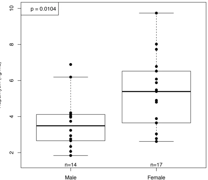
Blood levels of rapamycin were comparable to those measured in our previous study of C57BL/ 6 receiving enteric rapamycin started at 19 months of age and measured 6 months later
(old-fed mice, OF) [11]. Unlike our previous results in OF mice, the YF females showed significantly
higher blood levels of rapamycin than YF males (Fig 1). The sex difference in rapamycin blood
levels of YF mice was not due to differences in food consumption, as rapamycin-fed females did not consume more food per gram of body mass at testing age than did rapamycin-fed
males (p= 0.83). This is in contrast to our observations in old-fed mice, where OF females
con-sumed significantly more food per gram body mass than males (p= 0.004) but showed no
dif-ferences in rapamycin levels [11]. These results are consistent with those obtained by Fok et al
[35] in a companion study previously reported here. In this study, females also showed
signifi-cantly higher blood levels of rapamycin than males at 10 months of age; however, rapamycin levels in liver tissue measured at 25 months of age were no different between males and females
[35]. Fok et al reported that mTOR activity in the liver, as measured by the ratio of
phosphory-lated S6 Kinase 1 to total S6 Kinase 1, did not differ between control and rapamycin-fed ani-mals at 21 months of age; however, mTOR transcripts in the liver were significantly increased
in rapamycin-fed females and a subset of the rapamycin-fed males [35].
Body Mass and Composition
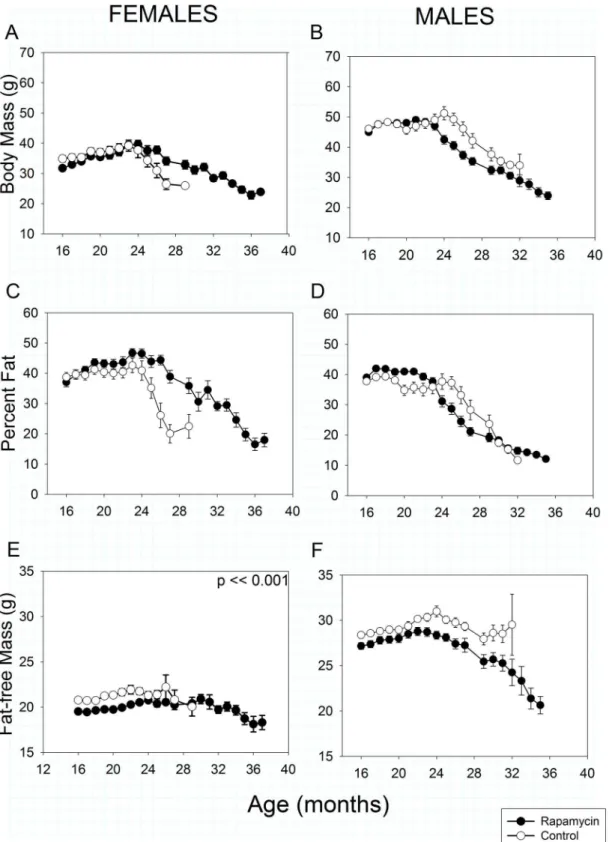
In both sexes and treatments, total body mass and percent fat declined progressively beginning
at 20–25 months of age (Fig2A–2D). At 16 months of age, after one year of treatment,
rapamy-cin-fed females weighed slightly less than controls (Treatment effect,p= 0.04) due to lower
fat-free mass. However, once weight loss began rapamycin-fed females lost body and fat mass
sig-nificantly more slowly with age than controls due to a slower loss of fat (Fig2Aand2C:
Treat-ment X Age interaction,p<<0.001 in both cases). Rapamycin treated females had less fat-free
mass than controls at all ages measured (Treatment effect,p<<0.001,Fig 2E). Fat-free mass
also declined significantly with age in both rapamycin-treated and control-fed females
(Treat-ment X Age interaction,p<0.001), although this loss is obscured somewhat by the scaling in
Fig 2, which is designed to allow easy comparison between sexes. The degree of fat-free mass
Control and rapamycin-fed males did not differ in body mass or composition at 16 months of age, but lost mass with aging significantly differently (Treatment X Age interaction,
p<0.001). Rapamycin-fed males began losing body mass about one month earlier than
con-trols (Fig 2B), although once it began to decline, mean body mass in both groups fell at similar
rates with age such that from about 25 months of age rapamycin-fed males consistently weighed less than controls. For both rapamycin-fed and control males, trajectories of age-relat-ed changes in both fat mass and fat-free mass were similar to those in total body mass.
Metabolism and Spontaneous Activity
A meaningful interpretation of metabolism requires accounting for body mass and composi-tion because fat-free mass is significantly more metabolically active than fat mass. Therefore, our metabolic rate measures are adjusted to amount of fat-free mass. According to this mea-sure, all mice showed an age-related decline in total, mass-specific metabolic rate irrespective Fig 1. Rapamycin concentration in whole blood at 10 months of age (after 6 months of rapamycin feeding).Blood concentrations of rapamycin were significantly higher in females than males after 6 months of rapamycin feeding (n = 18 and 14 respectively).
Fig 2. Body mass and composition in rapamycin-fed mice (filled circles) versus controls (hollow circles).P-values shown on individual panels only if there is a significant treatment effect independent of age. Sample sizes varied, depending on age, control females, n = 32–2; rapamycin females, n = 30–4;
control males, n = 33–2; rapamycin males, n = 36–3.A, B: Total body mass.Highly significant differences (p<<0.001) exist in treatment x age effects in
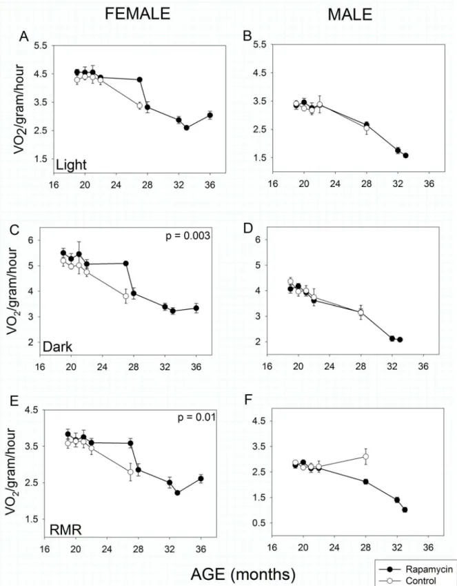
of sex or treatment from about 20 months of age (Age effect,p<<0.001 in all cases). In
fe-males, there was no significant effect of rapamycin treatment during the light (= inactive)
phase of the 24 hour light:dark cycle (Fig 3A), but rapamycin-fed females exhibited highertotal
mass-specific metabolic rate during the dark (i.e., active) phase (Fig 3C: Treatment effect,
p= 0.003) and a higher mass-specificrestingmetabolic rate (Fig 3E: Treatment effect,p= 0.01)
irrespective of light:dark cycle.
By contrast, males showed no differences in total metabolic rate as a function of treatment in either phase of the 24 hour light:dark cycle. Interestingly, control males showed no decline in restingmetabolic rate with age, whereas it declined significantly in the rapamycin-fed group (Fig
3F), thus there was a highly significant Treatment X Age interaction (p<<0.001) in males.
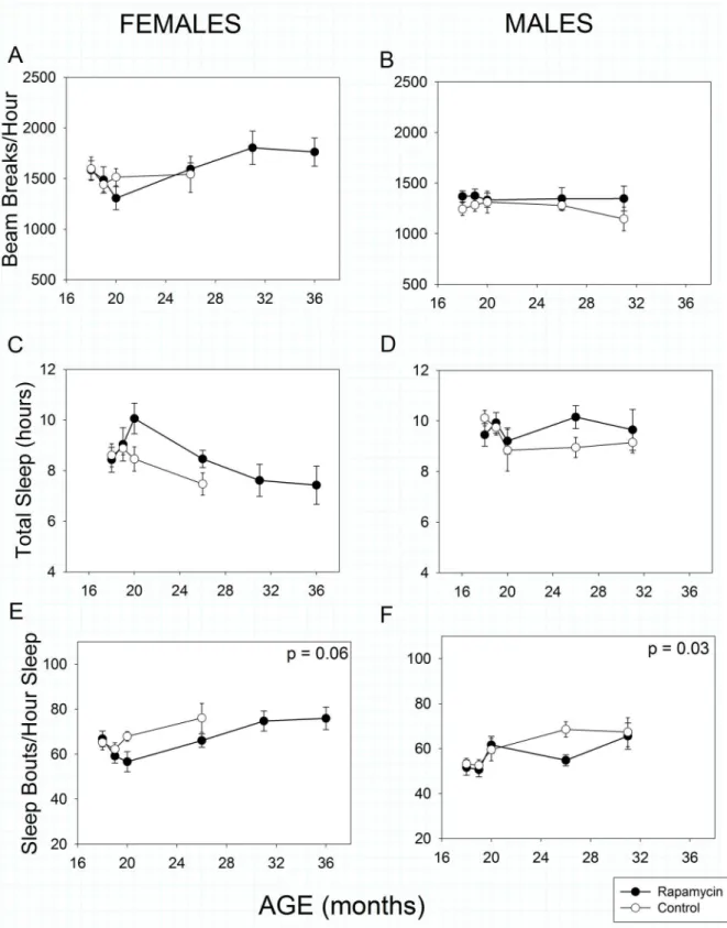
Total spontaneous activity over 24 hours was statistically greater in females than males (Fig
4Avs4B, Sex effect, p<<0.001). Surprisingly, spontaneous activity as we measured it increased
with age in females (Age effect, p = 0.008) but decreased as expected in males (Age effect,
p= 0.003). Rapamycin feeding had no impact on total activity in females (Treatment effect,
p= 0.54) or males (Treatment effect,p= 0.39).
Temporal patterns of sleep were monitored by analyzing bouts of inactivity greater than 40
seconds in length as previously validated for male C57BL/6 mice [11,41]. Using this metric,
fe-males, but not fe-males, slept less as they aged (Fig 4C, Age effect,p< <0.001).
Rapamycin-feed-ing resulted in a marginally significant increase in total sleep time (Treatment effect,p= 0.05)
when the sexes were combined, but not when each sex was treated separately (females:
Treat-ment effect,p= 0.10, males: Treatment effect,p= 0.26). Sleep fragmentation, as assessed by the
number of sleep bouts per hour of sleep, increased with age in all animals (Fig4Eand4F, Age
effect,p<<0.001) as has been previously reported for both humans and mice. However,
con-sistent with our OF results, rapamycin treatment reduced sleep fragmentation in males relative
to controls (Treatment effect,p =0.03). Unlike our OF results, rapamycin treatment reduced
sleep fragmentation marginally in females (Treatment effect,p= 0.06) compared to controls.
Because this high-throughput sleep assessment protocol has only been validated in C57BL/6 males, the above results for females should be interpreted with caution.
Strength and Movement
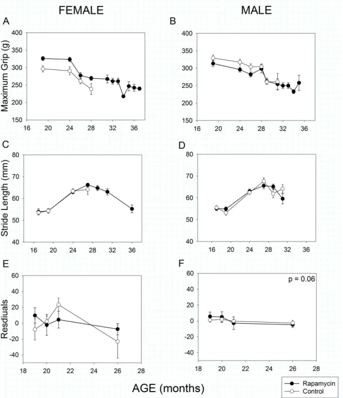
As in humans, mouse grip strength declined with age in both male and female mice (Fig5Aand
5B, Age effect,p<<0.001). As with a number of other parameters, rapamycin-feeding had a
sex-specific effect. Rapamycin-fed females’grip was stronger than that of female controls at all
ages measured (Treatment effect,p= 0.005), whereas grip strength of control and rapamycin-fed
males did not differ (Treatment effect,p= 0.21). Although there were sex- and treatment-specific
differences in body mass, these did not explain the grip strength results as body mass did not
cor-relate with grip strength in either male (r2= 0.07,p= 0.07) or female mice (r2= 0.01,p= 0.90).
Stride length has previously been reported to decline with age in C57BL/6 mice [11,42]. In
both sexes stride length actuallyincreaseduntil 27 months of age, after which it declined with
increasing age (Fig5Cand5D, Age effect,p<<0.001). However, rapamycin treatment had no
effect on age-related changes in stride length in either sex (Treatment X Age effect, females:
p= 0.88, males:p= 0.48).
Rotarod performance in mice is well known to be strongly affected by body mass [11,43,
44] as it was in this study (Mass effect,p<0.001 for both sexes). Therefore, we used body mass
body fat more slowly than age-matched controls. In contrast, rapamycin-fed males initially had a higher percentage of body fat, but lost fat mass earlier than controls.E, F: Fat-free mass, sometimes referred to as lean mass. Although obscured by the scaling, rapamycin-fed females had lower fat-free mass than controls at all ages measured. Fat-free mass declined more slowly with age in rapamycin-fed females than males.
Fig 3. Metabolic activity in rapamycin-fed mice (filled circles) compared to controls (hollow circles).P-values shown on individual panels only if there is a significant treatment effect independent of age. Sample sizes varied, depending on age, control females, n = 8–4; rapamycin females, n = 11–3; control
males, n = 9–5; rapamycin males, n = 16–2.A, B: Mass-specific metabolic rate during the light (= inactive) phase. Males and females showed no effects
of rapamycin treatment on mass-specific metabolic rate during the inactive phase of their daily 24-hour cycle, although both sexes showed highly significant (p<<0.001) sex x age treatment effects.C, D: Mass-specific metabolic rate during the dark (= active) phase. Aging rapamycin-fed females, but not
as a covariate in our analysis. There was no clear pattern of change with age in either females (Fig 5E) or males (Fig 5F). Rapamycin treatment had no effect on female performance
(Treat-ment effect,p= 0.42), but had a marginally significant negative effect on male rotarod
perfor-mance (Treatment effect,p= 0.06). If body mass was ignored, there was still no rapamycin
effect in females (Treatment effect,p= 0.65) and a significant negative effect in males
(Treat-ment effect,p= 0.01).
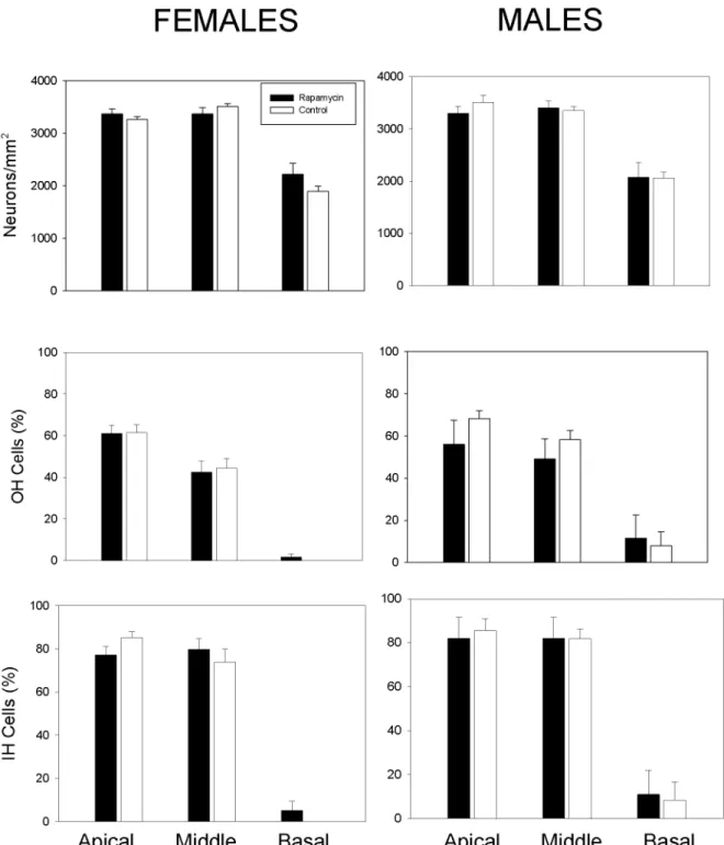
Cochlear Histology
Age-related hearing loss is a common feature of aging and associated with loss of spiral gangli-on neurgangli-ons and sensory inner and outer hair cells in the cochlea. Cochlear histology in rapa-mycin-treated and control mice was assessed upon natural death or health-related euthanasia. There were no statistically significant differences in the ages of animals of either sex examined
as a function of treatment (Mann-Whitney Rank Sum Test: males:p= 0.63, females:p= 0.45).
Median ages examined were 24.4 and 25.4 months for control and rapamycin-fed males, re-spectively, and 24.5 and 25.5 months form control and rapamycin-fed females. No statistical
differences between rapamycin-fed and control mice were observed (Fig 6). Collectively,
rapa-mycin treatment had no impact on age-related cochlear pathology in males or females.
Discussion
This study administered enteric rapamycin to C57BL/6J mice of both sexes andlongitudinally
monitored their phenotypes for a longer time—from 4 months of age until death—than any previous study. Therefore, individual, life-long trajectories of phenotypic change could be
ana-lyzed. Although survival was increased in both sexes [35], only some presumptive metrics of
health were improved (female grip strength, reduced sleep fragmentation, body mass), a num-ber showed no significant change, and at least one (male rotarod performance) was negatively affected. Age-related improvements in total sleep and greater sleep consolidation with rapamy-cin-feeding were observed in both sexes here, this is contrast to our previous OF study where
only rapamycin treated males showed improvements in sleep [11]. These are the first reports of
rapamycin affecting sleep.
At least 7 previous studies in an array of genotypes have reported enhanced longevity in mice
administered rapamycin [2,3,5,11,15,21,45]. However, reports of rapamycin’s effects on
health have been less numerous, none have made repeated assessments of the same animals lon-gitudinally, and results have been considerably less uniform than the survival results. The health effects described here differ from those reported in our own previous study—performed in the same facility with the same equipment. Compared to our previous study, in which enteric
rapa-mycin treatment was initiated at 19 months and continued throughout life (OF mice) [11], the
YF mice in this study exhibited considerable differences in phenotype, and not just in the ex-pected direction one might associate with earlier age and longer duration of rapamycin treatment (Table 1). For instance, we previously found rapamycin feeding significantly decreased
mass-spe-cific food consumption and resting metabolic rate in OF females relative to controls [11]. By
con-trast, YF females showed no difference in mass-specific food consumption and anincreased
resting metabolic rate compared to controls (Table 1). The pattern of age-related body mass and
composition changes also differs between the two studies. In OF females, there was no significant phase of the 24-hour light cycle. Both males and females showed highly significant (p<<0.001) decline dark-phase metabolic rate with age irrespective of
treatment.E, F: Resting mass-specific metabolic rate. Resting metabolic rate declined with age in females, but aging rapamycin-fed females had higher resting metabolic rates compared to matched controls. Resting metabolic rate declined significantly in aging rapamycin-fed males but not in age-matched controls (treatment x age, p<<0.001).
Fig 4. Spontaneous activity and sleep in rapamycin-fed mice (filled circles) compared to controls (hollow circles).P-values shown on individual panels only if there is a significant treatment effect independent of age. Sample sizes varied, depending on age, control females, n = 14–2; rapamycin
females, n = 18–5; control males, n = 13–4; rapamycin males, n = 22–7.A, B: Spontaneous 24-hour activitywas greater in females than males (p<<
Fig 5. Strength, coordination and movement in rapamycin-fed mice (filled circles) compared to controls (hollow circles).P-values shown on individual panels only if there is a significant treatment effect independent of age.A, B: Grip strengthdeclined significantly with age in all animals, regardless of treatment (p<<0.001); however rapamycin treatment affected males and females differently (treatment x sex, p = 0.003). Rapamycin-fed females had greater grip strength than controls at all ages measured; whereas grip strength in control and rapamycin treated males did not differ. Sample sizes varied, depending on age, control females, n = 17–7; rapamycin females, n = 27–4; control males, n = 22–4; rapamycin males, n = 31–3.C, D: Stride length
increased in males and females until 27 months of age and then declined with increasing age (p<<0.001). Rapamycin treatment had no effect on stride length in either sex. Sample sizes varied, depending on age, control females, n = 15–6; rapamycin females, n = 21–5; control males, n = 19–4; rapamycin
difference in total body mass, percent body fat, or their rate of decline with age compared with controls; however YF females were significantly better than controls at maintaining body and fat
mass into late life (Fig 2). Given its well-known role in growth and previous, shorter-term studies
of its effects on body mass [3,21,45], the better maintenance of body and fat mass with age in
our YF females was a substantial surprise. OF females showed no difference in fat-free mass com-pared with controls, whereas YF females had less fat-free mass than controls. Thus, the impact of
rapamycin-feeding on various metabolic parametersin femalesappears to depend on the age of
initiation and/or the duration of feeding in sometimes counterintuitive ways.
OF and YF male phenotypic changes also differed in response to rapamycin treatment, but mainly in easily understood directions. Several parameters such as percent body fat or mass-specific food consumption that did not achieve statistical significance in the shorter duration, OF study, did achieve significance in the longer, YF study. Unlike the current study, there was no statistically significant difference between the sexes in blood concentration of rapamycin in the OF study. However, both studies showed differences in the same direction, with females showing higher blood levels of rapamycin than males.
Sex-specific phenotypic effects of enteric rapamycin treatment in mice have been widely
re-ported; in this study we report sex differences in thedirectionof phenotypic change. While
males and females on enteric rapamycin were both significantly different from controls, they exhibited changes in the opposite direction in measures of body mass, percent fat and mass-specific resting metabolic rate. While this pattern of sex differences has not been previously
re-ported, few phenotypic studies have been performed on both sexes (although see [15,45]) and
none have been performed longitudinally in both sexes over as long a time course as the pres-ent study. In the few phenotypic studies of both sexes that have been reported, it is not uncom-mon to find a statistically significant difference associated with enteric rapamycin treatment in
one sex but not the other. Both this study and Miller, et al. [45] found significantly greater
blood concentrations of rapamycin in females compared with males. In this study, the pattern was not a result of greater mass-specific food consumption in females, suggesting that such dif-ferences could be a consequence of greater bioavailability of rapamycin in females compared with males. Still, interpretation of differences in blood concentration should be treated with
caution as they do not necessarily reflect tissue concentration of rapamycin [11,35]. The
mech-anistic basis of these sex-specific differences certainly warrants further study.
A number of studies have previously reported health effects of rapamycin administration in shorter-term experiments. For instance, rapamycin treatment by injection in 22–24 month old male C57BL/6 mice improved several measures of health, including hematopoietic stem cell
function [5] and 3 months of enteric rapamycin treatment in even older female C57BL/6 mice
improved cardiac function and some metrics of behavioral, motor and skeletal phenotypes [1].
Similarly, chronically administered enteric rapamycin started at 9 months of age at a range of doses (4.7–42 ppm) maintained activity and blunted the expression of aging-related changes in heart, liver, adrenal glands, endometrial tissue and tendons in 20–22 month-old male and
fe-male UM-HET3 mice [15]. Likewise, chronic, enteric rapamycin treatment enhanced cognitive
function in young C57BL/6 adult mice and reduced age-associated cognitive decline in older
mice of both sexes [20] and Neff et al. [21] reported improved hepatic, immune and endocrine
function in male mice of the same strain.
was included as a covariate in the analysis. With the effects of body size removed, females showed no effects of rapamycin treatment and males showed a marginally significant negative effect of rapamycin treatment on rotarod performance. The y-axis shows the residuals of rotarod performance (latency to fall) regressed against body mass. Sample sizes varied, depending on age, control females, n = 11–6; rapamycin females, n = 19–6; control males, n = 13–7;
rapamycin males, n = 21–8.
Conclusions
In sum, longitudinal measures of health in C57BL/6J mice treated with enteric rapamycin (14 ppm) from 4 months of age revealed a number of previously unreported patterns. Although Fig 6. Age-related changes in inner ear histology was not altered by rapamycin treatment. A, B: The number of cochlear neuronsin male and female mice were not statistically different between control and rapamycin-fed animals.C,D: The number of outer hair cellsin male and female mice were not statistically different between control and rapamycin-fed animals.E,F: The number of inner hair cellsin male and female mice were not statistically different between control and rapamycin-fed animals.
a number of health parameters were improved and a number unchanged, at least one (male rotarod performance) was marginally worse under rapamycin treatment. Other deleterious side effects including testicular atrophy, accelerated cataract formation, and glucose
insensitivi-ty have been reported in shorter-term (~1 year) rapamycin feeding studies [15,45]. Whether
some of these effects, such as glucose insensitivity abate with longer-term treatment remains an intriguing question. We also observed considerable sex-specificity in the effects of enteric rapamycin treatment, including not only significant effects in one sex that were absent from the other, but also opposite phenotypic effects between the sexes. This phenomenon may pro-vide insights into mechanisms underlying sex differences in aging and deserves further inquiry.
Acknowledgments
John Ramos, Vivian Diaz and the staff at the Barshop Institute provided exemplary animal care that made this study possible. Dr. Alex Bokov offered expert advice on analytical issues. Prof. Walter Ward and Vivian Diaz provided data on food consumption; Drs. Lauren Sloane and Steven Treaster, and Samantha Rendon and Keith Maslin assisted with data collection; Prof. Martin Javors analyzed blood levels of rapamycin. Logan K. Walker assisted with cochlear neuron counting. We thank the editor and an anonymous reviewer for their helpful
sugges-tions. Figures were prepared using Scientifig (https://grr.gred-clermont.fr/labmirouse/
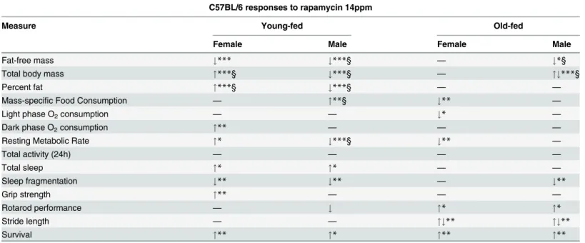
software/index.html) [46]. Table 1. Differences in two rapamycin studies.
C57BL/6 responses to rapamycin 14ppm
Measure Young-fed Old-fed
Female Male Female Male
Fat-free mass #*** #***§ — #*§
Total body mass "***§ #***§ — "#***§
Percent fat "***§ #***§ — —
Mass-specific Food Consumption — "**§ #** —
Light phase O2consumption — — #* —
Dark phase O2consumption "** — — —
Resting Metabolic Rate "* #***§ #** —
Total activity (24h) — — — —
Total sleep "* "* — —
Sleep fragmentation #** #** — #**
Grip strength "** — — —
Rotarod performance — # "* "*
Stride length — — "#** "#**
Survival "** "* "** "**
"Increased#Decreased,"#Maintained with age,—No difference from controls
*p<0.05
**p<0.01
***p<0.001 §
Treatment X Age effect
Healthspan measures: young-fed measured between ages 16 and 29 months; old-fed measured at 25, 31 and 32 months. Young-fed mice (rapamycin feeding begun at 4 mo, this study) compared with old-fed (rapamycin feeding begun at 19 mo) [11].
Author Contributions
Conceived and designed the experiments: KEF AR SNA. Performed the experiments: KEF VYS SS CH. Analyzed the data: KEF JALG SNA. Contributed reagents/materials/analysis tools: JALG SS. Wrote the paper: KEF SNA JALG. Revised manuscript: VYS AR SS CH.
References
1. Flynn JM, O'Leary MN, Zambataro CA, Academia EC, Presley MP, Garrett BJ, et al. Late-life rapamycin treatment reverses age-related heart dysfunction. Aging Cell. 2013; 12(5):851–62. doi:10.1111/acel.
12109PMID:23734717.
2. Harrison DE, Strong R, Sharp ZD, Nelson JF, Astle CM, Flurkey K, et al. Rapamycin fed late in life ex-tends lifespan in genetically heterogeneous mice. Nature. 2009; 460(7253):392–5. doi:10.1038/
nature08221PMID:19587680
3. Miller RA, Harrison DE, Astle CM, Baur JA, Boyd AR, de CR, et al. Rapamycin, but not resveratrol or simvastatin, extends life span of genetically heterogeneous mice. JGerontolA BiolSciMedSci. 2011; 66 (2):191–201. doi: glq178 [pii];doi:10.1093/gerona/glq178
4. Anisimov VN, Zabezhinski MA, Popovich IG, Piskunova TS, Semenchenko AV, Tyndyk ML, et al. Rapamycin increases lifespan and inhibits spontaneous tumorigenesis in inbred female mice. Cell Cycle. 2011; 10(24):4230–6. doi: 18486 [pii];doi:10.4161/cc.10.24.18486PMID:22107964
5. Chen C, Liu Y, Liu Y, Zheng P. mTOR regulation and therapeutic rejuvenation of aging hematopoietic stem cells. SciSignal. 2009; 2(98):ra75.
6. Medvedik O, Lamming DW, Kim KD, Sinclair DA. MSN2 and MSN4 link calorie restriction and TOR to sirtuin-mediated lifespan extension in Saccharomyces cerevisiae. PLoS Biol. 2007; 5(10):e261. doi: 06-PLBI-RA-0346 [pii];doi:10.1371/journal.pbio.0050261PMID:17914901
7. Powers RW III, Kaeberlein M, Caldwell SD, Kennedy BK, Fields S. Extension of chronological life span in yeast by decreased TOR pathway signaling. Genes Dev. 2006; 20(2):174–84. PMID:16418483
8. Kapahi P, Chen D, Rogers AN, Katewa SD, Li PW, Thomas EL, et al. With TOR, less is more: a key role for the conserved nutrient-sensing TOR pathway in aging. Cell Metab. 2010; 11(6):453–65. doi:
S1550-4131(10)00153-1 [pii];doi:10.1016/j.cmet.2010.05.001PMID:20519118
9. Kaeberlein M, Powers RW III, Steffen KK, Westman EA, Hu D, Dang N, et al. Regulation of yeast repli-cative life span by TOR and Sch9 in response to nutrients. Science. 2005; 310(5751):1193–6. PMID:
16293764
10. Katewa SD, Kapahi P. Role of TOR signaling in aging and related biological processes in Drosophila melanogaster. Experimental gerontology. 2011; 46(5):382–90. Epub 2010/12/07. doi:10.1016/j.exger.
2010.11.036PMID:21130151; PubMed Central PMCID: PMCPmc3058120.
11. Zhang Y, Bokov A, Gelfond J, Soto V, Ikeno Y, Hubbard G, et al. Rapamycin extends life and health in C57BL/6 mice. The journals of gerontology Series A, Biological sciences and medical sciences. 2014; 69(2):119–30. doi:10.1093/gerona/glt056PMID:23682161; PubMed Central PMCID: PMC4038246.
12. Wullschleger S, Loewith R, Hall MN. TOR signaling in growth and metabolism. Cell. 2006; 124(3):471–
84. doi: S0092-8674(06)00108-5 [pii];doi:10.1016/j.cell.2006.01.016PMID:16469695
13. Caccamo A, Majumder S, Richardson A, Strong R, Oddo S. Molecular interplay between mammalian target of rapamycin (mTOR), amyloid-beta, and Tau: effects on cognitive impairments. JBiolChem. 2010; 285(17):13107–20. doi: M110.100420 [pii];doi:10.1074/jbc.M110.100420PMID:20178983
14. Spilman P, Podlutskaya N, Hart MJ, Debnath J, Gorostiza O, Bredesen D, et al. Inhibition of mTOR by rapamycin abolishes cognitive deficits and reduces amyloid-beta levels in a mouse model of Alzhei-mer's disease. PLoSOne. 2010; 5(4):e9979. doi:10.1371/journal.pone.0009979PMID:20376313 15. Wilkinson JE, Burmeister L, Brooks SV, Chan CC, Friedline S, Harrison DE, et al. Rapamycin slows
aging in mice. Aging Cell. 2012; 11(4):675–82. doi:10.1111/j.1474-9726.2012.00832.xPMID:
22587563
16. Komarova EA, Antoch MP, Novototskaya LR, Chernova OB, Paszkiewicz G, Leontieva OV, et al. Rapamycin extends lifespan and delays tumorigenesis in heterozygous p53+/- mice. Aging. 2012; 4 (10):709–14. Epub 2012/11/06. PMID:23123616; PubMed Central PMCID: PMCPmc3517941.
17. Livi CB, Hardman RL, Christy BA, Dodds SG, Jones D, Williams C, et al. Rapamycin extends life span of Rb1+/- mice by inhibiting neuroendocrine tumors. Aging. 2013; 5(2):100–10. PMID:23454836;
PubMed Central PMCID: PMC3616197.
19. Majumder S, Caccamo A, Medina DX, Benavides AD, Javors MA, Kraig E, et al. Lifelong rapamycin ad-ministration ameliorates age-dependent cognitive deficits by reducing IL-1beta and enhancing NMDA signaling. Aging Cell. 2011. doi:10.1111/j.1474-9726.2011.00791.x
20. Halloran J, Hussong SA, Burbank R, Podlutskaya N, Fischer KE, Sloane LB, et al. Chronic inhibition of mammalian target of rapamycin by rapamycin modulates cognitive and non-cognitive components of behavior throughout lifespan in mice. Neuroscience. 2012; 223C:102–13. doi: S0306-4522(12)00672-0
[pii];doi:10.1016/j.neuroscience.2012.06.054
21. Neff F, Flores-Dominguez D, Ryan DP, Horsch M, Schroder S, Adler T, et al. Rapamycin extends mu-rine lifespan but has limited effects on aging. The Journal of clinical investigation. 2013; 123(8):3272–
91. Epub 2013/07/19. doi:10.1172/jci67674PMID:23863708; PubMed Central PMCID: PMCPmc3726163.
22. Fang Y, Westbrook R, Hill C, Boparai RK, Arum O, Spong A, et al. Duration of rapamycin treatment has differential effects on metabolism in mice. Cell Metab. 2013; 17(3):456–62. doi:10.1016/j.cmet.2013.
02.008PMID:23473038; PubMed Central PMCID: PMC3658445.
23. Spong A, Bartke A. Rapamycin slows aging in mice. Cell Cycle. 2012; 11(5):845. Epub 2012/02/24. doi:10.4161/cc.11.5.19607PMID:22356747.
24. Lamming DW, Ye L, Katajisto P, Goncalves MD, Saitoh M, Stevens DM, et al. Rapamycinduced in-sulin resistance is mediated by mTORC2 loss and uncoupled from longevity. Science. 2012; 335 (6076):1638–43. doi: 335/6076/1638 [pii];doi:10.1126/science.1215135PMID:22461615
25. Lamming DW, Ye L, Astle CM, Baur JA, Sabatini DM, Harrison DE. Young and old genetically hetero-geneous HET3 mice on a rapamycin diet are glucose intolerant but insulin sensitive. Aging Cell. 2013; 12(4):712–8. doi:10.1111/acel.12097PMID:23648089; PubMed Central PMCID: PMC3727050.
26. Smith JM, Nemeth TL, McDonald RA. Current immunosuppressive agents: efficacy, side effects, and utilization. PediatrClinNorth Am. 2003; 50(6):1283–300. PMID:14710781
27. Welzl K, Kern G, Mayer G, Weinberger B, Saemann MD, Sturm G, et al. Effect of different immunosup-pressive drugs on immune cells from young and old healthy persons. Gerontology. 2014; 60(3):229–
38. Epub 2014/01/18. doi:10.1159/000356020PMID:24434865.
28. Hinojosa CA, Mgbemena V, Van RS, Austad SN, Miller RA, Bose S, et al. Enteric-delivered rapamycin enhances resistance of aged mice to pneumococcal pneumonia through reduced cellular senescence. ExpGerontol. 2012; 47(12):958–65. doi: S0531-5565(12)00250-1 [pii];doi:10.1016/j.exger.2012.08.
013PMID:22981852
29. Turner AP, Shaffer VO, Araki K, Martens C, Turner PL, Gangappa S, et al. Sirolimus enhances the magnitude and quality of viral-specific CD8+ T-cell responses to vaccinia virus vaccination in rhesus macaques. American journal of transplantation: official journal of the American Society of Transplanta-tion and the American Society of Transplant Surgeons. 2011; 11(3):613–8. Epub 2011/02/24. doi:10.
1111/j.1600-6143.2010.03407.xPMID:21342450; PubMed Central PMCID: PMCPmc3076606. 30. Dickinson JM, Fry CS, Drummond MJ, Gundermann DM, Walker DK, Glynn EL, et al. Mammalian
tar-get of rapamycin complex 1 activation is required for the stimulation of human skeletal muscle protein synthesis by essential amino acids. JNutr. 2011; 141(5):856–62. doi: jn.111.139485 [pii];doi:10.3945/
jn.111.139485PMID:21430254
31. Aoyagi T, Kusakari Y, Xiao CY, Inouye BT, Takahashi M, Scherrer-Crosbie M, et al. Cardiac mTOR pro-tects the heart against ischemia-reperfusion injury. AmJPhysiol Heart CircPhysiol. 2012; 303(1):H75–
H85. doi: ajpheart.00241.2012 [pii];doi:10.1152/ajpheart.00241.2012PMID:22561297
32. Stoica L, Zhu PJ, Huang W, Zhou H, Kozma SC, Costa-Mattioli M. Selective pharmacogenetic inhibition of mammalian target of Rapamycin complex I (mTORC1) blocks long-term synaptic plasticity and mem-ory storage. ProcNatlAcadSciUSA. 2011; 108(9):3791–6. doi: 1014715108 [pii];doi:10.1073/pnas.
1014715108PMID:21307309
33. Parsons RG, Gafford GM, Helmstetter FJ. Translational control via the mammalian target of rapamycin pathway is critical for the formation and stability of long-term fear memory in amygdala neurons. JNeur-osci. 2006; 26(50):12977–83. doi: 26/50/12977 [pii];doi:10.1523/JNEUROSCI.4209-06.2006PMID:
17167087
34. Larsen JL, Bennett RG, Burkman T, Ramirez AL, Yamamoto S, Gulizia J, et al. Tacrolimus and siroli-mus cause insulin resistance in normal sprague dawley rats. Transplantation. 2006; 82(4):466–70. doi:
10.1097/01.tp.0000229384.22217.15;00007890-200608270-00005 [pii]. PMID:16926589
35. Fok WC, Chen Y, Bokov A, Zhang Y, Salmon AB, Diaz V, et al. Mice fed rapamycin have an increase in lifespan associated with major changes in the liver transcriptome. PLoS One. 2014; 9(1):e83988. Epub 2014/01/11. doi:10.1371/journal.pone.0083988PMID:24409289; PubMed Central PMCID:
36. Ikeno Y, Hubbard GB, Lee S, Richardson A, Strong R, Diaz V, et al. Housing density does not influence the longevity effect of calorie restriction. JGerontolA BiolSciMedSci. 2005; 60(12):1510–7. PMID:
16424282
37. Fried LP, Tangen CM, Walston J, Newman AB, Hirsch C, Gottdiener J, et al. Frailty in older adults: evi-dence for a phenotype. The journals of gerontology Series A, Biological sciences and medical sci-ences. 2001; 56(3):M146–56. Epub 2001/03/17. PMID:11253156.
38. Someya S, Yu W, Hallows WC, Xu J, Vann JM, Leeuwenburgh C, et al. Sirt3 mediates reduction of oxi-dative damage and prevention of age-related hearing loss under caloric restriction. Cell. 2010; 143 (5):802–12. Epub 2010/11/26. doi:10.1016/j.cell.2010.10.002PMID:21094524; PubMed Central
PMCID: PMCPmc3018849.
39. Kaeberlein M, Kennedy BK. Ageing: A midlife longevity drug? Nature. 2009; 460(7253):331–2. Epub
2009/07/17. doi:10.1038/460331aPMID:19606132.
40. Someya S, Xu J, Kondo K, Ding D, Salvi RJ, Yamasoba T, et al. Age-related hearing loss in C57BL/6J mice is mediated by Bak-dependent mitochondrial apoptosis. Proc Natl Acad Sci U S A. 2009; 106 (46):19432–7. Epub 2009/11/11. doi:10.1073/pnas.0908786106PMID:19901338; PubMed Central
PMCID: PMCPmc2780799.
41. Pack AI, Galante RJ, Maislin G, Cater J, Metaxas D, Lu S, et al. Novel method for high-throughput phe-notyping of sleep in mice. Physiol Genomics. 2007; 28(2):232–8. doi: 00139.2006 [pii];doi:10.1152/
physiolgenomics.00139.2006PMID:16985007
42. Judge JO, Davis RB III, Ounpuu S. Step length reductions in advanced age: the role of ankle and hip ki-netics. JGerontolA BiolSciMedSci. 1996; 51(6):M303–M12. PMID:8914503
43. McFadyen MP, Kusek G, Bolivar VJ, Flaherty L. Differences among eight inbred strains of mice in motor ability and motor learning on a rotorod. Genes Brain Behav. 2003; 2(4):214–9. PMID:12953787
44. Schellinck HMC DP, Brown RE. How many ways can mouse behavioral experiments go wrong? Con-founding variables in mouse models of neurodegenerative diseases and how to control them. In: Brock-man HJ, editor. Advances in the Study of Behavior. Burlington, VT: Academic Press; 2010. p. 255–
366.
45. Miller RA, Harrison DE, Astle CM, Fernandez E, Flurkey K, Han M, et al. Rapamycin-mediated lifespan increase in mice is dose and sex dependent and metabolically distinct from dietary restriction. Aging Cell. 2014; 13(3):468–77. Epub 2013/12/18. doi:10.1111/acel.12194PMID:24341993; PubMed
Cen-tral PMCID: PMCPmc4032600.