1. Research Laboratories and Academic Division of Clinical Rheumatology - Department of Internal Medicine and Specialities – IRCCS San Martino – University of Genova, Italy
2. Department of Rheumatology, Ghent University Hospital; Department of Internal Medicine, Ghent University – Ghent, Belgium
Systemic sclerosis: markers and targeted treatments
ACTA REUMATOL PORT. 2016;41:18-25
abStract
Systemic sclerosis (SSc) is characterized by autoanti-body production, progressive microvasculopathy, and aberrant extracellular matrix protein (ECM) synthesis in tissues. The disease presents two major clinical hall-marks: Raynaud’s phenomenon (RP) and skin involve-ment, followed by varying prevalences of internal or-gan involvement. Despite significant advances in the management of certain organ-specific involvements and symptoms, the research for efficient markers and targets, to be used for an optimized treatment, is still ongoing. Therapies targeting the vasculature (i.e. ET-1 receptor antagonists, phosphodiesterase-5 (PDE-5) inhi bitor, angiotensin-converting enzyme inhibition, prostacyclins), the immune system and/or the fibrotic process (i.e. traditional disease modifying antirheu matic drugs DMARDs such as methotrexate, cyclospo rine or mycophenolate mofetil, biologicals like rituxi -mab, tocilizumab or abatacept) have been or are being eva luated in SSc. Advanced approaches, reserved to unres ponsive SSc patients, include autologous haema -topoietic stem cell transplantation (HSTC) and intra-venous immunoglobulins (IVIG). Interestingly, it is ex-pected that new and future possible diagnostic and therapeutical approaches in SSc will come from epige-netic studies (MicroRNAs).
Ideally, combination therapy in SSc seems the best approach, together with the early intervention on the major hallmarks of the disease in “at risk” patients, that consists of the microvascular damage/altered function and the autoimmune reaction, followed by the pro-gressive and systemic fibrotic process.
Keywords: Systemic sclerosis; Capillaroscopy; Ray-naud phenomenon; Targeted therapies; Connective tis-sue diseases; Autoimmune diseases.
Cutolo M1, Sulli A1, Pizzorni C1, Paolino S1, Smith V2
IntroductIon
Systemic sclerosis (SSc) is a connective tissue disease (CTD) with autoimmune reactivity, chronic vascular and tissue stromal progressive alterations. These pro-cesses lead to autoantibody production, progressive mi-crovasculopathy, and aberrant extracellular matrix pro-tein (ECM) deposition with subsequent severe organ damage in skin, lungs, gastrointestinal tract and seve -ral other internal organs.
Research is still ongoing to define the initiating cause of the pathophysiological cascade and despite signifi-cant advances in the management of certain organ-spe-cific involvements and symptoms, SSc remains the CTD with the higher morbidity and mortality1,2.
Systemic sclerosis is an orphan disease with an an-nual incidence of 19 per million and prevalence of 19--75 per 100,000. The female:male ratio is 3:1, and 8:1 in mid to late childbearing years. Interestigly, juvenile SSc (children < 16 years old) is even more rare and one of the worst rheumatological conditions in children3.
Based on the argument that the past SSc classifica-tion criteria did not meet current standards for clini-metric properties, recently, new classification criteria based on a collaboration between the American Col-lege of Rheumatology (ACR) and the European League Against Rheumatism (EULAR) have been introduced4.
The sensitivity and specificity of the classification criteria have been significantly improved after this revision, and by introducing the analysis of the micro vascular damage as detected by the presence of telean -ge ctasia and capillarscopic features.
Additionally, to overcome the issue of properly classifying subjects with subclinical disease, Very Early Di -sease Onset Systemic Sclerosis (VEDOSS) criteria have been proposed5.
SyStemIc ScleroSIS hallmarkS
ma-jor hallmarks: Raynaud’s phenomenon (RP) and skin involvement, followed by varying prevalences of in-ternal organ involvement.
Based on the extension on skin involvement pa-tients with SSc can be classified into limited cutaneous (lcSSc) or diffuse cutaneous (dcSSc) involvement. Pa-tients with dcSSc with severe organ involvement may have the worst prognosis.
The vast majority of patients with SSc have RP (bi or tri-phasic colour changes of extremities upon ex-posure to cold or stress)6. It is noteworthy that the RP may occur in a healthy population (without any asso-ciation with CTD) or in secondary Raynaud's phe-nomenon (SRP) (mostly due SSc). The key point as a clinical medical doctor or specialist is to be able to make this differential diagnosis. The combination of capillaroscopy and serology allows to do so.
Moreover, this combination also allows to pinpoint those patients who only have RP at baseline, without any sign of another CTD, who will progress to a secon -dary over long time follow up (13.6% of patients with isolated RP at baseline)7.
Next to RP and skin involvement, other clinical complications are frequent in SSc.
Digital ulcers (DU) and gastrointestinal involvement occur frequently. DU occur in a major part of SSc pa-tients in their disease course. They are associa ted with an important morbidity (pain, reduced qua lity of life, disability and disfigurement) and may evolve into tis-sue necrosis and amputation8.
Gastrointestinal involvement is frequent in sclero-derma (almost 90%) and in 10% of cases is the pre-senting feature of the disease9. Of note, several patients are asymptomatic, ranging from 20% for small bowel to half for esophageal involvement. Most frequent gas-trointestinal organs affected in SSc are the oesophagus, followed by the anorectum and the small bowel, whe -reas severe gastrointestinal involvement luckily affects only 8% of the patients, but is associated with a high mortality, with a 9 year survival of only 15%. Also, the risk of malnutrition is high in SSc, with over 28% of patients being at medium or high risk of malnutrition. Other complications are less frequent but linked with high mortality in SSc and should therefore be screened for. Among those pulmonary arterial hyper-tension (PAH), severe lung fibrosis, myocardial in-volvement and scleroderma renal crisis are associated with high mortality10.
Screening for these afflictions is recommended. In this way screening for PAH is standardly incorporated
into daily SSc management and screening according to highly performant the DETECT algorythm which encompasses echocardiography, pulmonary functio -nal tests and optio-nally BNP or NT-proBNP.
Even though, at present, no therapies have attested to be able to stop the natural progression of the disease, still there are organ specific effective treatments. Both for PAH and digital trophic lesions therapies have attested treatment effect through randomised con-trolled trials.
Therapies targeting the vasculature (i.e. ET-1 re-ceptor antagonists, phosphodiester-ase-5 (PDE-5) inhi bitor, ACE inhibition, prostacyclins), the immune system and/or the fibrotic process (i.e. traditional DMARDs such as methotrexate, cyclosporine or my-cophenolate mofetil, biologicals like rituximab or tocilizumab and abatacept) have been or are being evaluated in SSc.
Rather than waiting for the clinical overt disease to set in, eyes are geared to diagnose the disease “early” or “very early”, with the aim to futurely be able to treat selected SSc patients before the clinical complications occur.
early markerS of SyStemIc ScleroSIS
Key pathways and mediators involved in the SSc pathophysiology can be identified within the skin and blood vessels, and several new molecules are now being examined in early stage clinical trials.
However, early clinical symptoms (i.e. RP) and biomarkers (i.e. serum autoantobodies) seem to repre -sent the best “early signals” of possible SSc to be con-sidered.
Among them, RP may be present in 5% of healthy subjects, with a higher prevalence for females, where-as appro ximately 90% of SSc patients and 85% of mixed connective tissue disease (MCTD) patients, pre-sent RP as an early symptom.
As matter of fact, the best system for the early diffe ren -tial diagnosis between primary and secondary RP (PRP, SRP) and prediction of organ involvement in SSc is con-sidered the nailfold videocapillaroscopy (NVC)11-14. Of course, the presence of defined and validated criteria for the definition of NVC SSc pattern should be considered in order to better distiguish specific from non-specific microvascular abnormatities.
Therefore, several authors stated that NVC abnor-malities could be observed in healthy individuals as
well as in primary RP (PRP) subjects15,16.
Indeed, NVC abnormalities in a subject with isola -ted RP are known to predict for evolution to a CTD within 2 years17. A prospective study on 3029 consecu -tive patients with RP, showed predictivity of sclero-derma type capillary changes during follow-up of pa-tients transitioned to scleroderma spectrum diseases18.
A further prospective study on 412 patients fol-lowed up for 5 years, individuated the percentage of patients initially diagnosed with PRP and later transi-tioned to SRP associated to SSc19. The study offereded a hint at the increased probability of transition in RP patients into SRP presenting at baseline observation with capillary dilations and slight reduction of capil-lary number compared to those which did not present such alterations19.
In a more recent study, a significant increased base-line frequency of average, arterial and venous capillary dilations, was found in SRP patients, and a threshold value with high negative predictive value for reassu rance of patients that probably are not going to deve -lop SSc20. Indeed, the very early diameter threshold value (over 30 μm) here observed, might represent the structural alteration preceding the uniform capillary loop dilation, that underlies the formation of the giant capillaries (over 50 μm), pathognomonic for the “Ear-ly” SSc pattern21.
Together with the specific NVC alterations, speci fic autoantibodies (ANA, ACA and TOPO-I) seem to play an important role in the pathophysiology of SSc and in SRP patient they seem to represent another reliable ear-ly biomarker to monitor the evolution towards SSc21. More specifically, the combined presence of SScspeci -fic NVC patterns together with the presence of SSc--spe cific autoantibodies (anti-centromere, antitopoisomerase I as well as antiTh/To and antiRNA polyme -rase III), in a patient group with isolated RP and no other sign of CTD, has a positive predictive value of 79% to predict which patients will develop SSc over ti -me22. This study, as first, prospectively attested that the com bination of a SSc pattern on capillaroscopy, in com bination with an SSc-specific antibody, is highly pre di cti ve of a patient prone to develop SSc. On the other hand, the combination of a normal capillaros co py together with the absence of one of the SScspeci -fic antibodies has a negative predictive value of 93%.
More recently, non-specific antibodies against the angiotensin receptor type-1 (AT1R) together with antien dothelin1 (ET) receptor type1 (ETAR) autoan -tibo dies have been studied in patients with SSc and
about 85% of patients fulfilling the former ACR crite-ria showed autoantibodies against AT1R and ETAR23,24.
The antibodies are agonistic for endothelin-1 (ET1) and therefore exhibit functional properties, which may contribute to SSc pathogenesis25,26. Both antibody le -vels seem to correlate with each other suggesting a strong interrelationship, which is possible since both the angiotensin as well as the endothelin system me-diate similar effects23.
In addition, high levels of AT1R/ETAR anti-bodies were found to be associated with vascular and fibrotic SSc complications23. In particular, a recent stu -dy showed their capacity to predict the development of PAH and of PAH-related mortality and, as conse-quence, failure of response to PAH therapy26. Anti--AT1R antibodies below 15.8 units and anti-ETAR antibodies below 18.3 units revealed a negative predicti -ve value of 76.9% and 77.1%, respecti-vely.
targeted therapIeS In SyStemIc ScleroSIS
Therefore, the early detection of microvascular da mage (NVC pattern) together with RP and autoimmune biomarkers (autoantibodies) might be used to stratify patients for the risk of SSc development and subse-quently start early-targeted therapies27,28.
For example, combination treatment with im-munosuppressive and vasoactive drugs, should represent an important early intervention with possible di -sease modifying activity29,30. Key question here is when to start early intervention. In order to be able to reply to this question indices are needed that evaluate which patients with early (scleroderma pattern and SScspeci -fic antibody) or very early (VEDOSS criteria) will de-velop a severe disease course5.
Additionally, the concept of targeted therapy for SSc include at least two different approaches31. One approach to targeted therapy in SSc is the possible ear-ly treatment of individual disease mechanisms such as immune activation/inflammation, vascular disease or fibrosis. The other classical approach is to targeting specific organ based complications in overt disease, such pulmonary arterial hypertension, or specific symptoms such as RP, gastrooesophageal reflux or scle-roderma renal crisis.
Interestinlgly, final result of the local SSc immuneinflammatory process is the activation of a sort of ne -ver ending “wound healing” mechanism (tissue repair),
that is finally characterized by the recruitment of cir-culating fibrocytes, resident fibroblasts and their pre-cursors (pericytes) and activation into myofibroblasts, with high production of extra ECM proteins32,33.
Furthermore, microvascular alterations and re-duced capillary density, reduce blood flow, impair tis-sue oxygenation and the generated tistis-sue hypoxia, fur-ther enhances the production of ECM proteins by SSc fibroblasts34,35.
In relation to the “tissue repair”, the endothelial-to-mesenchymal cell transition (EndoMT) process has been recently recognized a strongly implicated process and a potentially target for the early diseases modi fying intervention in SSc36.
In fact, the endothelial/microvascular injury and the myofibroblast activation, are crucial events that seem to contribute to the development of fibrosis in CTD
such as SSc, and it seems mainly related to the local in-creased production and influence of several growth factor molecules including TGF-β and ET-133,37.
Interestingly, several fibrotizing conditions seem to involve the EndoMT process together with high ET-1 local concentrations, such as the cardiac fibrosis in dia -betic hearts, the idiopathic pulmonary fibrosis or the scleroderma renal crisis in both glomeruli and arterio-lar lesions38-40.
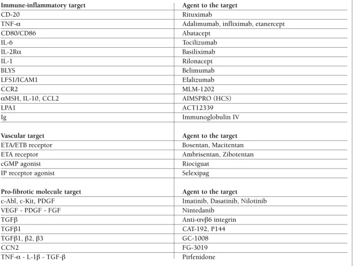
Treatments targeting in SSc potential mediators of fi-brosis and potential pathways (TGF-β, ET-1, VEGF, PDGF, FGF) have been tested or are under evaluation in clinical trials, with different results (Table I).
Very recently a further clinical trial has been des -cribed that blocks TGFbeta more effectively then ear-lier temptatives, and although this was an open label study, it does suggest that the skin score may be
im-table I. poSSIble targeted therapeutIc approacheS In SyStemIc ScleroSIS (IncludIng agentS under evaluatIon)
Immune-inflammatory target Agent to the target
CD-20 Rituximab
TNF-α Adalimumab, infliximab, etanercept
CD80/CD86 Abatacept IL-6 Tocilizumab IL-2Rα Basiliximab IL-1 Rilonacept BLYS Belimumab LFS1/ICAM1 Efalizumab CCR2 MLM-1202 αMSH, IL-10, CCL2 AIMSPRO (HCS) LPA1 ACT12339 Ig Immunoglobulin IV
Vascular target Agent to the target
ETA/ETB receptor Bosentan, Macitentan
ETA receptor Ambrisentan, Zibotentan
cGMP agonist Riociguat
IP receptor agonist Selexipag
Pro-fibrotic molecule target Agent to the target
c-Abl, c-Kit, PDGF Imatinib, Dasatinib, Nilotinib
VEGF - PDGF - FGF Nintedanib TGFβ Anti-αvβ6 integrin TGFβ1 CAT-192, P144 TGFβ1, β2, β3 GC-1008 CCN2 FG-3019 TNF-α - L-1β - TGF-β Pirfenidone
proved and that gene expression signatures may be atte nuated with this approach41. The durability and clinical meaning of this therapeutic strategy remains to be determined in future studies, but this recently publi shed clinical trial provides strong support to the rationale for targeting TGF-β in SSc, especially for skin disease42.
In this perspective of targeted therapy in SSc, two very recent clinical studies have shown that long-term treatment with ET-1 receptor antagonist modifies the progression and morphology of the nailfold microvas-cular damage, as assessed by NVC, in patients with SSc, and respectively affected by pulmonary arterial hypertension and DU (in combination with iloprost), over a 1-3 year follow-up period43,44.
Interestingly, both studies showed, at the nailfold capillaroscopy, at least after 1 year, a significant increased number of capillaries with reduced avasculari -ty and increased neoangiogenesis, suggesting the pos-sible achievement of a miscrovascular/tissue de-re-modelling in SSc. The effects might be related to in-terferences exerted by ET-1 receptor antagonists on the already mentioned process of EndoMT and on the TGF-β activities in SSc45-47.
An efficient system to target the autoantibody pro-ducing B cells in SSc is emerging from the successfull use of rituximab (RTX) in treatment of SSc patients48. As matter of fact, a recent clinical open-study by de-pleting B cells with RTX on a 2-treatment course (months 0/6), was found to be well tolerated and to have potential efficacy for skin disease and stabilization of internal organ status in early dcSSc49. Moreover, sta-bilisation of microcirculation (number of capillaries) has also been attested by this regimen50.
In a more recent study, the comparison of RTX trea-ted SSc patients versus untreatrea-ted matched-controls showed improvement of skin fibrosis and prevention of worsening lung fibrosis, and further supports the therapeutic concept of B cell depletion in order to re-duce autoantibody production/effects in SSc pa-tients51,52.
advanced and future therapIeS for SyStemIc ScleroSIS
A possible therapeutic option to be considered in SSc patients who are refractory to conventional treatments is the autologous haematopoietic stem cell transplan-tation (HSTC)53.
The mechanisms responsible for the benefits of HSTC are not already fully understood, and possibly it induces a reestablishment of immunological tole -rance together with non-specific immunosuppressive activities. However, HSTC seems to cause an impro -ve ment of vasculopahy, modified Rodnan skin thick-ness score (MRSS) and lung function, in patients with dcSSc and mild-moderate internal organ involvement (maximum disease duration of 4–5 years) or dcSSc pa-tients with progressive internal organ involvement54. However, despite the potential benefits, HSTC is, dangerous therapeutic option with a high risk of death and a higher morbidity rate and new trials try to opti-mize this approach especially in dcSSc55.
A further therapeutic option in SSc patients resistant to conventional therapies, seems be represented by in-travenous immunoglobulins (IVIG), but their mecha-nisms of action remain unclear56. Recent clinical data about the effects of IVIG on SSc patients report that this approach may improve several clinical manifesta-tions such as skin, joint and lung involvement al-though optimal dosages and timing of administration are not yet defined. A double blind, randomised, pla -cebo-controlled study is in progress to assess the safe-ty and efficacy of IVIG in scleroderma patients57.
Interestingly, new and future possible diagnostic and therapeutical approaches in SSc will come from epigenetic studies58.
In fact, the interest about microRNAs (miRNAs) that are non-coding RNAs regulating a large variety of bio-logical functions in plants and animals, is growing59. Each miRNA expressed in a cell may target about 100 to 200 messenger RNAs that it downregulates. It appears that about 60% of human protein coding ge-nes are regulated by miRNAs, whilst many miRNAs are epigenetically regulated.
There is currently good evidence for the role of miR NAs in fibrotic diseases, either organ-specific or sys temic fibrosis, such as in SSc60. Whereas the exact targets of these miRNAs are unknown, for some of them it is know, since they are regulating key downstream pathways in pathogenesis of diseases such as miRNA--29, which is a key mediator of fibrosis61. Gene therapy with the restoration of miRNA-29, at least in an animal model of fibrosis, appears to reduce fibrosis62.
Therefore, pharma companies are searching for miRNA technologies for modulating various fibrotic conditions and recent data are emerging on the role of increasing miRNAs in vivo, especially miRNA-29a, to restore its function and thus suppress fibrosis without
the need for viral vectors63.
As matter of fact, in complex diseases such as SSc, it may be that the clinical course of the disease is in-fluenced by the expression of miRNAs, which them-selves are epigenetically altered by body environmenal factors (i.e. estrogens).
concluSIonS
The fast and progressive increase in knowledge about the mechanisms underlying the SSc pathogenesis should offer new possible targets for therapeutical approaches adressed to an efficient and early disease-modifying strategy (Table I).
Combination therapy in SSc seems the best approach, as well as the early intervention on the major hallmarks of the disease in “at risk” patients that con-sists of the microvascular damage/altered function and the autoimmune reaction, followed by the progressi-ve and systemic fibrotic process.
correSpondence to
Prof. Maurizio Cutolo, MD Research Laboratories and
Academic Division of Clinical Rheumatology Department of Internal Medicine and Specialities University of Genova Italy
Viale Benedetto XV, 6 IRCCS San Martino 16132 Genova Italy E-mail: [email protected]
referenceS
1. Allanore Y, Distler O. Systemic sclerosis in 2014: Advances in cohort enrichment shape future of trial design. Nat Rev Rheu-matol. 2015;11:72-74.
2. Khanna D, Furts DE, Allanore Y et al. Twenty-two points to consider for clinical trials in systemic sclerosis, based on EU-LAR standards. Rheumatology 2015; 54;144-151.
3. Cutolo M, Smith V. Novel insights into systemic sclerosis ma-nagement: foreword. In: Cutolo M, Smith V editors. Novel insights into systemic sclerosis management. London:Future Medicine Ltd; 2013. p. 2-5.
4. van den Hoogen F, Khanna D, Fransen J et al. 2013 classifi-cation criteria for systemic sclerosis: an American college of rheumatology/European league against rheumatism collabo-rative initiative. Ann Rheum Dis 2013;72:1747-1755. 5. Avouac J , Fransen J, Walkerua et al. Preliminary criteria for
the very early diagnosis of systemic sclerosis: results of a Del -phi Consensus Study from EULAR Scleroderma Trialsand Research Group. Ann Rheum Dis 2011;70:476-481. 6. Herrick AL. The pathogenesis, diagnosis and treatment of
Raynaud phenomenon. Nat Rev Rheumatol 2012;8:469-479. 7. Cutolo M, Pizzorni C, Sulli A. Identification of transition from
primary Raynaud’s phenomenon to secondary Raynaud’s phe-nomenon by nailfold videocapillaroscopy. Arthritis Rheum 2007;56:2102-2103.
8. Landewé R, van der Heijde D, Walker UA et al EUSTAR co-au thors. Digital ulcers predict a worse disease course in pa-tients with systemic sclerosis. Mihai C, Ann Rheum Dis 2015 Feb 16. pii: annrheumdis-2014-205897.
9. Gyger G, Baron M. Systemic Sclerosis: Gastrointestinal Di-sease and Its Management. Rheum Dis Clin North Am 2015;41: 459-473.
10. Highland KB. Recent advances in scleroderma-associated pul-monary hypertension. Curr Opin Rheumatol 2014;26:637--645.
11. Heidrich H. Functional vascular diseases: Raynaud’s syndro-me, acrocyanosis and erythromelalgia. VASA 2010;39:33-41. 12. Kallenberg CG. Early detection of connective tissue disease in patients with Raynaud’s phenomenon. Rheum Dis Clin North Am 1990;16:11-30.
13. Smith V, Riccieri V, Pizzorni C et al. Nailfold capillaroscopy for prediction of novel future severe organ involvement in syste-mic sclerosis. J Rheumatol. 2013;40:2023-2027.
14. Smith V, Decuman S, Sulli A et al. Do worsening scleroderma capillaroscopic patterns predict future severe organ involve-ment? a pilot study. Ann Rheum Dis 2012;71:1636-1639. 15. Ingegnoli F, Gualtierotti R, Lubatti C et al. Nailfold capillary
patterns in healthy subjects: a real issue in capillaroscopy. Mi-crovasc Res 2013;90:90-95.
16. Anderson ME, Allen PD, Moore T et al. Computerized nail-fold video capillaroscopy-a new tool for assessment of Ray-naud’s phenomenon. J Rheumatol 2005;32:841-848. 17. Wigley FM. Clinical practice: Raynaud’s phenomenon. N Engl
J Med 2002;347:1001-1008.
18. Pavlov-Dolijanovic S, Damjanov NS, Stojanovic RM et al. Scle-roderma pattern of nailfold capillary changes as predictive va-lue for the development of a connective tissue disease: a fol-low--up study of 3,029 patients with primary Raynaud’s phe-nomenon. Rheumatol Int 2012;32:3039-3045.
19. Bernero E, Sulli A, Ferrari G et al. Prospective capillaroscopy-ba-sed study on transition from primary to secondary Raynaud’s phenomenon: preliminary results. Reumatismo 2013;65:186--191.
20. Trombetta AC , Smith V, Pizzorni C et al. Quantitative alte-rations of capillary diameter have a predictive value for deve-lopment of the capillaroscopic scleroderma pattern. J Rheu-matol 2016 (in press).
21. Cutolo M, Sulli A. Therapy. Optimized treatment algorithms for digital vasculopathy in SSc. Nat Rev Rheumatol 2015;11:569-571.
22. Koenig M, Joyal F, Fritzler MJ et al. Autoantibodies and mi-crovascular damage are independent predictive factors for the progression of Raynaud’s phenomenon to systemic sclerosis: A twenty-year prospective study of 586 patients, with valida-tion of proposed criteria for early systemic sclerosis. Arthritis Rheum 2008;58:3902-3912.
23. Riemekasten G, Philippe A, Näther M et al. Involvement of functional autoantibodies against vascular receptors in syste-mic sclerosis. Ann Rheum Dis 2011;70:530-536.
an-giotensin and endothelin receptors in systemic sclerosis in-duce cellular and systemic events associated with disease pat-hogenesis. Arthritis Res Ther 2014;16:R29.
25. Günther J, Kill A, Becker M et al. Angiotensin receptor type 1 and endothelin receptor type A on immune cells mediate mi-gration and the expression of IL-8 and CCL18 when stimula-ted by autoantibodies from systemic sclerosis patients. Arth-ritis Res Ther 2014;16:R65.
26. Becker M, Kill A, Kutsche M et al. Vascular receptor autoan-tibodies in pulmonary arterial hypertension associated with systemic sclerosis. Am J Respir Crit Care Med 2014;190:808--817.
27. Cutolo M, Smith V. State of the art on nailfold capillaroscopy: a reliable diagnostic tool and putative biomarker in rheuma-tology? Rheumatology (Oxford). 2013;52:1933-1940. 28. Ingegnoli F, Ardoino I, Boracchi P et al. EUSTAR co-authors.
Nailfold capillaroscopy in systemic sclerosis: data from the EULAR scleroderma trials and research (EUSTAR) database. Microvasc Res 2013;89:122-128.
29. Cutolo M. Disease modification in systemic sclerosis. Do in-tegrated approaches offer new challenges? Z Rheumatol. 2013;72:326-328.
30. Cutolo M, Sulli A. Optimized treatment algorithms for digi-tal vasculopathy in SSc. Nat Rev Rheumatol. 2015;11:569--571.
31. Denton CP. Systemic sclerosis: from pathogenesis to targeted therapy. Clin Exp Rheumatol 2015;33(4 Suppl 92):S3-7. 32. Dulmovits BM, Herman IM. Microvascular remodeling and
wound healing: a role for pericytes. Int J Biochem Cell Biol 2012;44:1800-1812.
33. Bhattacharyya S, Wei J, Varga J. Understanding fibrosis in sys-temic sclerosis: shifting paradigms, emerging opportunities. Nat Rev Rheumatol. 2011;8:42-54.
34. Distler JH, Jüngel A, Pileckyte M et al. Hypoxia-induced in-crease in the production of extracellular matrix proteins in systemic sclerosis. Arthritis Rheum 2007;561:4203-415. 35. Müller-Ladner U, Distler O, Ibba-Manneschi L et al.
Mecha-nisms of vascular damage in systemic sclerosis. Autoimmunity 2009;42:587-595.
36. Manetti M, Guiducci S, Matucci-Cerinic M. The origin of the myofibroblast in fibroproliferative vasculopathy: does the en-dothelial cell steer the pathophysiology of systemic sclerosis? Arthritis Rheum 2011;63:2164-2167.
37. Wynn TA. Cellular and molecular mechanisms of fibrosis. J Pathol. 2008;214:199-210.
38. Widyantoro B, Emoto N, Nakayama K et al. Endothelial cell--derived endothelin-1 promotes cardiac fibrosis in diabetic hearts through stimulation of endothelial-to-mesenchymal transition. Circulation 2010;121:2407-2418.
39. Nataraj D, Ernst A, Kalluri R. Idiopathic pulmonary fibrosis is associated with endothelial to mesenchymal transition. Am J Respir Cell Mol Biol 2010;43:129-30.
40. Mouthon L, Mehrenberger M, Teixeira L et al. Endothelin-1 ex-pression in scleroderma renal crisis. Hum Pathol 2011;42:95--102.
41. Rice LM, Padilla CM, McLaughlin SR et al. Fresolimumab treatment decreases biomarkers and improves clinical symp-toms in systemic sclerosis patients. J Clin Invest 2015;125:
2795-2807.
42. Denton CP. Systemic sclerosis: from pathogenesis to targeted therapy. Clin Exp Rheumatol 2015;33(4 Suppl 92):S3-7. 43. Guiducci S, Bellando Randone S, Bruni C et al. Bosentan
fos-ters microvascular de-remodelling in systemic sclerosis. Clin Rheumatol 2012;31:1723-1725.
44. Cutolo M, Zampogna G, Vremis L et al. Longterm effects of endothelin receptor antagonism on microvascular damage evaluated by nailfold capillaroscopic analysis in systemic scle-rosis. J Rheumatol 2013;40:40-45.
45. Cutolo M, Montagna P, Brizzolara R et al. Effects of maciten-tan and its active metabolite on cultured human systemic scle-rosis and control skin fibroblasts. J Rheumatol 2015;42:456--463.
46. Cipriani P, Di Benedetto P, Ruscitti P et al. Macitentan inhibits the transforming growth factor- profibrotic action, blocking the signaling mediated by the ETR/T RI complex in systemic sclerosis dermal fibroblasts. Arthritis Res Ther 2015;17:247--252.
47. Cipriani P, Di Benedetto P, Ruscitti P et al. The Endothelial-me-senchymal Transition in Systemic Sclerosis Is Induced by En-dothelin-1 and Transforming Growth Factor- and May Be Blocked by Macitentan, a Dual Endothelin-1 Receptor Anta-gonist. J Rheumatol 2015;42:1808-1816.
48. Sakkas LI, Bogdanos DP. Systemic sclerosis: New evidence re-enforces the role of B cells. Autoimmun Rev 2016;15:155--161.
49. Smith V, Piette Y, van Praet J et al. Two-year results of an open pilot study of a 2-treatment course with rituximab in patients with early systemic sclerosis with diffuse skin involvement. J Rheumatol 2013;40:52-57.
50. Smith V, Pizzorni C, Riccieri V et al. Stabilisation of micro-circulation damage in early systemic sclerosis patients with diffuse skin involvement following rituximab treatment: an open label study. J Rheumatol 2016 (in press).
51. Jordan S, Distler JH, Maurer B et al. Effects and safety of ritu-ximab in systemic sclerosis: an analysis from the European Scleroderma Trial and Research (EUSTAR) group. EUSTAR Rituximab study group. Ann Rheum Dis 2015;74:1188-1194. 52. Smith V, De Keyser F. Advances in therapies for systemic scle-rosis. In: Van Vollenhoven R, editor. Clinical Therapy Research in the Inflammatory Diseases. Singapore: World Scientific Pu-blishing Co. Pte. Ltd; 2015. p. 165-198.
53. Van Laar JM, Naraghi K, Tyndall A. Haematopoietic stem cell transplantation for poor-prognosis systemic sclerosis. Rheu-matology 2015;54: doi: 10.1093/rheuRheu-matology/kev117. 54. Khanna D, Georges GE, Couriel DR. Autologous
hemato-poietic stem cell therapy in severe systemic sclerosis: ready for clinical practice? JAMA 2014;311: 2485-2487. 55. Van Laar JM, Farge D, Sont JK, et al. Autologous
hemato-poietic stem cell transplantation vs intravenous pulse cy-clophosphamide in diffuse cutaneous systemic sclerosis: a randomized clinical trial. JAMA 2014;311:2490–2498. 56. Cantarini L, Rigante D, Vitale A et al. Intravenous
immuno-globulins (IVIG) in systemic sclerosis: a challenging yet pro-mising future. Immunol Res 2015;61:326-337.
57. Georgetown University, CSL Behring. IVIG Treatment in Sys-temic Sclerosis In: Clinical Trials.gov [Internet]. Bethesda
(MD): National Library of Medicine (US). 2013- [cited 2015 Jul 16]. Available from: https:// clinicaltrials.gov/ct2/show/ NCT01785056 NLM Identifier: NCT01785056.
58. O’Reilly S. MicroRNAs in fibrosis: opportunities and challen-ges Arthritis Res Ther. 2016; 18: 11. Published online 2016 Jan 13. doi: 10.1186/s13075-016-0929-x.
59. Montgomery RL, Yu G, Latimer PA et al. MicroRNA mimicry blocks pulmonary fibrosis. EMBO Mol Med 2014;6:1347--1356.
60. Altorok N, Almeshal N, Wang Y et al. Epigenetics, the holy grail in the pathogenesis of systemic sclerosis. Rheumatology 2015;54:1759–1770.
61. Cushing L, Kuang PP, Qian J et al. miR-29 is a major regula-tor of genes associated with pulmonary fibrosis. Am J Respir Cell Mol Biol 2011;45:287–294.
62. Chen H-Y, Zhong X, Huang XR et al. MicroRNA-29b inhibits diabetic nephropathy in db/db mice. Mol Ther 2014;22:842-853.
63. Millar NL, Gilchrist DS, Akbar M et al. MicroRNA29 a regu-lates IL-33-mediated tissue remodelling in tendon disease. Nat Commun 2015;6:6774.