into Pancreatic Islet and Conversion into Insulin-Positive
Cells
In Vivo
LuGuang Luo1*, John Z. Q. Luo1,2, Fang Xiong1, Mehrdad Abedi1, Deborah Greer1
1Center for Stem Cell Biology, Roger Williams Hospital, Providence, Rhode Island, United States of America,2PLME, Brown University, School of Medicine, Providence, Rhode Island, United States of America
Abstract
We hypothesize that specific bone marrow lineages and cytokine treatment may facilitate bone marrow migration into islets, leading to a conversion into insulin producing cells in vivo. In this study we focused on identifying which bone marrow subpopulations and cytokine treatments play a role in bone marrow supporting islet function in vivo by evaluating whether bone marrow is capable of migrating into islets as well as converting into insulin positive cells. We approached this aim by utilizing several bone marrow lineages and cytokine-treated bone marrow from green fluorescent protein (GFP) positive bone marrow donors. Sorted lineages of Mac-1+, Mac-12, Sca+, Sca2, Sca2/Mac-1+and Sca+/Mac-12 from GFP
positive mice were transplanted to irradiated C57BL6 GFP negative mice. Bone marrow from transgenic human ubiquitin C promoter GFP (uGFP, with strong signal) C57BL6 mice was transplanted into GFP negative C57BL6 recipients. After eight weeks, migration of GFP positive donor’ bone marrow to the recipient’s pancreatic islets was evaluated as the percentage of positive GFP islets/total islets. The results show that the most effective migration comes from the Sca+/Mac2lineage and
these cells, treated with cytokines for 48 hours, were found to have converted into insulin positive cells in pancreatic islets in vivo. This study suggests that bone marrow lineage positive cells and cytokine treatments are critical factors in determining whether bone marrow is able to migrate and form insulin producing cells in vivo. The mechanisms causing this facilitation as well as bone marrow converting to pancreatic beta cells still need to be investigated.
Citation:Luo L, Luo JZQ, Xiong F, Abedi M, Greer D (2009) Cytokines Inducing Bone Marrow SCA+
Cells Migration into Pancreatic Islet and Conversion into Insulin-Positive CellsIn Vivo. PLoS ONE 4(2): e4504. doi:10.1371/journal.pone.0004504
Editor:Kathrin Maedler, University of Bremen, Germany
ReceivedAugust 5, 2008;AcceptedDecember 9, 2008;PublishedFebruary 19, 2009
Copyright:ß2009 Luo et al. This is an open-access article distributed under the terms of the Creative Commons Attribution License, which permits unrestricted use, distribution, and reproduction in any medium, provided the original author and source are credited.
Funding:JDRF, NIH COBRE This publication was made possible, in part, by Research Grant Number 1-2007-180 for Dr. LuGuang Luo by Juvenile Diabetes Research Foundation International (JDRF), Roger Williams Hospital Research Funding for Dr. Luo and NIH COBRE Grant award Number 2P20RR018757-06 from the National Center for Research Resources and. Its contents are solely the responsibility of the authors and do not necessarily represent the official views of the NIH. The funders had no role in study design, data collection and analyss, decision to publish, or preparation of the manuscript.
Competing Interests:The authors have declared that no competing interests exist. * E-mail: [email protected]
Introduction
Studies have shown that bone marrow transplantation contrib-utes to the recovery of isletbcell function through differentiation into isletbcells [1]. However, there are controversial arguments regarding a failure to document such transdifferentiation or a very low frequency of isletbcell differentiation [2]. Moreover, reports on bone marrow correcting experimental hyperglycemia in diabetic animals show that bone marrow initiates recipientbcell regeneration through donor derived endothelial cells in the pancreas rather than directly transdifferentiating intob cells [3], but others showed no improvement in hyperglycemia after bone marrow transplantation [4,5]. Although this controversy has yet been resolved, bone marrow as a candidate for diabetes cell therapy has been considered and explored [6–8].
In this study, we proposed to identify whether specific bone marrow subpopulations and cytokine treatments play a critical role in bone marrow migration to pancreatic islets and conversion into islet cells in vivo. Specific populations of bone marrow can be induced into forming pancreatic isletbcells [9–11] and cytokines can be major regulators in influencing bone marrow conversion into pancreatic islet cells [12]. However, it is important to know
which specific bone marrow population and cytokine treatment regulates bone marrow migration (homing) into islets after transplantation and whether migrated bone marrow can convert into islet cells. In this manuscript, we attempted to address these questions by testing GFP labeled bone marrow lineage cells in colocalized pancreatic islets with insulin positive cells under the following circumstances, a) GFP lineage cell populations from bone marrow cells only and, b) GFP lineage cells pre-cultured with cytokines, to evaluate whether these conditions facilitate bone marrow migration and conversion into pancreatic islet cells after transplantation.
Materials and Methods
Experimental Designs and Methods
Harbor, ME). Mice were certified to be pathogen free and housed in our animal facility with given ad libitum access to food and water. The eGFP mice were pancreatic donors and the uGFP mice were GFP labeled bone marrow donors. GFP negative C57BL mice were the recipients. Animal study protocols were approved by the Institutional Animal Care and Use Committee at Roger Williams Hospital.
Bone Marrow Transplantation. 6–8 week old mice were used as donors or recipients. After sacrificing the mice and dissecting the femur, tibia and pelvic bones, bone marrow was obtained by flushing the bones using a syringe and a 22-gauge needle with PBS containing 5% heat-inactivated fetal calf serum (HI-FCS). After re-suspension in PBS without HI-FCS, cells were passed through a 40mm cell strainer. Cell numbers were counted in crystal violet and viability was assessed by trypan blue staining. Whole bone marrow cells or selected populations of marrow cells, based on their surface markers, were injected intravenously by tail vein into each recipient. The dose of the cells infused was different in individual experiments and is mentioned in the results section. A photon producing linear accelerator (Elekta) was used for the radiation of recipient animals before each transplant. Radiation was given at a dose rate of 100 cGy per minute. The recipients received 500 cGy of whole body.
Pancreas Collection and Immunohistochemistry Staining. Specimens were collected after sacrificing the
anesthetized mice by cervical dislocation. Excised pancreatic specimens were placed in freshly prepared PLP fixative solution (balanced phosphate solution with 2% paraformaldehyde, sodium m-periodate and L-lysine) for 2 hours at 4uC, with frequent agitation. Samples were then washed in a 7% sucrose buffer overnight followed by a 15% sucrose buffer wash for 2–3 hours and a 25% sucrose plus 10% glycerol buffer wash for another 2 hours, all at 4uC. They were then rinsed in PBS and embedded in tissue freezing medium (OCT), frozen and stored at 270uC until sectioning. Immunofluorescent staining was performed on 5 micron cryosections. For intracellular antigens, permeabilization was performed with 0.2% Triton X-100 for 20 minutes, followed by two PBS rinses. Sections were then blocked for 30 minutes with a 20% normal serum buffer. Sections were rinsed with PBS then incubated with anti-proinsulin, insulin and biotinylated anti-mouse CD45, rat monoclonal anti-CD34 (Abcam, Cambridge, MA) or Alexa Fluor 488 conjugated anti GFP antibodies (Molecular Probes, Eugene, OR) for 2 hours at room temperature, followed by 1 hour incubations with respective secondaries (Texas Red anti-guinea pig for insulin, Rhodamine anti-mouse for proinsulin and Alexa Flour streptavidin for CD45). GFP expression and antigens labeled by different fluorescence-conjugated antibodies were visualized by fluorescent microscopy (Axiovert w 135, Carl Zeiss, Oberko-chen, Germany). Additional staining with non-fluorescent DAB and Blue
Figure 1.Weak GFP signals in pancreatic islet of eGFP transgenic C57BL6 mice.GFP signals in pancreatic islets of eGFP transgenic positive mice were examined. A. Light microscopy image with the arrow indicating the tissue difference between a potential islet and the rest of the pancreatic tissue. B. The area indicated by the arrow in islet shows weak green fluorescence vs. surrounding area. C. Immunohistochemistry with insulin specific antibody indicated positive insulin cells (red) in islet but not yellow (mixture of green and red) and there is a CD45 cell (anti CD45 antibody blue) in the islet indicated by arrow. D. shows insulin positive cells only. E. shows weak GFP signals in islet areas vs. surrounding area. F. shows CD 45 positive cell only. A and B image magnification:65; C image magnification:620; D–F image magnification:640.
doi:10.1371/journal.pone.0004504.g001
conjugated anti insulin, CD34 antibodies (Molecular Probes) in parallel sections were performed to confirm the fluorescent signals. Cell Separation. Bone marrow was isolated from iliac bones, femur, and tibiae of GFP transgenic mice 6 to 8 weeks of age. Bone marrow cells were incubated with anti CD45-APC, anti c-Kit APC and anti-Sca-1-biotin SA- APC (Pharmingen, San Diego, CA). Cells positive and negative for individual markers were then sorted into different tubes with a high speed MoFlow cell sorter (Cytomation). For lineage negative separation, a low-density fraction (,1.077 g/cm2) was isolated on Nycoprep 1.077A (Accurate Chemical and Scientific Corporation). These cells were lineage depleted using magnetic beads from a Lineage Depletion kit (Miltenyi Biotec Inc. Auburn, CA). Cells were washed and counted after depletion. The population of sorted cells are displayed in Appendix S1.
Counting and Statistics. To count GFP positive islets in the pancreas, 12 sections, 5 microns thick and 250 microns apart, were made of each pancreatic specimen. The number of GFP positive islets and the total islets in each section were counted. The ratio between GFP positive islets and the total number of islets were determined as percentage of GFP positive islets in total islets. Data are presented as mean6one standard error of the mean. In the text, table, and figures, all data are presented as means6SEM. Most described experiments have been repeated three times. However, for figures and tables, only one experiment of the results is shown. Data used for graphical presentation and statistical analysis are expressed as per experiment. Data were analyzed by the ANOVA statistics program using a two factor analysis of variance of repeated measures. Post hoc comparisons among individual means were made by Tukey’s t-test.
Results
GFPs are expressed in the pancreatic islets of uGFP transgenic mice and hematopoietic cells distribution in the pancreas
We detected weak signals of GFP in the pancreatic islet area of eGFP transgenic mice (Figure 1.). Un-homomorphy of GFP signals were found in (A) and (B) and these insulin positive areas were confirmed by fluorescent immunohistochemistry. There are significantly strong GFP positive expression in areas surrounding the insulin positive islet and CD45+
(C). The images further display individual channels for insulin (D), GFP (E) and CD45+
(F). GFP signals in uGFP transgenic mice are much stronger (Figure 2.) than GFP signals in pancreatic islet from eGFP transgenic mice (Figure 1). Since the pancreas is a more bone marrow resistant organ, there are low numbers of hematopoietic cells in the pancreas (about 4,5/ 1000 pancreatic cells) as shown in Figure 3 (A) and (B), compared with an abundance of hematopoietic cells found in spleen (C) and (D). This suggests that bone marrow is more likely to home into the spleen than to the pancreas.
Bone marrow cells migrate to pancreatic islets after transplantation
Utilization of uGFP transgenic mice as bone marrow donors allowed for tracking the labeled bone marrow migration into the recipient’s pancreatic islets after transplantation (bone marrow 256106cells) after 500 cGy (TBI). We found that infused bone marrow homes to the pancreatic islets, as indicated by anti insulin immunohistochemistry. These cells are also CD45 negative (Figure 4 A) and CD34 positive (B).
Figure 2. GFP expresses in pancreatic islet in uGFP transgenic C57BL6 mice.Clear GFP signals in pancreatic islets have been shown via immunohistochemistry of pancreatic sections from uGFP transgenic mice. A. Isletbcells have been identified by anti-insulin antibody (red, large arrow indicated) and the positive insulin islet mixture with green GFP fluorescence (small arrow indicated) combination color image; B. Isletbcells (red, arrows indicated) in red color image only; C. Nuclear staining with DAPI blue in blue color only; D. Positive GFP green in islet area (arrow indicated) in green color only, image magnification:640.
Bone marrow subpopulation affects their migration into pancreatic islets
Lineage GFP positive bone marrow cells were isolated as described above in methods and 2.56105/ml cells were infused into recipients after 500 cGy irradiation. The positive GFP cells in
pancreatic islets were analyzed 8 weeks after transplantation. There were six different cell lineages tested and whole bone marrow (WBM) was used as a control. The results show that the most favorable bone marrow population for migration to the islets is Mac-12/Sca+vs. WBM (Figure 5, * = p,0.01).
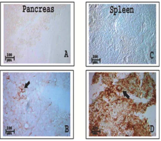
Figure 3. Distribution of CD45 cells in the pancreas and spleen of GFP negative mice.Identification of CD45+cell migration into the pancreas and spleen was performed in GFP negative C57BL6 mice. A. Pancreatic tissue as negative control for immunohistochemistry with anti-CD45 specific antibody. B. CD45+cells were found in pancreas (brown indicated by black arrow). CD45+population in pancreas was about 4,5/1000
pancreatic cells. C. Negative control of spleen. D. Spleen contains a large number of CD45+cells 600,700/1000 spleen cells while the amounts of
bone marrow cells in pancreatic tissue (B) were low. Image magnification:65.
doi:10.1371/journal.pone.0004504.g003
Figure 4. uGFP positive cell distribution in islet is CD 45 negative, but CD34 positive.A. Fluorescent immunohistochemistry with anti CD45 (blue) and insulin (red) antibodies were used to identify mice pancreatic GFP positive and CD45+donor cells in GFP negative C57BL6 mice, which were transplanted with 256106ml GFP positive WBM via tail vein after recipients had 500 cGy irradiation. GFP positive cells (indicated by
narrow arrow) are CD45 negative. CD45 positive cell (indicated by large arrow) is GFP negative. B. CD34 positive cells were further confirmed by double labeling color immunohistochemistry with anti CD34 antibody (dark blue indicated by black arrow) in islet (brown) stained with anti-insulin antibody. Image magnification:640.
doi:10.1371/journal.pone.0004504.g004
Cytokines affect Sca+lineage bone marrow
differentiation into insulin positive cells in the recipient’s islets
Subpopulations of Sca+cells were pre-cultured with cytokines IL-3 (50 U/ml), IL-6 (50 ng/ml), IL-11 (50 ng/ml) and steel (50 ng/ml) for 0, 24 and 48 hours. Cells positive for both GFP and insulin were only found in the recipient’s pancreas from the 48 hour culture group as shown in Figure 5 immunohistochem-istry images. Immunohistochemimmunohistochem-istry with specific anti-insulin antibody under de-confocal microscopy further confirmed that insulin positive cells were derived from positive GFP donor marrow. As viewed in Figure 6 A–D, cultures exposed to cytokines for 48 hours stimulated bone marrow Sca+ cells to differentiate into insulin positive cells in the pancreas. The amplified detail in the cytosol of insulin positive cells shows GFP staining colocalized with cytosol positive insulin granule staining in (E) and (F) (arrows indicate GFP positive and insulin granules in cytosol). The insulin positive granules can be clearly observed in the cellular cytosol of both GFP positive- insulin positive cells and insulin positive cells.
Discussion
In this study, we found that GFP signals in eGFP animal pancreatic islets cells were particularly weak. This may be one reason that there were controversial reports for GFP in islets [3,13,14] . Therefore, in this study, we used uGFP animals as a bone marrow donor to avoid the weak GFP signal and focused on how bone marrow specific lineages, with the combination of cytokine treatment, influence bone marrow migration to islets and conversion into insulin positive cells in vivo.
We found that bone marrow cell migration into pancreatic tissue was restricted vs. migration into the spleen, as shown figure 3 (about 120 folds difference). More GFP positive cells were found in the outer perimeter of the islets than within the islet, it was 8.5:1 or
about 12% of total population, suggesting that bone marrow migration into islets is a critical step for bone marrow to contribute to isletbcell function and regeneration.
After testing six different sorted subpopulations of GFP positive bone marrow on GFP negative animals, we found that the bone marrow cell population Sca+/Mac-12 can migrate into islets more efficiently than WBM and the other five cell subpopulations. The mechanisms involved may rely on cellular surface chemokine receptor expression and pancreatic islet releasing factors, in which activation of chemokine receptors in bone marrow cells promotes cell migration into the islets [15–17]. On the other hand, Sca+cell lineage has the potential to differentiate into insulin positive cells in vitro and in vivo [18–20]. Although we only found Sca+/Mac-12 favorable in this migration study, we are not excluding the possibility that other sub populations from bone marrow may also play a role in islet homing.
It has been reported that cytokine enriched bone marrow cultures alters bone marrow surface markers and improves bone marrow repopulation [21]. In this study, the Sca+/Mac-1 cell population was cultured with cytokines before transplantation for 0, 24 and 48 hours, revealing that Sca+cells treated by cytokines for 48 hours induced differentiation into insulin positive cells in vivo. This suggests that cytokines are critical for bone marrow’s participation in isletbcell function recovery andbcell regeneration. Cytokines tested in this study include IL-3, 6, 11 and steel factors in vitro. However, the levels of these factors, or others in vivo, could be different for individual responses to bone marrow transplantation, especially in damaged pancreatic animal models.
In summary, the current study provides evidence that transgenic mouse GFP gene control promoter, bone marrow subpopulation, and cytokines are critical factors to stimulate successful bone marrow migration and conversion into insulin-positive cells in islets in vivo. The phenomenon of cytokine treated bone marrow Sca+subpopulations having the ability to migrate and potentially
differentiate into insulin-positive islet cells still requires additional mechanistic studies to identify the factors behind this effect.
Supporting Information
Appendix S1
Found at: doi:10.1371/journal.pone.0004504.s001 (0.11 MB DOC)
Acknowledgments
Dr. LG Luo designed, conducted and performed most of the experiments and wrote the manuscript. John ZQ Luo participated in experiments and assisted in writing and editing the manuscript. Dr. Abedi provided animals, sorted bone marrow cells and transplanted cells into animals. D Greer and F Xiong provided technical help.
The authors would like to thank Dr. Peter Quesenberry for his kind help in preparing and reviewing this manuscript.
Figure 6. Cultured in presence of cytokines for 48 hours initiated BM differentiation to insulin positive cells in islet.A. Insulin positive cells were identified by insulin fluorescent immunohistochemistry as GFP positive (arrow indicated); B. Blue nucleus staining (DAPI) indicated by arrow; C. GFP positive cell indicated by arrow; D. The arrow indicates same cell with insulin positive staining. Image magnification:640; E. Anti-insulin
antibody immunohistochemistry imaged under de-confocal microscopy to further identify differentiated cells. The arrow indicates the GFP positive cell is insulin positive with clear nucleus staining and F. insulin staining indicated by arrow and DAPI nuclear staining is blue. Image magnification:
6100.
doi:10.1371/journal.pone.0004504.g006
Author Contributions
Conceived and designed the experiments: LL JZQL. Performed the experiments: LL FX MA DG. Analyzed the data: LL JZQL FX.
Contributed reagents/materials/analysis tools: MA. Wrote the paper: LL JZQL.
References
1. Ianus A, Holz GG, Theise ND, Hussain MA (2003) In vivo derivation of glucose-competent pancreatic endocrine cells from bone marrow without evidence of cell fusion. J Clin Invest 111: 843–850.
2. Choi JB, Uchino H, Azuma K, Iwashita N, Tanaka Y, et al. (2003) Little evidence of transdifferentiation of bone marrow-derived cells into pancreatic beta cells. Diabetologia 46: 1366–1374.
3. Mathews V, Hanson PT, Ford E, Fujita J, Polonsky KS, et al. (2004) Recruitment of bone marrow-derived endothelial cells to sites of pancreatic beta-cell injury. Diabetes 53: 91–98.
4. Taneera J, Rosengren A, Renstrom E, Nygren JM, Serup P, et al. (2006) Failure of transplanted bone marrow cells to adopt a pancreatic beta-cell fate. Diabetes 55: 290–296.
5. Lavazais E, Pogu S, Sai P, Martignat L (2007) Cytokine mobilization of bone marrow cells and pancreatic lesion do not improve streptozotocin-induced diabetes in mice by transdifferentiation of bone marrow cells into insulin-producing cells. Diabetes Metab 33: 68–78.
6. Lee RH, Seo MJ, Reger RL, Spees JL, Pulin AA, et al. (2006) Multipotent stromal cells from human marrow home to and promote repair of pancreatic islets and renal glomeruli in diabetic NOD/scid mice. Proc Natl Acad Sci U S A 103: 17438–17443.
7. Voltarelli JC, Couri CE, Stracieri AB, Oliveira MC, Moraes DA, et al. (2007) Autologous nonmyeloablative hematopoietic stem cell transplantation in newly diagnosed type 1 diabetes mellitus. Jama 297: 1568–1576.
8. Banerjee M, Kumar A, Bhonde RR (2005) Reversal of experimental diabetes by multiple bone marrow transplantation. Biochem Biophys Res Commun 328: 318–325.
9. Ai C, Todorov I, Slovak ML, Digiusto D, Forman SJ, et al. (2007) Human marrow-derived mesodermal progenitor cells generate insulin-secreting islet-like clusters in vivo. Stem Cells Dev 16: 757–770.
10. Sun Y, Chen L, Hou XG, Hou WK, Dong JJ, et al. (2007) Differentiation of bone marrow-derived mesenchymal stem cells from diabetic patients into insulin-producing cells in vitro. Chin Med J (Engl) 120: 771–776.
11. Tang DQ, Cao LZ, Burkhardt BR, Xia CQ, Litherland SA, et al. (2004) In vivo and in vitro characterization of insulin-producing cells obtained from murine bone marrow. Diabetes 53: 1721–1732.
12. Naselli G, Deaizpurua HJ, Thomas HE, Johnston AM, Kay TW (2001) Lack of expression of Gp-130 makes pancreatic beta cell lines unresponsive to the IL-6 family of cytokines. Int J Exp Diabetes Res 1: 239–248.
13. Kang EM, Zickler PP, Burns S, Langemeijer SM, Brenner S, et al. (2005) Hematopoietic stem cell transplantation prevents diabetes in NOD mice but does not contribute to significant islet cell regeneration once disease is established. Exp Hematol 33: 699–705.
14. Hasegawa Y, Ogihara T, Yamada T, Ishigaki Y, Imai J, et al. (2007) Bone marrow (BM) transplantation promotes beta-cell regeneration after acute injury through BM cell mobilization. Endocrinology 148: 2006–2015.
15. Ehses JA, Perren A, Eppler E, Ribaux P, Pospisilik JA, et al. (2007) Increased number of islet-associated macrophages in type 2 diabetes. Diabetes 56: 2356–2370.
16. Zanone MM, Favaro E, Ferioli E, Huang GC, Klein NJ, et al. (2007) Human pancreatic islet endothelial cells express coxsackievirus and adenovirus receptor and are activated by coxsackie B virus infection. FASEB J 21: 3308–3317. 17. Sordi V, Malosio ML, Marchesi F, Mercalli A, Melzi R, et al. (2005) Bone
marrow mesenchymal stem cells express a restricted set of functionally active chemokine receptors capable of promoting migration to pancreatic islets. Blood 106: 419–427.
18. Chang KH, Chan-Ling T, McFarland EL, Afzal A, Pan H, et al. (2007) IGF binding protein-3 regulates hematopoietic stem cell and endothelial precursor cell function during vascular development. Proc Natl Acad Sci (USA) 104: 10595–10600.
19. Zhang CC, Lodish HF (2004) Insulin-like growth factor 2 expressed in a novel fetal liver cell population is a growth factor for hematopoietic stem cells. Blood 103: 2513–2521.
20. Baddoo M, Hill K, Wilkinson R, Gaupp D, Hughes C, et al. (2003) Characterization of mesenchymal stem cells isolated from murine bone marrow by negative selection. J Cell Biochem 89: 1235–1249.