e38
Myocardial Homing after Intrapericardial Infusion of Bone Marrow
Mononuclear Cells
Érika Branco
1, Emerson Ticona Fioretto
2, Rosa Cabral
3, Carlos Alberto Sarmento, Palmera
4, Guilherme Buzon
Gregores
4, Angelo João Stopiglia
4, Paulo César Maiorka
4, Pedro Alves Lemos
5, Carlos Campos
5, Celso Takimura
5,
José Antônio Franchini Ramires
5, Maria Angelica Miglino
4Faculdade de Medicina Veterinária da Universidade Federal Rural da Amazônia – UFRA1, Belém, PA; Universidade Federal do Sergipe – UFS2, Aracajú, SE; Universidade Feral do Piauí – UFPI3, Teresina, PI; Faculdade de Medicina Veterinária e Zootecnia FMVZ/USP4, São Paulo, SP; Instituto do Coração – Incor5, São Paulo, SP, Brasil
Introduction
Stem cells have been explored as a treatment for cardiac diseases, being related to possible neovascularization and tissue formation1. Stem cell transplantation has been performed through intracoronary, intramyocardial and retrograde infusion techniques2-4; however, it presents limitations and has reduced applications in clinical practice.
Previous studies tested the pericardial space for drug administration5. Theoretically, the transpericardial approach presents potential advantages for cell administration, including the pericardial sac’s non-vascular characteristic that may permit repeated procedures. The pericardial space may serve as a depot compartment, thereby enhancing local bioavailability of infused cells. However, the transpericardial approach for stem cell therapy has been poorly explored.
This preliminary study evaluates whether the administration of autologous Bone Marrow Mononuclear Cells (BMMC) into the pericardial space is followed by myocardial cell homing in control and myocardial infarction-induced cases.
Methods
Study Design
This study aimed at evaluating whether myocardial cell homing occurs after the injection of autologous BMMC into pericardial space. Seven domestic female swine were divided into two groups: induced myocardial infarction (n=3) and non-myocardial infarction (n=3). One animal from the induced myocardial infarction group died before the BMMC injection. Animals were euthanized for heart examination 21 days after BMMC injection. Protocols were approved by the local Bioethics Committee.
Mailling Address: Érika Branco •
Av. Pte. Tancredo Neves, 2501 – Montese - 66077-530 – Caixa Postal: 917- Belém – PA - Brasil
E-mail: [email protected]
Manuscript received July 09, 2008; revised manuscript received October 21, 2008; accepted October 21, 2008.
Key Words
Stem Cells, Myocardial Infarction; Pericardium.
Bone Marrow Sampling & Cell Separation and Labeling
Animals were pre-medicated with intramuscular chloridrate of ketamine (5mg/kg) and midazolam (0.5 mg/kg). Induction was performed with intravenous propofol (5mg/kg) followed by maintenance with isofluorane. A 60-ml bone marrow aspirate was obtained using a bone puncture needle introduced in iliac crest.
The aspirated bone marrow was diluted into saline and BMMC were separated by density gradient using Ficoll-PaqueTM Plus (Amersham Pharmacia). After 20-minute
centrifugation at 2000 rpm (20ºC), a halo of BMMC cells was formed. Bone marrow cells were aspirated and added to saline for further centrifugation (7 minutes at 2000 rpm and 20ºC). Agglomerated cells were suspended in saline and filtrated in a 150 Mesh stainless steel mesh. The filtrate was re-suspended in saline and re-centrifuged as described above. An aliquot of the solution was Trypan-blue stained and evaluated in a Neubauer chamber to determine viability. We separated 1x10⁸ cells and DNA stained with 1µl Hoechst 33342 (Invitrogen/H1399, Brazil) incubated in water-bath during 30 minutes at 37ºC.
Intrapericardial Infusion Technique
Pericardial space was accessed through the subxiphoid region using an epidural needle (Figure 1). The needle was advanced under fluoroscopic guidance past the parietal pericardium. The position of the needle tip at the pericardial sac was radiographically confirmed by infusion of diluted iodinated contrast. Once at the pericardial space, a total of 108 of BMMC stained with Hoechst® diluted in 10ml of saline were injected.
Experimental Acute Myocardial Infarction
Following the previously described procedures 6, an acute myocardial infarction was induced in 3 animals immediately before BMMC injection. After accessing the femoral artery by blunt dissection, a 6F right Judkins guiding catheter was manipulated under fluoroscopic guidance up to the left coronary and selectively cannulated. A 0.014” guide-wire was inserted into the left anterior descending artery, over which a balloon catheter was positioned just distal to the origin of the first diagonal branch. An acute myocardial infarction of anterior wall was induced by a 45-minute total occlusion of artery.
Animals were euthanized after 21 days. The heart was collected and the ventricles were separated from the atria at the level of the atrioventricular groove. Ventricles were sliced and the fragments were collected for analysis from the basal, medial, and apical regions. Specimens were processed for standard optical microscopy, and immunohistochemical analysis. Immunohistochemistry fragments were embedded in a solution of 3 parts of PBS and 7 parts of glycerol, and cryopreserved in nitrogen, and stored in a -80ºC freezer. Analyses were performed on frozen slices using an epifluorescence microscope (Leica® DM 50 microscope). BMMC myocardial homing was estimated by total number of BMMC stained with Hoechst® present in the ventricular myocardium.
Statistical analysis
Continuous variables are presented as averages ± standard deviation and discrete variables as counts and percentages. Student’s T test was used to compare cell counting between the study groups.
Results
Acute results
One experimental death in the infarction group occurred due to ventricular fibrillation during the balloon inflation. The animal died before the administration of BMMC and was not considered for the analysis. All other 6 animals completed the procedure without complications.
Pericardial space access was attained without accidents and the full dose of BMMC was administered in all cases. No puncture complications, heart chamber perforation, arrhythmia, or hemodynamic instability occurred during intrapericardial manipulation and cell infusion.
21-Day Follow-up
There were no post-procedure deaths. All animals survived to index procedure completing 21 study-days protocol without adverse events or signs of heart failure.
In all animals, pericardial surface seemed normal at visual inspection, without fibrin deposition, fibrosis, adherences or visible neovascularization. In animals with induced infarction, a clear myocardial scar was identified at the anteroapical wall.
In both groups (infarcted and non-infarcted pigs), microscopic evaluation showed an average of 109494 ±
46505 BMMC homing in the myocardium. Furthermore, myocardial homing was significantly pronounced in the infarction group than in animals without infarction (150342±22456 vs. 68645±19421; p<0.01). Degrees of penetration of BMMC into the myocardium varied throughout the heart. The largest penetration of BMMC in the myocardium occurred in the infarcted area. BMMC were observed adhered to the wall epithelia of small vessels in the infarcted area (Figure 2). Small amounts of BMMC were seen in the epicardium and myocardium in animals from the control group (Figure 2).
Discussion
In our preliminary study, we demonstrated that the intrapericardial administration of BMMC is able to induce myocardial homing in swine. Moreover, after 21 days, cell homing was more extensive in animals with induced acute myocardial infarction.
Our results suggest that BMMC are able to penetrate the myocardium through the visceral pericardium, following the administration at the pericardial space. Our findings are similar to those described for the intracoronary, retrograde and intramyocardial techniques6,7. However, the intrapericardial injection presents characteristics that may translate into advantages for clinical use, related to its minimally invasive nature and non-necessity of arterial entry. The safety of transpericardial access has been reported previously for other applications, even though training in cardiac punction is highly necessary. Nevertheless, the transpericardial approach might theoretically be at least as safe as the commonly reported intramyocardial cell administration8. Moreover, costs of application of the transpericardial technique are lower than intracoronary, retrograde and intramyocardial ones3,9.
Pericardial fluid has been reported to present a low turnover rate, indicating the potential of the pericardial space to function as a reservoir for delayed delivery of pharmacological agents, which can be administered at high concentrations without systemic effects5,10. Accordingly, the depot ability of the pericardial space may prove beneficial for cell therapy, as it may prolong the bioavailability of viable cells for myocardial penetration.
In our study, we observed a diffuse penetration of BMMC across the myocardium, which contrasts with the more localized nature of intracoronary, intramyocardial, or retrograde techniques3,7,8. It is possible, therefore, to foresee possible application of transpericardial approach to diffuse forms of cardiomyopathy. On the other hand, cell Figure 1 - Place of infusion of the bone marrow mononuclear cells according
to (pericardial space - *) visualization by luoroscopy. Needle epidural (red
arrow) and iodized contrast (β).
Arq Bras Cardiol 2009; 93(3) : e38-e41
Branco et al Intrapericardial Administration of BMMC
e39
1. Bourassa MG, Detre KM, Johnston JM, Vlachos HA, Holubkov R. Effect of prior revascularization on outcome following percutaneous coronary intervention; NHLBI Dynamic Registry. Eur Heart J. 2002; 23: 1546-55. 2. Leor J, Patterson M, Quinones MJ, Kedes LH, Kloner RA. Transplantation
of fetal myocardial tissue into infarcted myocardium of rat: a potential method for repair of infarcted myocardium? Circulation. 1996; 94 (Suppl II): 332-6.
3. Hou DM, Cates P, Bekkers S, Miller MA, Carl L. Rouch CL, et al. Efficient myocardial delivery of microspheres and endothelial cells via selective retrograde coronary venous delivery [abstract]. J Am Coll Cardiol. 2002; 39: A76.
References
4. Hou DM, McLaughlin F, Thiesse M, Rogers P, Johnson R, Wang J, et al. Widespread regional myocardial transfection by plasmid encoding Del-1 after retrograde coronary venous delivery. Cathet Cardiovasc Interv. 2003; 58: 207-11.
5. Kolettis TM, Kazakos N, Katsouras CS, Niokou D, Pappa L, Koulouras V, et al. Intrapericardial drug delivery: pharmacologic properties and long-term safety in swine. Int J Cardiol. 2005; 99: 415-21.
6. Hou DM, Youssef EA, Brinton TJ, Zhang P, Rogers P, Prince ET, et al. Radiolabeled cell distribution after intramyocardial, intracoronary, and interstitial retrograde coronary venous delivery – Implications for current clinical trials. Circulation. 2005; 112: 150-6.
concentration was significantly higher in regions with acute myocardial infarction. This finding suggests that local factors at the infarcted region may interact to increase either cell migration and/or cell survival at the injured myocardium.
Potential Conflict of Interest
No potential conflict of interest relevant to this article was reported.
Sources of Funding
This study was funded by FAPESP and Incor-SP.
Study Association
This article is part of the thesis of doctoral submitted by Érika Renata Branco, from Faculdade de Medicina Veterinária e Zootecnia da Universidade de São Paulo - FMVZ/USP - Departamento de Cirurgia.
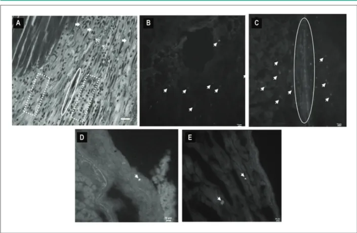
Figure 2 : Photomicrograph of heart of animal 21 days post acute myocardium infarction, showing in A: Note granular tissue in cardiac tissue containing partially intact
longitudinal cardiac ibers (circle). Observe an immature ibrotic organization with neo-vase formation (thin arrow), extracellular matrix deposition (non-polymerized vertical collagen ibers) (thick arrow), ibroblasts and moderate mononuclear inlammatory iniltrate (doted square). Stain: Masson’s trichrome. Bar scale: 20µm. B and
C: Infarcted animals: Hoechst positive staining on bone marrow mononuclear cells in the interstitial space of the myocardium (arrows) and BMMC homing in vessel wall (ellipse). Stain: Evans’ blue. Bar scale: 10µm (A), 20µm (B). D and E: Control animals: Masson’s trichrome positive stain in the epicardium, interstitial space of myocardium (arrows). Stain: Evans’ blue. Bar scale: 10µm (A)
Arq Bras Cardiol 2009; 93(3) : e38-e41
Branco et al
Intrapericardial Administration of BMMC
e40
7. Vulliet PR, Greeeley SM, Macdonald KA, Kittelson M. Intra-coronary arterial injection of mesenchymal stromal cells and microinfaction in dogs. Lancet. 2004; 363: 783-4.
8. Fuchs S, Baffour R, Zhou YF, Shou M, Pierre A, Tio FO, et al. Transendocardial delivery of autologous bone marrow enhances collateral perfusion and regional function in pigs with chronic experimental myocardial ischemia. J Am Coll Cardiol. 2003; 37: 1726-32.
9. Mannam AP, Kalon KK, Cultip DE, Carrozza, JP, Cohen DJ, Lorell BH, et al. Safety of subxyphoid pericardial access using a blunt-tip needle. Am J Cardiol. 2002; 89: 891-3.
10. Baek SH, Hrabie JA, Keefer LK, Hou D, Fineberg N, Rhoades R, et al. Augmentation of intrapericardial nitric oxide level by a prolonged-release nitric oxide donor reduces luminal narrowing after porcine coronary angioplasty. Circulation. 2002; 105: 2779-84.
Arq Bras Cardiol 2009; 93(3) : e38-e41
Branco et al Intrapericardial Administration of BMMC