Chronic Bronchitis and Current Smoking Are
Associated with More Goblet Cells in
Moderate to Severe COPD and Smokers
without Airflow Obstruction
Victor Kim1*, Michelle Oros2, Heba Durra2, Steven Kelsen1, Mark Aksoy1, William D. Cornwell3, Thomas J. Rogers3, Gerard J. Criner1
1Division of Pulmonary and Critical Care Medicine, Temple University School of Medicine, Philadelphia, Pennsylvania, United States of America,2Department of Pathology, Temple University School of Medicine, Philadelphia, Pennsylvania, United States of America,3Center for Inflammation, Translational and Clinical Lung Research, Temple University School of Medicine, Philadelphia, Pennsylvania, United States of America
Abstract
Background
Goblet cell hyperplasia is a classic but variable pathologic finding in COPD. Current litera-ture shows that smoking is a risk factor for chronic bronchitis but the relationship of these clinical features to the presence and magnitude of large airway goblet cell hyperplasia has not been well described. We hypothesized that current smokers and chronic bronchitics would have more goblet cells than nonsmokers or those without chronic bronchitis (CB), in-dependent of airflow obstruction.
Methods
We recruited 15 subjects with moderate to severe COPD, 12 healthy smokers, and 11 healthy nonsmokers. Six endobronchial mucosal biopsies per subject were obtained by bronchoscopy and stained with periodic acid Schiff-Alcian Blue. Goblet cell density (GCD) was quantified as goblet cell number per millimeter of basement membrane. Mucin volume density (MVD) was quantified as volume of mucin per unit area of
basement membrane.
Results
Healthy smokers had a greater GCD and MVD than nonsmokers and COPD subjects. COPD subjects had a greater GCD than nonsmokers. When current smokers (healthy smokers and COPD current smokers, n = 19) were compared with all nonsmokers (non-smoking controls and COPD ex-smokers, n = 19), current smokers had a greater GCD and MVD. When those with CB (n = 12) were compared to those without CB (n = 26), the CB group had greater GCD. This finding was also seen in those with CB in the COPD group
OPEN ACCESS
Citation:Kim V, Oros M, Durra H, Kelsen S, Aksoy M, Cornwell WD, et al. (2015) Chronic Bronchitis and Current Smoking Are Associated with More Goblet Cells in Moderate to Severe COPD and Smokers without Airflow Obstruction. PLoS ONE 10(2): e0116108. doi:10.1371/journal.pone.0116108
Received:August 26, 2014
Accepted:December 4, 2014
Published:February 3, 2015
Copyright:© 2015 Kim et al. This is an open access article distributed under the terms of theCreative Commons Attribution License, which permits unrestricted use, distribution, and reproduction in any medium, provided the original author and source are credited.
Data Availability Statement:Data are available in the supplementary files.
Funding:Funding for this work came from the National Heart, Lung, and Blood Institute (National Institute of Health) K23HL094696. The funders had no role in study design, data collection and analysis, decision to publish, or preparation of the manuscript.
alone. In multivariate analysis, current smoking and CB were significant predictors of GCD using demographics, lung function, and smoking pack years as covariates. All other covari-ates were not significant predictors of GCD or MVD.
Conclusions
Current smoking is associated with a more goblet cell hyperplasia and number, and CB is associated with more goblet cells, independent of the presence of airflow obstruction. This provides clinical and pathologic correlation for smokers with and without COPD.
Background
Chronic Obstructive Pulmonary Disease (COPD) is characterized by persistent, progressive
airflow limitation.[1] COPD encompasses a spectrum of clinical and pathological phenotypes,
including emphysema on one end of the spectrum with chronic bronchitis (CB) on the other. CB has been associated with multiple adverse outcomes, including increased exacerbations,
lung function decline, and mortality.[2–6] The pathologic correlate is goblet cell hyperplasia
(GCH), which has been shown in several studies to be present in COPD.[7,8] Mucus burden
has prognostic significance; one study in lung volume reduction surgery patients found that small airway mucous metaplasia inversely correlated with changes in lung function after
sur-gery,[9] whereas another study found that the degree of small airway mucus luminal occlusion
correlated with mortality.[10]
However, there is a growing recognition of the disconnect between respiratory symptoms and magnitude of GCH. Few studies have addressed the correlation between the clinical phe-notype and pathology, and the ones that do address it have not shown much of a relationship.
Prior studies have examined subjects with mild airflow obstruction only,[7] and another
exam-ined those with CB only.[8] One study in advanced emphysema patients found no relationship
between cough and sputum symptoms and degree of small airway mucus impaction,[11] and
an established pathologic measure of mucous gland hyperplasia has little to no correlation with
clinical symptoms.[12] We sought to quantify goblet cell density and mucin volume density in
moderate to severe COPD subjects and in those with and without CB. It is also recognized that smoking is the greatest risk factor for CB. We hypothesized that those with CB would have greater goblet cell density and mucin volume density compared to those without CB. We also hypothesized that current smokers would have more goblet cell density and mucin volume density independent of the degree of airflow obstruction.
Methods
Patient Selection
We recruited subjects with moderate to severe COPD, current smokers without airflow ob-struction (heretofore referred to as healthy smokers), and healthy nonsmokers. This study was conducted in accordance with the amended Declaration of Helsinki. Institutional Review Board approval was obtained from the Temple University Institutional Review Board, protocol
number 20567, and all subjects signed written informed consent.Table 1summarizes the
in-clusion and exin-clusion criteria. COPD subjects needed to have an FEV1between 30–60%
pre-dicted. Healthy smokers needed to be currently smoking, have no airflow obstruction, and have a smoking history of greater than 10 pack years. Subjects with allergic rhinitis, acute or
chronic sinusitis, upper respiratory tract infection, or COPD exacerbation within 6 weeks of the screening visit were excluded to negate the effect of acute or chronic infection on goblet cell hyperplasia. To eliminate the effects of steroids, subjects taking inhaled or oral steroids had them discontinued for 4 weeks prior to enrollment.
Subjects also were asked the following questions:“Do you cough on most days for 3
conse-cutive months or more during a 12-month period?”and“Do you bring up phlegm on most
days for 3 consecutive months or more during a 12-month period?”Subjects were classified as
having CB if they answered yes to both questions for 2 consecutive years.
Goblet cell density and mucin volume density quantification
The subjects underwent bronchoscopy and 6 endobronchial mucosal biopsies were obtained, from the right lower, middle, and upper lobe bronchi. The specimens were embedded in paraf-fin and stained with periodic acid Schiff-Alcian blue. Goblet cells from all 6 specimens were
counted and related to the length of basement membrane using Image J.[13] The resultant
val-ues were expressed as goblet cell density (cells/mm) as previously described.[8] Two
investiga-tors performed the measurements in a double blinded fashion (intraclass correlation
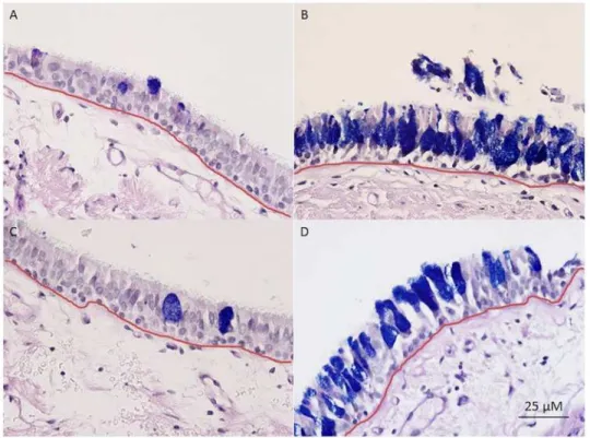
coefficient of 0.744, p<.0001).Fig. 1shows examples of a healthy nonsmoker and a COPD
sub-ject with CB. In order to assess goblet cell volume, mucin volume was measured using a
modi-fied model described by us ([14] using Image J. Length of basement membrane (LBM) and total
area of mucin granules (MA) were measured. Mucin volume density (μL/mm2) was calculated
using stereologic techniques as described previously:[15,16] Mucin volume density = MA/
(LBM)(4/π).
Statistics
Statistics were performed using SPSS v21 (Cary, NC). Differences between groups (nonsmok-ers, healthy smok(nonsmok-ers, COPD) were assessed by one way ANOVA for continuous variables and Chi squared test for categorical variables. Individual groups were compared with Bonferroni test in a post hoc analysis. The correlation between goblet cell density and pack year history of
smoking was assessed with Pearson’s correlation. Multivariate linear regression was performed
Table 1. Inclusion and Exclusion Criteria.
Inclusion Criteria
Age between 40 and 70 years Diagnosis of COPD or at risk for COPD Smoking History>10 pack years
FEV130%–60% (GOLD II/III group), normal FEV1(GOLD 0)
English speaking
Exclusion Criteria
Diagnosis of chronic sinusitis or allergic rhinitis Presence of other lung disease (including asthma) Pregnancy
Sinusitis or URI within the last 6 weeks
COPD Exacerbation within 6 weeks of screening visit Presence of infiltrate or mass on CT scan
Anticoagulation or antiplatelet therapy within 6 half lives of bronchoscopy Known allergy to lidocaine
with goblet cell density and mucin volume density as separate outcomes with current smoking, smoking pack year history, CB, demographic factors, and lung function as covariates. A p value
of<0.05 was considered statistically significant.
Results
The study population consisted of 38 subjects (11 nonsmokers, 12 healthy smokers, and 15
COPD subjects). Baseline characteristics are summarized inTable 2. The nonsmokers and
healthy smokers were younger than COPD subjects. There were no differences in gender, race, or body mass index. Lung function, current smoking, and smoking history were by definition different between groups. There were no subjects with CB in the nonsmoker group, 2 (16.7%) in the healthy smoker group, and 10 (66.7%) in the COPD group. There were no differences in the presence of CB between active smokers and non- or ex-smokers.
Healthy smokers (9.80±3.49 cells/mm) had a greater goblet cell density than nonsmokers
(2.31±1.81 cells/mm, p<.0001). Healthy smokers (26.35±10.96μL/mm2) also had a greater
mucin volume density than nonsmokers (5.77±4.34μL/mm2, p<.0001). Of considerable
inter-est was that healthy smokers had a greater goblet cell density and mucin volume density
com-pared to the COPD subjects (6.57±3.29 cells/mm, p = .020 and 14.83±13.63μL/mm2, p = .027,
respectively). SeeFig. 2. When all smokers (healthy smokers and COPD current smokers
com-bined, n = 19) were compared with all nonsmokers (nonsmokers and COPD ex-smokers, n =
19), the smokers had a greater goblet cell density (8.69±3.69 vs. 4.02±3.23 cells/mm, p<.0001)
and mucin volume density (22.49±13.73 vs. 8.08±7.46μL/mm2, p<.0001). All COPD
ex-Fig 1. Examples of mucosal biopsies from A) a healthy nonsmoker, B) a healthy smoker, C) a COPD subject without chronic bronchitis, and D) a COPD subject with chronic bronchitis, taken at 40x.
Specimens stained with periodic acid Schiff-Alcian Blue, staining goblet cells blue/purple. Basement membrane measured is outlined in red.
smokers quit smoking greater than 5 years prior to enrollment. SeeFig. 3. In addition, there was a significant correlation between pack-year history of smoking with goblet cell density (r = .417, p = .010). The COPD group was analyzed separately, and there was no
difference in GCD (7.19±3.97 vs. 6.74±3.21 gc/mm, p = 0.828) or MVD (12.57±11.54 vs. 16.71
±16.10μL/mm2, p = 0.629) between COPD smokers and COPD non-smokers.
When those with CB (healthy smokers and COPD subjects included, n = 12) were com-pared to those without CB (in all 3 groups combined, n = 26), the CB group had greater goblet
Table 2. Baseline Characteristics.
Nonsmokers Healthy Smokers COPD
n = 11 n = 12 n = 15 p
Age (years) 49.36±12.64 49.58±5.57 58.53±4.88 0.007
Gender (male, n(%)) 6 (54.5) 4 (33.3) 12 (80) 0.053
BMI (kg/m2) 30.85±5.07 30.78±4.74 29.66±5.60 0.800
Race*(White, n (%)) 5 (45.5) 2 (16.7) 2 (13.3) 0.136
FEV1(% pred) 94.00±11.46 101.17±16.30 45.40±8.99 <0.0001
FVC (%pred) 93.00±15.28 103.67±16.87 77.73±16.26 0.001
FEV1/FVC 93.27±13.37 96.67±6.14 47.13±11.51 <0.0001
Smoking History (pack years) 0 25.58±10.66 29.00±14.77 <0.0001
Current Smoking (n, %) 0 (0) 12 (100) 7 (46.7) <0.0001
Chronic Bronchitis (n, %) 0 (0) 2 (16.7) 10 (66.7) 0.001
Goblet Cells (n) 19.18±17.10 84.08±31.78 57.13±36.07 <0.0001
Length BM (mm) 7.04±2.87 9.47±3.52 10.02±3.89 0.101
Fields Measured (n) 7.09±2.81 8.83±3.01 8.00±3.48 0.426
GCD (cells/mm) 2.31±1.81 9.80±3.49 6.57±3.29 <0.0001
MVD (μL/mm2) 5.77±4.34 26.35±10.96 14.83±13.63 <0.0001
Definition of Abbreviations: BMI = body mass index, FEV1= forced expiratory volume in 1 second, FVC = forced vital capacity, GCD = goblet cell density,
MVD = mucin volume density.
*remainder of the cohort was African American.
doi:10.1371/journal.pone.0116108.t002
Fig 2. Goblet cell density in healthy nonsmokers, smokers without airflow obstruction, and COPD subjects.A. Data expressed as goblet cells per millimeter of basement membrane. Mucin volume density in the same three groups, B. Data expressed as mean±SE. Normal = healthy nonsmoking group, Healthsmoke = healthy smoker group, COPD = chronic obstructive pulmonary disease group, GC/MM = goblet cells per millimeter basement membrane, MVD = mucin volume density.
cell density (8.55±3.41 vs. 5.46±4.16 cells/mm, p = .036) but not mucin volume density (19.89
±14.59 vs. 14.17±12.65μL/mm2, p = .238). The COPD group was then analyzed separately.
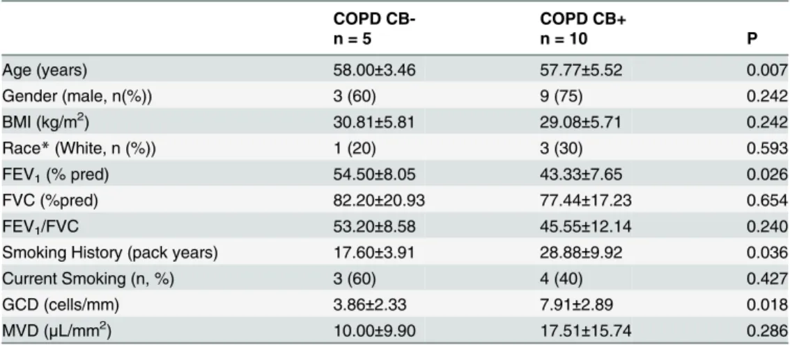
The differences between COPD CB+ and COPD CB- groups are summarized inTable 3. In
comparison to the COPD CB- group (n = 5), the COPD CB+ group (n = 10) was older (57.77 ±5.52 vs. 58.00±3.46 years, p = .007), had a greater smoking history (28.88±9.92 vs. 17.60±3.91
pack years, p = 0.036), and had a lower FEV1(43.33±7.65 vs. 54.40±8.05%predicted, p = .026).
There were no significant differences in gender, racial distribution, or percentage of subjects currently smoking. The COPD CB+ group had a greater goblet cell density compared to the COPD CB- group (7.91±2.89 vs. 3.88±2.33 cells/mm, p = .018), but there was not a significant
difference in mucin volume density (17.51±15.74 vs. 10.00±9.90μL/mm2, p = .286). SeeFig. 4.
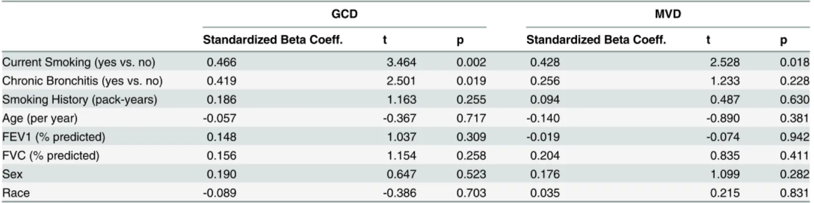
In multivariate linear regression, both CB and current smoking were significant determi-nants of goblet cell density (standardized beta coefficients. 419, p = .019 and. 466, p = .002, re-spectively). The variables pack year history of smoking, age, gender, race, and lung function
Fig 3. Goblet cell density in all smokers (smokers without airflow obstruction and COPD subjects that currently smoke) compared with all nonsmokers (healthy nonsmokers and COPD subjects who quit smoking).A. Data expressed as goblet cells per millimeter of basement membrane. Mucin volume density in all smokers compared with all nonsmokers, B. Data expressed as mean±SE. GC/MM = goblet cells per millimeter basement membrane, MVD = mucin volume density.
doi:10.1371/journal.pone.0116108.g003
Table 3. Characteristics of COPD patients with and without chronic bronchitis.
COPD CB- COPD CB+
n = 5 n = 10 P
Age (years) 58.00±3.46 57.77±5.52 0.007
Gender (male, n(%)) 3 (60) 9 (75) 0.242
BMI (kg/m2) 30.81±5.81 29.08±5.71 0.242
Race*(White, n (%)) 1 (20) 3 (30) 0.593
FEV1(% pred) 54.50±8.05 43.33±7.65 0.026
FVC (%pred) 82.20±20.93 77.44±17.23 0.654
FEV1/FVC 53.20±8.58 45.55±12.14 0.240
Smoking History (pack years) 17.60±3.91 28.88±9.92 0.036
Current Smoking (n, %) 3 (60) 4 (40) 0.427
GCD (cells/mm) 3.86±2.33 7.91±2.89 0.018
MVD (μL/mm2) 10.00±9.90 17.51±15.74 0.286
Definition of Abbreviations: BMI = body mass index, FEV1= forced expiratory volume in 1 second, FVC =
forced vital capacity, GCD = goblet cell density, MVD = mucin volume density.
*remainder of the cohort was African American.
did not reach statistical significance. For mucin volume density, the only statistically significant
covariate was current smoking (standardized beta coefficient. 428, p = .018). SeeTable 4.
Discussion
We used two different measures of goblet cell hyperplasia (goblet cell density for goblet cell number and mucin volume density to assess for hyperplasia) to show that smokers without air-flow obstruction and COPD subjects had a greater goblet cell density compared to nonsmokers, and that smokers without airflow obstruction had a greater goblet cell density and mucin vol-ume density compared to COPD subjects. We also showed that active smokers and chronic bronchitics had a greater goblet cell density compared to nonsmokers and those without chronic bronchitis, respectively. This difference was observed regardless of the presence or
Fig 4. Goblet cell density between those with chronic bronchitis and those without chronic bronchitis in the entire cohort (left) and in the COPD subjects alone (right).Data expressed as goblet cells per millimeter of basement membrane. GC/MM = goblet cells per millimeter basement membrane, CB+ = chronic bronchitis, CB- = no chronic bronchitis, COPD CB+ = COPD subjects with chronic bronchitis, COPD CB- = COPD subjects without chronic bronchitis.
doi:10.1371/journal.pone.0116108.g004
Table 4. Multivariate Linear regression for Goblet Cell Density and Mucin Volume Density.
GCD MVD
Standardized Beta Coeff. t p Standardized Beta Coeff. t p
Current Smoking (yes vs. no) 0.466 3.464 0.002 0.428 2.528 0.018
Chronic Bronchitis (yes vs. no) 0.419 2.501 0.019 0.256 1.233 0.228
Smoking History (pack-years) 0.186 1.163 0.255 0.094 0.487 0.630
Age (per year) -0.057 -0.367 0.717 -0.140 -0.890 0.381
FEV1 (% predicted) 0.148 1.037 0.309 -0.019 -0.074 0.942
FVC (% predicted) 0.156 1.154 0.258 0.204 0.835 0.411
Sex 0.190 0.647 0.523 0.176 1.099 0.282
Race -0.089 -0.386 0.703 0.035 0.215 0.831
absence of airflow obstruction, overall smoking burden, or demographic factors. This study is unique by virtue of the clinical correlation to large airway pathology with segregation of sub-jects by chronic bronchitis and current smoking.
Prior studies have established that GCH is greater in COPD subjects compared to those without airflow obstruction. Innes et al. performed a similar study of smokers with and without airflow obstruction and found significantly increased goblet cell density compared to non-smokers, and those with mild airflow obstruction had more goblet cells compared to the
smok-ers without airflow obstruction.[7] Saetta et al. found an increased number of goblet cells in the
peripheral airways of COPD subjects with CB compared to those without airflow obstruction.
[8] In addition, the expression of MUC5AC and MUC5B, two predominant airway mucins,
were increased in COPD subjects as compared to controls.[17] We have shown that mucin
volume density increased incrementally as COPD disease severity increased.[14] A large
patho-logic study revealed that the number of small airways occluded by mucus increased with
great-er degrees of airflow obstruction.[18] However, literature on the differences between
nonsmokers and current smokers and on the differences between those with CB and those without is sparse.
Our study differs from prior bronchoscopic studies by the inclusion of COPD subjects (cur-rent and ex-smokers) with moderate to severe airflow obstruction and those with and without CB. Other studies of large airway goblet cell hyperplasia in COPD included subjects with mild
to moderate disease.[8] We chose subjects with moderate to severe disease because this group
is more prone to COPD exacerbations, their goblet cell pathology has been poorly described in
other bronchoscopic studies, and they can safely undergo bronchoscopy.[19] Although we
showed that the COPD subjects had a greater goblet cell density compared to nonsmokers, we also showed that smokers without airflow obstruction, in comparison to the COPD subjects and normal controls, had the highest goblet cell density and mucin volume density, which dif-fers from prior studies. Based on these results and previously reported data,(7) our data sug-gests that large airway epithelial mucin stores increase initially before the onset of mild airflow obstruction and then decrease as more severe disease develops. In our cohort, this observation is predominantly driven by the presence of active smoking; active smokers had an increased goblet cell density and mucin volume density, and a weak but significant linear relationship ex-isted between goblet cell density and smoking history.
What also sets this study apart from prior ones is the demonstration of greater goblet cell density in chronic bronchitics versus those without CB, in the COPD group but also in the smokers without airflow obstruction. Thurlbeck et al. demonstrated that peripheral airway mu-cous gland hyperplasia and airway mucus were increased in CB but failed to show an increase
in goblet cell metaplasia in chronic bronchitics with little airflow obstruction.[12,20] The
study by Saetta et al. included subjects with CB alone, making the detection of pathologic
dif-ferences between those with and without CB impossible. [8] Other studies failed to show any
differences in the Reid Index, an established measure of submucosal gland hypertrophy,
be-tween those with CB and those without.[12,21]
Smoking has been associated with CB in many prior studies.[3,5,22,23] We have shown
that in subjects with moderate to severe COPD, those with CB were more likely to be current
smokers.[3] A thirty-year observational study of 1,711 Finnish men found that smoking
re-sulted in a cumulative incidence of CB of 42%.[5] A meta-analysis pooling numerous studies
showed that current smoking conferred a relative risk of 3.41 for the development of chronic
bronchitis.[22] However, few studies have addressed differences in goblet cell pathology
be-tween smokers and nonsmokers. Whereas some studies have shown increases in goblet cell
hy-perplasia in smokers compared with nonsmokers,[7,21] others have not.[8] In this study, we
cell density and mucin volume density compared to nonsmokers or ex-smokers, a rather novel finding. This finding remained significant on multivariate analysis for both measures of goblet
cell hyperplasia. Indeed, lung function, as measured by FEV1and FVC, did not have significant
bearing on GCD or MVD in multivariate analysis. This suggests that goblet cell hyperplasia does not contribute to airflow obstruction but rather that active smoking is its
primary determinant.
Similarly, goblet cell hyperplasia has been shown in CB,[7,8,24] but few studies have
com-pared goblet cell density in COPD subjects with CB comcom-pared to those without it. Many studies
about COPD pathology have either studied COPD undifferentiated by phenotype[14,18] or
chronic bronchitics alone.[8,24] To make matters worse, the existing literature showing the
link between symptoms of cough and phlegm and large airway pathology is weak at best.[25]
This study improves our current understanding of the clinico-pathologic link between CB and airway pathology.
We feel that the cohort used in this study adds strength to our findings. We purposefully ex-cluded individuals with conditions linked or thought to be associated with lower airway goblet cell hyperplasia (upper airway disease, recent exacerbations, etc.). In addition, we did not in-clude subjects actively treated with inhaled steroids to eliminate the confounding nature of this treatment on goblet cells. Finally, bronchoscopic literature on those with moderate to severe disease is rare, and these results offer a greater contribution our current understanding.
While this study has its strengths, there are also some limitations. One can argue that the study subgroup populations are small, particularly when dividing the COPD subjects into CB+ vs. CB-. By the nature of the study, we only sampled large airways, and therefore could not comment on small airway disease. We did not include subjects with mild to moderate airflow obstruction, as in prior literature. We excluded subjects on inhaled or oral steroids whom we considered unsafe to have them discontinued, thereby removing frequent exacerbators from the study. In addition, exacerbation history was not collected prior to enrollment in a systemat-ic fashion. Finally, while both measures of goblet cell hyperplasia were signifsystemat-icantly different between current smokers and non- or ex-smokers, only one was found to be different between those with and without CB. These observed differences are up for speculation, but suggest that goblet cell number and volume are greater as a result of active smoking, where goblet cell num-ber alone is greater in chronic bronchitics. The reasons for these differences are unknown. Fi-nally, it is unclear if the goblet cell density causes the clinical phenotype of CB or if they are purely associated.
Conclusions
Our study offers insight into the correlation between airway pathology and clinical phenotype. We were able to demonstrate in this small sample that current smoking and CB, and not air-flow obstruction, were independent factors associated with goblet cell hyperplasia. These find-ings have significant clinical implications, underscoring the importance of smoking cessation and identifying a phenotype at higher risk for poor outcomes. However, more subjects need to be analyzed in order to make a more conclusive determination of the links between airflow ob-struction and smoking on goblet cell hyperplasia.
Author Contributions
References
1. Global initiative for chronic obstructive lung disease (GOLD) guidelines, global strategy for the diagno-sis, management and prevention of chronic obstructive lung disease (2006) NHLMI/WHO workshop re-port. Available:www.goldcopd.com.
2. Kim V, Garfield JL, Grabianowski CL, Krahnke JS, Gaughan JP, et al. (2011) The effect of chronic spu-tum production on respiratory symptoms in severe COPD. COPD 8(2):114–20. doi:10.3109/15412555. 2011.558546PMID:21495839
3. Kim V, Han MK, Vance GB, Make BJ, Newell JD, et al. (2011) The chronic bronchitic phenotype of COPD: An analysis of the COPDGene study. Chest 140(3):626–33. doi:10.1378/chest.10-2948PMID: 21474571
4. Burgel PR, Nesme-Meyer P, Chanez P, Caillaud D, Carre P, et al. (2009) Cough and sputum produc-tion are associated with frequent exacerbaproduc-tions and hospitalizaproduc-tions in COPD subjects. Chest 135 (4):975–82. doi:10.1378/chest.08-2062PMID:19017866
5. Pelkonen M, Notkola IL, Nissinen A, Tukiainen H, Koskela H (2006) Thirty-year cumulative incidence of chronic bronchitis and COPD in relation to 30-year pulmonary function and 40-year mortality: A follow-up in middle-aged rural men. Chest 130(4):1129–37. PMID:17035447
6. Vestbo J, Prescott E, Lange P (1996) Association of chronic mucus hypersecretion with FEV1 decline and chronic obstructive pulmonary disease morbidity. copenhagen city heart study group. Am J Respir Crit Care Med 153(5):1530–5. PMID:8630597
7. Innes AL, Woodruff PG, Ferrando RE, Donnelly S, Dolganov GM, et al. (2006) Epithelial mucin stores are increased in the large airways of smokers with airflow obstruction. Chest 130(4):1102–8. PMID: 17035444
8. Saetta M, Turato G, Baraldo S, Zanin A, Braccioni F, et al. (2000) Goblet cell hyperplasia and epithelial inflammation in peripheral airways of smokers with both symptoms of chronic bronchitis and chronic air-flow limitation. Am J Respir Crit Care Med 161(3 Pt 1):1016–21. PMID:10712357
9. Kim V, Criner GJ, Abdallah HY, Gaughan JP, Furukawa S, et al. (2005) Small airway morphometry and improvement in pulmonary function after lung volume reduction surgery. Am J Respir Crit Care Med 171(1):40–7. PMID:15477494
10. Hogg JC, Chu FS, Tan WC, Sin DD, Patel SA, et al. (2007) Survival after lung volume reduction in chronic obstructive pulmonary disease: Insights from small airway pathology. Am J Respir Crit Care Med 176(5):454–9. PMID:17556723
11. Sciurba F, Martinez FJ, Rogers RM, Make B, Criner GJ, et al. (2006) The effect of small airway patholo-gy on survival following lung volume reduction surgery (LVRS). [abstract]. Proc Am Thorac Soc 3: A712.
12. Mullen JB, Wright JL, Wiggs BR, Pare PD, Hogg JC (1985) Reassessment of inflammation of airways in chronic bronchitis. Br Med J (Clin Res Ed) 291(6504):1235–9. PMID:3933614
13. Image J (2006) Available:http://rsb.info.nih.gov.
14. Kim V, Kelemen SE, Abuel-Haija M, Gaughan J, Sharafkhaneh A, et al. (2008) Small airway mucous metaplasia and inflammation in chronic obstructive pulmonary disease. J COPD 5(6):329–38. doi:10. 1080/15412550802522445PMID:19353346
15. Harkema JR, Plopper CG, Hyde DM, St George JA (1987) Regional differences in quantities of histo-chemically detectable mucosubstances in nasal, paranasal, and nasopharyngeal epithelium of the bon-net monkey. J Histochem Cytochem 35(3):279–86. PMID:2434556
16. Weibel ER (1979) Stereological methods. London: Academic Press Inc. Ltd. PMID:25121236
17. Caramori G, Casolari P, Di Gregorio C, Saetta M, Baraldo S, et al. (2009) MUC5AC expression is in-creased in bronchial submucosal glands of stable COPD patients. Histopathology 55(3):321–31. doi: 10.1111/j.1365-2559.2009.03377.xPMID:19723147
18. Hogg JC, Chu F, Utokaparch S, Woods R, Elliott WM, et al. (2004) The nature of small-airway obstruc-tion in chronic obstructive pulmonary disease. N Engl J Med 350(26):2645–53. PMID:15215480
19. Hattotuwa K, Gamble EA, O’Shaughnessy T, Jeffery PK, Barnes NC (2002) Safety of bronchoscopy, bi-opsy, and BAL in research patients with COPD. Chest 122(6):1909–12. PMID:12475825
20. Thurlbeck WM, Malaka D, Murphy K (1975) Goblet cells in the peripheral airways in chronic bronchitis. Am Rev Respir Dis 112(1):65–9. PMID:1147385
22. Forey BA, Thornton AJ, Lee PN (2011) Systematic review with meta-analysis of the epidemiological evidence relating smoking to COPD, chronic bronchitis and emphysema. BMC Pulm Med 11:36, 2466–11–36. doi:10.1186/1471-2466-11-36PMID:21672193
23. Martinez CH, Kim V, Chen Y, Kazerooni EA, Murray S, et al. (2013) The clinical impact of non-obstructive chronic bronchitis in current and former smokers. Respir Med 15.
24. Saetta M, Turato G, Facchini FM, Corbino L, Lucchini RE, et al. (1997) Inflammatory cells in the bron-chial glands of smokers with chronic bronchitis. Am J Respir Crit Care Med 156(5):1633–9. PMID: 9372687