Evaluation of conjunctival bacterial flora in patients
with Stevens-Johnson Syndrome
Luciana Frizon, Marı´lia Cavalcante Arau´jo, Larissa Andrade, Maria Cecı´lia Zorat Yu, Tais Hitomi Wakamatsu, Ana Luisa Ho¨fling-Lima, Jose´ A´lvaro Pereira Gomes
Federal University of Sa˜o Paulo (UNIFESP), Vision Institute, Ophthalmology Department, Sa˜o Paulo/SP, Brazil.
OBJECTIVE:To determine the conjunctival bacterial flora present in patients with Stevens-Johnson syndrome. METHODS:A prospective study of the conjunctival bacterial flora was performed in 41 eyes of 22 patients with Stevens-Johnson syndrome. The information gathered included the patient’s sex and age, the duration of disease, the cause of Stevens-Johnson syndrome, and treatments. Scrapings of the inferior conjunctival fornix were performed in both eyes. Fourteen days before scraping, the patients were asked to interrupt all topical medication and start using 0.5% nonpreserved methylcellulose. The microbiological evaluation included microorganism identification and determination of antibiotic sensitivity.
RESULTS:Of 22 patients (41 eyes), 14 (64%) were females, and eight (36%) were males. The mean age was 33.2 years, and the mean duration of disease was 15.6 years. Visual acuity ranged from light perception to 20/25 (1.57 logMar). The treatment received by most patients consisted of tear substitutes, topical antibiotics, and contact lenses. Bacterial identification was positive in 39 eyes (95%) and negative in two eyes (5%). Gram-positive cocci accounted for 55.5% of the microorganisms, whereas gram-Gram-positive bacilli and gram-negative bacilli accounted for 19% and 25.5%, respectively.
Half of the patients (54%) had multiple bacterial species in their flora, and only one bacterial species was identified in the other half. Resistant bacteria were isolated from four eyes. The antibiotic sensitivity results for theStreptococcusgroup showed the lowest sensitivity and the highest microbial resistance identified. CONCLUSION: Patients with Stevens-Johnson syndrome have a diverse conjunctival flora that includes many pathogenic species.
KEYWORDS: Conjunctival Flora; Microbial Sensitivity Tests; Stevens-Johnson Syndrome.
Frizon L, Arau´jo MC, Andrade L, Yu MC, Wakamatsu TH, Ho¨fling-Lima AL, et al. Evaluation of conjunctival bacterial flora in patients with Stevens-Johnson Syndrome. Clinics. 2014;69(3):168-172.
Received for publication onJune 25, 2013;First review completed onJuly 15, 2013;Accepted for publication onAugust 15, 2013 E-mail: [email protected]
Tel.: 55 11 5085-2003
& INTRODUCTION
Stevens-Johnson syndrome (SJS) is an acute, self-limiting disorder that affects the skin and two or more mucosal membranes. SJS is frequently accompanied by systemic involvement and can be lethal in certain cases. Although its incidence is low, SJS is life threatening, and its mortality is estimated at 5%. Despite certain idiopathic cases, SJS is usually triggered by medications or associated with an infectious process (1-3). Acute and chronic ocular complica-tions of SJS can be severe, involving the conjunctiva, the eyelids, and the cornea (1-3).
The normal microbial flora of the eyelids and conjunctiva is composed of a heterogeneous bacterial assemblage that is controlled by intrinsic mechanisms (nutrient competition, metabolic inhibition, and enzyme production) and by host mechanisms (anatomical barriers and the immunological system). Coagulase-negativeStaphylococci(CNS),Staphylococcus aureus, andCorynebacterium spare the most common micro-organisms isolated from a healthy patient’s conjunctiva (4-8).
Alteration of the conjunctival flora depends on seasonal variation, the host’s age, and environmental exposure, as well as ocular trauma, surgical procedures, and changes in local or systemic immunity (9). Topical or systemic antibiotics can also decrease the number of positive cultures (4,6,7).
Nouri et al. have demonstrated that patients with SJS and cicatricial pemphigoid ocular-surface diseases are at high risk of postoperative ocular infection compared with patients with noncicatrizing corneal disease (10). One of the hypotheses for this observation is that eyelid anatomical alterations (trichia-sis and entropion), conjunctival cicatricial disorders (sym-blepharon and keratinization), corneal alterations (limbal
Copyrightß2014CLINICS– This is an Open Access article distributed under the terms of the Creative Commons Attribution Non-Commercial License (http:// creativecommons.org/licenses/by-nc/3.0/) which permits unrestricted non-commercial use, distribution, and reproduction in any medium, provided the original work is properly cited.
DOI:10.6061/clinics/2014(03)04
deficiency), and severe dry eye are associated, which creates a propitious environment for the colonization of the ocular surface in these patients.
Although ocular surface alterations have been extensively studied in patients with SJS, PubMed and Medline searches with the key words ‘‘Stevens Johnson Syndrome’’ and ‘‘bacterial flora’’ revealed little information in the literature. To provide a better understanding of the components of the conjunctival flora and relevant information that can guide us in the treatment of ocular infections in patients with SJS, we performed a microbiological evaluation of inferior conjunctival scrapings, including microorganism identifica-tion and determinaidentifica-tion of antibiotic sensitivity.
& MATERIALS AND METHODS
This prospective nonrandomized study consisted of sampling the inferior conjunctival eyelids of patients with SJS who were selected at the Cornea and External Ocular Diseases Clinic of the Ophthalmology Department at the Federal University of Sa˜o Paulo (UNIFESP) between February and July, 2007. The protocol was approved by the Research Ethics Committee of UNIFESP. Informed consent was obtained from each subject studied. All of the experiments in this study followed the tenets of the Declaration of Helsinki.
The patients were submitted to ophthalmological exam-ination, and information regarding gender, age, the dura-tion of disease, the probable cause of SJS, previous surgical treatments, and current clinical treatment were collected.
After an initial evaluation, the patients received non-preserved 0.5% methylcellulose eye drops (Ophthalmos, SP, Brazil) for use q.i.d. for 2 weeks, up to the sampling day. Any other types of eye drops and systemic antibiotics, anti-inflammatories, and antifungals were discontinued for this period. Patients with a current diagnosis of any ocular or systemic infection or with a history of surgical procedures in the ocular region in the last 3 months were excluded from the study, as were patients who could not have their medications suspended during the study period.
Specimens were obtained with a sterile cotton swab moistened with brain-heart infusion (BHI) medium, scrap-ing the lower conjunctival sac and inferior tarsal without touching the eyelid margins or cilia, during slit-lamp observation and without the use of any anesthetic eye drops. We repeated the same technique in the other eye.
The scrapings were performed in the same examination room, with the air conditioner turned off for 20 minutes before the standard technique procedure. The average room temperature was 20
˚
C, and the average humidity was 70%. The scrapings were seeded on two solid media (a blood agar plate and a chocolate agar plate) and one liquid medium (thioglycolate broth). The collected specimens were immediately spread on the solid media, incubated at 37˚
C, and examined daily for bacterial growth for 1 week and weekly for up to 1 month. Any transportation media were used to transport the samples to the laboratory.Cultures showing growth of bacteria for at least two strains in any plate or changes in the color or turbidity of the broth medium were considered positive. Liquid broth specimens that were considered positive were subcultured on blood agar plates to identify the microorganism. Specimens with fungi and bacteria growing outside the
seeded area were considered contaminated and were thus discarded.
Laboratory analyses consisted of culture, microorganism identification, and antimicrobial sensitivity tests. The isolated microorganisms were submitted to sensitivity testing by the agar diffusion method using the subsequent antibiotics: amikacin, tobramycin, ceftriaxone, penicillin, gatifloxacin, sulfazotrim, gentamicin, cefalotin, chloramphe-nicol, polymyxin B, moxifloxacin, vancomycin, neomycin, cefoxitin, oxacillin, ciprofloxacin, and ofloxacin (adapted from CLSI M100-S20, Vol. 30, No. 1, January 2010).
& RESULTS
Patient description
We evaluated 41 eyes of 22 patients, consisting of 14 females (64%) and eight males (36%) with a mean age of 33.2 years (range of 11 to 63 years) and an average disease duration of 15.6 years (range of 6 months to 55 years). Three patients had one of their eyes enucleated because of complications during disease development and were using an acrylic ocular prosthesis.
Regarding the probable causes triggering SJS, 20 patients (90%) reported drug use before the disease, and in two patients (10%), no causes were identified. The main causative drugs involved were analgesics, mainly dipyrone (eight patients), anticonvulsants (seven patients), and antibiotics such as penicillin (four patients).
The majority of the patients had been submitted to surgical procedures, including eyelid correction (seven patients), lacrimal point occlusion (five patients), penetrat-ing keratoplasty (five patients), symblepharon correction (five patients), limbal transplant (four patients), and enucleation (three patients). Several patients were sub-mitted to multiple surgical procedures.
Clinical treatment before the conjunctival sampling (up to 14 days before sampling) consisted of lubricants (eye drops, gel, or ointment) for every patient, vitamin A ointment for three patients and topical glucocorticoids for four patients (the majority using antibiotic-associated ointment).
The use of topical antibiotics was noted in seven patients. Chloramphenicol was being used by four patients (one alone and three as ointment combined with glucocorti-coids); ofloxacin, by two patients; and ciprofloxacin, by one patient, all for prophylaxis against recurrent infections or because of contact lens (CL) use. Four patients were using CLs: two eyes with scleral lenses and the other two eyes with soft CLs. One patient had acquired immune deficiency syndrome (AIDS) and had been receiving highly active antiretroviral therapy (HAART) for 6 years, which was not suspended for this study.
The best corrected visual acuity ranged from light perception and 20/25 (average 1.57 logMar). By slit-lamp examination, corneal conjunctivalization (19 patients); lim-bal deficiency (18 patients); punctate keratitis (16 patients); symblepharon (10 patients); entropion, trichiasis, and occluded lacrimal points (five patients); ocular surface keratinization (four patients); tarsorrhaphy (three patients); an ocular prosthesis (three patients after enucleation); and ocular atrophy in one eye were observed. Several patients had associated ocular surface alterations.
suspension of topical medication, none of the subjects needed drug reintroduction, and thus, we had no exclu-sions.
Smears and cultures
Smears from 39 eyes (95%) yielded positive results. For the two eyes (5%) of one patient, smears were negative for the presence of bacteria.
The following bacteria were identified: gram-positive cocci (CNS,Streptococcus spandStaphylococcus aureus)in 35 eyes (55.5%), gram-positive bacilli (Corynebacterium sp) in 12 eyes (19%), and gram-negative bacilli (Enterobacter sp, Serratia nonliquefaciens, Escherichia coli, Morganella morganii, Proteus mirabilis and Haemophilus sp) in 16 eyes (25.5%) (Table 1).
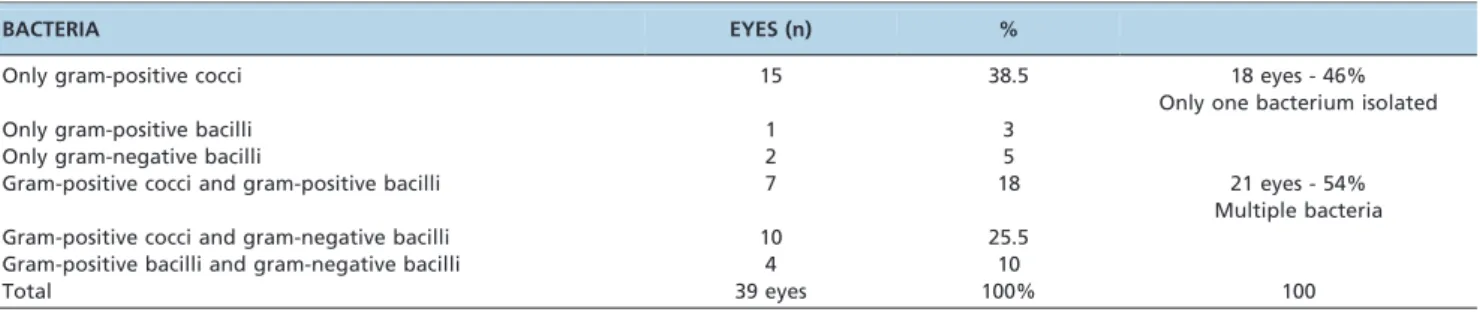
According to microorganism association analysis, cul-tures showed only one species of bacterium in 18 eyes (46%) and more than one species of bacterium in 21 eyes (54%). In this last group, two bacterial species were detected in 20 eyes, and three bacterial species were identified in only one eye (Table 2).
Positive cultures with only one species of bacterium in the conjunctiva showed gram-positive cocci in 15 eyes (38.5%), gram-positive bacilli in one eye (3%), and gram-negative bacilli in two eyes (5%). A combination of gram-positive cocci and gram-positive bacilli occurred in seven eyes (18%), and an association between positive cocci and gram-negative bacilli was found in 10 eyes (25.5%). Additionally, a combination of gram-positive bacilli and gram-negative bacilli was identified in four eyes (10%) (Table 2).
The respective conjunctival cultures of the three eyes with ocular prostheses demonstrated CNS (one eye), CNS associated withCorynebacterium sp(one eye), and no bacteria (one eye). One of the patients had AIDS, and his conjunctival flora showed the presence of Staphylococcus aureusandCorynebacterium spin both eyes.
Cultured conjunctival samples from two patients with soft CLs were positive for CNS andCorynebacterium sp.In the two patients who were users of scleral CLs, the bacteria wereProteus mirabilisand CNS.
In topical glucocorticoid users (four patients), we found
Proteus mirabilis(one patient), CNS (two patients), and CNS associated withStreptococcus sp(one patient).
Regarding the antibiotics used before sampling, 12 eyes of seven patients were treated with chronic antibiotic use.
Chloramphenicol was being used for seven eyes; ofloxacin, for three eyes; and ciprofloxacin, for two eyes. Patient 5 (two eyes), who previously used chloramphenicol, exhibited the presence ofProteus mirabilis that was resistant to chloram-phenicol. In patient 17 (two eyes), who used ciprofloxacin and showed the bacteria Corynebacterium sp and
Streptococcus spin culture, theStreptococcus spwas resistant to ciprofloxacin (Table 3).
In the general evaluation of antimicrobial sensitivity, we divided the microorganisms into the following groups, according to level of resistance: Staphylococcus (CNS and
Staphylococcus aureus), Streptococcus (Streptococcus sp), and gram-negative bacilli (Enterobacter sp,Serratia nonliquefaciens, Escherichia coli, Morganella morganii, Proteus mirabilis, and
Haemophilus sp). The sensitivity of the gram-negative bacilli (including Proteus mirabilis) to chloramphenicol was only 33%. In theStreptococcusgroup, the sensitivity was only 50% for ciprofloxacin (Table 4).
TheStaphylococcusgroup showed high sensitivity (.90%) to amikacin, tobramycin, ofloxacin, moxifloxacin, chloram-phenicol, vancomycin, oxacillin, polymyxin, and sulfazo-trim and low sensitivity (#50%) to neomycin and penicillin (Table 4).
TheStreptococcusgroup exhibited high sensitivity (.90%) to ofloxacin, gatifloxacin, cefalotin, and vancomycin and low sensitivity (#50%) to amikacin, tobramycin, gentamicin, neomycin, ciprofloxacin, chloramphenicol, polymyxin B, and sulfazotrim. This group had the lowest antimicrobial sensitivity and the highest index of resistance to antibiotics (Tables 3 and 4). In contrast, the group of gram-negative bacilli had high sensitivity (.90%) to amikacin, gentamicin, ofloxacin, moxifloxacin, gatifloxacin, ciprofloxacin, penicil-lin, and polymyxin B, whereas the group had low sensitivity (#50%) to the antibiotics ceftriaxone, cefalotin, chloramphe-nicol, vancomycin, and oxacillin.
& DISCUSSION
SJS is associated with many acute and chronic ocular complications. Patients with this illness are deficient in the ocular protection represented by tears, which prevent the entrance of pathogens and consequent infections (1,4).
The majority of SJS carriers show acute ocular involve-ment, varying from light conjunctivitis to serious corneal ulcers. Chronic complications, including conjunctival scars, symblepharon, entropion, and dry eye, can lead to corneal damage, which is a determinant of vision loss (1,3).
Different culture media can be used to isolate micro-organisms from ocular samples, and the use of certain types of media at the same time can increase the possibility of detection (4). In our study, we used two solid media (blood agar and chocolate agar) and one liquid medium (thiogly-colate).
Several studies have demonstrated that the flora of the eyelids and conjunctiva shows a high correlation with postoperative infection. CNS is the most frequently encoun-tered bacterium and is the most related to intraocular infections. Other microorganisms occurring in the ocular flora areCorynebacterium sp, which are normally found in the elderly (2,4-8,11-15).
Thiel et al. evaluated the normal conjunctival flora of patients of varying ages and reported the following bacteria as the most frequent: Corynebacterium sp, Megasphaera elsdenii, Bacteroides ureolyticus, Bacteroides pneumosintes,and Table 1 -Positive cultures according to bacterial
classification and number of eyes.
GROUP BACTERIA EYES (n) %
Gram-positive cocci
Coagulase-negative
Staphylococcus
19 55.5
Streptococcus sp 8
Staphylococcus aureus 8
Gram-positive bacilli Corynebacterium sp 12 19.0 Gram-negative
bacilli
Enterobacter sp 4 25.5
Serratia nonliquefaciens
4
Escherichia coli 2
Morganella morganii 2
Proteus mirabilis 2
Haemophilus sp 2
Total 63 eyes 100.0
Stomatococcus(13) Campos et al. demonstrated that 77.3% of normal patients were colonized by anaerobic bacteria; 63.3%, by Propionibacterium acnes; 13.6%, by species of
Lactobacillus; and 15.9%, by species of Veillonella, without significant differences in the conjunctival flora between patients with AIDS and patients with anophthalmia (9). Herde et al. studied the normal conjunctival flora of patients in the preoperative period before cataract surgery and demonstrated that the most frequent bacteria were
Staphylococcus sp and Proteus mirabilis (14). Hofling-Lima, analyzing bacteria isolated from the conjunctiva and eyelids of healthy patients prior to cataract and refractive surgeries in the Laboratory of Ocular Microbiology of the Escola Paulista de Medicina at UNIFESP, demonstrated that the most frequently found microorganisms were CNS,
Corynebacterium sp,andStaphylococcus aureus(11).
In our study, gram-positive cocci were the most common bacteria isolated, corresponding to 55.5% of the eyes. Gram-positive bacilli corresponded to 19% of the isolates, and gram-negative bacilli corresponded to 25.5%. Among all bacteria, CNS,Corynebacterium sp,Staphylococcus aureus, and
Streptococcus sp were the main microorganisms. As for gram-negative bacilli, we found pathogenic bacteria, includ-ing Enterobacter sp, Serratia nonliquefaciens, Escherichia coli, Morganella morganii, Proteus mirabilis, and Haemophilus sp. The flora was formed by multiple bacteria in 54% of the eyes and by only one bacterium in 46% of the eyes. Overall, the number of associated microorganisms was much higher than in previous studies involving healthy patients.
In our SJS patients who were using CLs (soft or hard scleral), the bacteria encountered were CNS, Proteus mirabilis, and Corynebacterium sp, agreeing with the study Table 3 -Bacterial cultures according to previous antibiotic use and antibiotic resistance.
Patient Antibiotic in Use Bacterial Culture Antibiotic Resistance
5 Chloramphenicol Proteus mirabilis TO, CHL, NEO, OXA
7 Chloramphenicol Corynebacterium Not tested
Enterobacter aerogenes CFT
12 Chloramphenicol Coagulase-negative Staphylococcus OXA, CIP
16 Ofloxacin Corynebacterium Not tested
Staphylococcus aureus GFX, GEN, OXA, CIP
17 Ciprofloxacin Corynebacterium Not tested
Streptococcus sp GEN,CIP
18 Chloramphenicol coagulase negative Staphylococcus
22 Ofloxacin Corynebacterium Not tested
CFT = cefalotin, OXA = oxacillin, TO = tobramycin, GEN = gentamicin, CHL = chloramphenicol, NEO = neomycin, FOX = cefoxotin, CIP = ciprofloxacin, GFX = gatifloxacin.
OBS: antibiotic abbreviations according to the Centers for Disease Control and Prevention (CDC).
Table 2 -Positive cultures isolated according to number of eyes and microorganism association.
BACTERIA EYES (n) %
Only gram-positive cocci 15 38.5 18 eyes - 46%
Only one bacterium isolated
Only gram-positive bacilli 1 3
Only gram-negative bacilli 2 5
Gram-positive cocci and gram-positive bacilli 7 18 21 eyes - 54%
Multiple bacteria
Gram-positive cocci and gram-negative bacilli 10 25.5
Gram-positive bacilli and gram-negative bacilli 4 10
Total 39 eyes 100% 100
Table 4 -Antibiotic sensitivity (%) of the bacterial groups in patients with SJS.
Bacterial Group Aminoglycoside Fluoroquinolone Cephalosporin Penicillin Sulfonamide Other
AK TO GEN OFL GFX MXF CIP CFR CFT FOX OXA PEN SZT NEO CHL VAN PB
Gram Positive-Cocci
Staphylococcus aureus 100% (2) 100% (4) 75% (8) 100% (2) 75% (8) 100% (8) 75% (8) 100% (2) 100% (8) 75% (8) 100% (2) 0% (2) 100% (2) 0% (2) 100% (2) 100% (2) 100% (2) Coagulase-Negative Staphylococcus 100% (2) 100% (2) 80% (16) 100% (2) 95% (18) 95% (18) 70% (16) 50% (4) 85% (18) 60% (18) 100% (2) 0% (2) 100% (2) 100% (2) 100% (2) 100% (2) 100% (2)
Streptococcus sp 0% (4) 0% (4) 50% (8) 100% (4) 100% (8) 75% (8) 50% (8) 50% (4) 100% (8) 75% (8) 50% (4) 50% (4) 0% (4) 50% (4) 100% (4) 0% (4) Gram-Negative Bacilli
Gram-Negative Bacilli 100% (6) 66% (6) 100% (16) 100% (6) 100% (16) 100% (16) 100% (16) 100% (6) 25% (16) 66% (12) 0% (6) 100% (2) 66% (6) 66% (6) 33% (6) 50% (4) 100% (6) AK = amikacin, TO = tobramycin, GEN = gentamicin, OFL = ofloxacin, GFX = gatifloxacin, MXF = moxifloxacin, CIP = ciprofloxacin, CFR = ceftriaxone, CFT = cefalotin, FOX = cefoxotin, OXA = oxacillin, PEN = penicillin, SZT = sulfazotrim, NEO = neomycin, CHL = chloramphenicol, VAN = vancomycin, PB = polymyxin B.
( ) = number of eyes tested.
of Erdogan et al., who demonstrated the lack of a difference between the normal conjunctival flora and the conjunctival flora of CL users (16).
In our study, four eyes of two patients who were receiving chronic antibiotic treatment until 2 weeks before sample collection, two eyes treated with chloramphenicol (with positive culture forProteus mirabilis), and another two eyes treated with ciprofloxacin (with positive cultures for
Streptococcus spandCorynebacteriumsp) showed antimicro-bial resistance to the antibiotics in use. The general sensitivity of the gram-negative bacilli to chloramphenicol was only 33%, and for the Streptococcus group, the sensitivity to ciprofloxacin was only 50%. We also found that the group with less sensitivity and a greater level of antimicrobial resistance included streptococci.
A high index of postoperative ocular infections is found in inflammatory conditions such as SJS and ocular cicatricial pemphigoid. Gomes et al. observed postoperative infection in 40% of patients with SJS who were submitted to ocular surface reconstruction surgery (17). Another study demon-strated a high level of ocular infections in patients with SJS who were submitted to keratoprosthesis implantation (10) Samson et al. reported a high incidence of infection in patients with chronic inflammatory diseases who under-went limbal stem cell transplantation (18).
In summary, we characterized the conjunctival bacterial flora in patients with SJS by describing the positive cultures according to bacterial classification and antibiotic sensi-tivity. This study revealed a diverse conjunctival flora, including many pathogenic species, in patients with SJS. The findings provide a guide for the treatment of ocular infections in SJS and provide additional information on necessary care in the preoperative and postoperative periods, mainly regarding broad-spectrum antibiotic pro-phylaxis.
& AUTHOR CONTRIBUTIONS
Frizon L conceived and designed the study, monitored the data collection, wrote the statistical analysis plan, synthesized and analyzed the data, and drafted the paper. Arau´jo MC and Andrade L monitored the data collection, wrote the statistical analysis plan, and synthesized and analyzed the data. Yu MC monitored the data collection and synthesized and analyzed the data. Wakamatsu TH synthesized and analyzed the data and revised the paper. Ho¨fling-Lima AL and Gomes JA conceived and designed the study, wrote the statistical analysis plan, synthesized and analyzed the data, and revised the paper.
& REFERENCES
1. Power WJ, Ghoraishi M, Merayo-Lloves J, Neves RA, Foster CS. Analysis of the acute ophthalmic manifestations of the erythema multiforme/ Stevens-Johnson syndrome/toxic epidermal necrolysis disease spec-trum. Ophthalmology. 1995;102(11):1669-76, http://dx.doi.org/10.1016/ S0161-6420(95)30811-1.
2. Holland EJ, Hardten DR. Stevens-Johnson syndrome. Pepose JS, Holland GN, Wilhelmus KR, editors. St. Louis: Mosby; 1996.
3. Frizon L, Santos MS, Ottaiano JAAea. Eritema Multiforme - Sı´ndrome de Stevens-Jonhson. Gomes JAP AM, editor. Rio de Janeiro: Cultura Me´dica; 2006.
4. Moeller CT, Branco BC, Yu MC, Farah ME, Santos MA, Hofling-Lima AL. Evaluation of normal ocular bacterial flora with two different culture media. Can J Ophthalmol. 2005;40(4):448-53.
5. Osato MS. Normal ocular flora. Pepose JS HG, Wilhelmus KR, editor. St. Louis: Mosby; 1995.
6. Liesegang TJ. Perioperative antibiotic prophylaxis in cataract surgery. Cornea. 1999;18(4):383-402, http://dx.doi.org/10.1097/00003226-199907000-00001.
7. Kramer A, Behrens-Baumann W. Prophylactic use of topical anti-infectives in ophthalmology. Ophthalmologica. 1997;211 Suppl 1:68-76, http://dx.doi.org/10.1159/000310889.
8. Khorazo D, Thompson R. The bacterial flora of the normal conjunctiva. Am J Ophthalmol 1935;18:1114-6.
9. Campos MS, Campos e Silva Lde Q, Rehder JR, Lee MB, O’Brien T, McDonnell PJ. Anaerobic flora of the conjunctival sac in patients with AIDS and with anophthalmia compared with normal eyes. Acta Ophthalmol (Copenh). 1994;72(2):241-5.
10. Nouri M, Terada H, Alfonso EC, Foster CS, Durand ML, Dohlman CH. Endophthalmitis after keratoprosthesis: incidence, bacterial causes, and risk factors. Arch Ophthalmol. 2001;119(4):484-9, http://dx.doi.org/10. 1001/archopht.119.4.484.
11. Ho¨fling-Lima AL, Farah ME, Montenegro L. Changes in conjunctival and lid flora after topical use of lomefloxacin and tobramycin in cataract and refractive surgery. Arq Bras Oftalmol. 2002;65(1):21-9, http://dx.doi. org/10.1590/S0004-27492002000100005.
12. Brinser J, Burd E. Principles of diagnostic ocular microbiology. Tobara KF HR, editor. Boston: Little Brown; 1996.
13. Thiel HJ, Schumacher U. [Normal flora of the human conjunctiva: examination of 135 persons of various ages]. Klin Monbl Augenheilkd. 1994;205(6):348-57, http://dx.doi.org/10.1055/s-2008-1045542. 14. Herde J, Tost M, Wilhelms D, Hohne C, Thiele T. [Perioperative
conjunctival flora]. Klin Monbl Augenheilkd. 1996;209(1):13-20, http:// dx.doi.org/10.1055/s-2008-1035270.
15. Mohan N, Gupta V, Tandon R, Gupta SK, Vajpayee RB. Topical ciprofloxacin-dexamethasone combination therapy after cataract sur-gery: randomized controlled clinical trial. J Cataract Refract Surg. 2001;27(12):1975-8, http://dx.doi.org/10.1016/S0886-3350(01)00863-X. 16. Erdogan H, Kemal M, Toker MI, Topalkara A, Bakici Z. Effect of
frequent-replacement contact lenses on normal conjunctival flora. CLAO J. 2002;28(2):94-5.
17. Gomes JA, Santos MS, Ventura AS, Donato WB, Cunha MC, Hofling-Lima AL. Amniotic membrane with living related corneal limbal/ conjunctival allograft for ocular surface reconstruction in Stevens-Johnson syndrome. Arch Ophthalmol. 2003;121(10):1369-74, http://dx. doi.org/10.1001/archopht.121.10.1369.