Published Ahead of Print 16 April 2007.
10.1128/AAC.01344-06.
2007, 51(7):2346. DOI:
Antimicrob. Agents Chemother.
Antoniana U. Krettli
Lopes, Virgílio E. do Rosário, Fernando de Pilla Varotti and
Valter F. de Andrade-Neto, Tito da Silva, Lucia M. Xavier
Holostylis reniformis
Lignans from
Antiplasmodial Activity of Aryltetralone
http://aac.asm.org/content/51/7/2346
Updated information and services can be found at:
These include:
REFERENCES
http://aac.asm.org/content/51/7/2346#ref-list-1
This article cites 27 articles, 2 of which can be accessed free at:
CONTENT ALERTS
more»
articles cite this article),
Receive: RSS Feeds, eTOCs, free email alerts (when new
http://journals.asm.org/site/misc/reprints.xhtml
Information about commercial reprint orders:
http://journals.asm.org/site/subscriptions/
To subscribe to to another ASM Journal go to:
on January 16, 2014 by UNESP - Universidade Estadual Paulista
http://aac.asm.org/
Downloaded from
on January 16, 2014 by UNESP - Universidade Estadual Paulista
http://aac.asm.org/
0066-4804/07/$08.00
⫹
0 doi:10.1128/AAC.01344-06
Copyright © 2007, American Society for Microbiology. All Rights Reserved.
Antiplasmodial Activity of Aryltetralone Lignans
from
Holostylis reniformis
䌤
Valter F. de Andrade-Neto,
1,2† Tito da Silva,
3Lucia M. Xavier Lopes,
3* Virgı´lio E. do Rosa´rio,
4Fernando de Pilla Varotti,
1and Antoniana U. Krettli
1Laborato
´rio de Malaria, Centro de Pesquisas Rene´ Rachou/FIOCRUZ, 30190-002, Belo Horizonte, MG, Brazil
1; Departamento de
Parasitologia, Universidade Federal de Minas Gerais, UFMG, Av. Augusto de Lima, 1715, 30190-002, Belo Horizonte,
MG, Brazil
2; Instituto de Quı´mica, Universidade Estadual Paulista, UNESP, C. P. 355, 14801-970, Araraquara,
SP, Brazil
3; and Instituto de Higiene e Medicina Tropical, Centro de Malaria e Doenc¸as Tropicais,
Universidade Nova de Lisboa, UNL, 1349-008, Lisboa, Portugal
4Received 26 October 2006/Returned for modification 12 November 2006/Accepted 3 April 2007
Extracts from
Holostylis reniformis
were tested in vivo against
Plasmodium berghei
and in vitro against a
chloroquine-resistant strain of
Plasmodium falciparum
. The hexane extract of the roots was the most active,
causing 67% reduction of parasitemia in vivo. From this extract, six lignans, including a new (7
ⴕ
R
,8
S
,8
ⴕ
S
)-3
ⴕ
,4
ⴕ
-methylenedioxy-4,5-dimethoxy-2,7
ⴕ
-cyclolignan-7-one, were isolated and tested in vitro against
P.
falcip-arum.
The three most active lignans showed 50% inhibitor concentrations of
<
0.32
M. An evaluation of
minimum lethal dose (30%) values showed low toxicity for these lignans in a hepatic cell line (Hep G2A16).
Therefore, these compounds are potential candidates for the development of antimalarial drugs.
Malaria is the most important parasitic disease in the world,
responsible for 500 million new cases and 2 to 3 million deaths
every year (http://www.who.int/malaria/epidemicsandemergencies
.html, http://www.who.int/tdr/diseases/malaria/diseaseinfo.html). The
number of clinical attacks due to
Plasmodium falciparum
seems to be 50% higher than WHO estimates (24). This
situ-ation, together with the progressive spread of
chloroquine-resistant strains of
P. falciparum
and, more recently,
Plasmo-dium vivax
, has caused an intensive search for novel blood
schizonticides to replace chloroquine, a cheap, safe, and,
for-merly effective therapeutic antimalarial drug (9, 20, 21). Many
natural products of various structural types have shown
anti-parasitic potency in the laboratory, and they represent
inter-esting lead structures for the development of new drugs (12).
The molecular diversity and efficacy of antiparasitic plants,
extracts, and herbal preparations have been intensively
dis-cussed in recent reviews (22, 26, 27).
More than 60 Brazilian plant species used in traditional
medicine to treat malaria and/or fever have been screened by
our group, and several species are active against
P. falciparum
in culture and
Plasmodium berghei
in mice (1, 13). In the
present study, we investigated the antimalarial activities and
toxicities of compounds isolated from
Holostylis reniformis
Duch. (Aristolochiaceae), which is used in traditional Brazilian
medicine as an antirheumatic, stomachic, and depurative (10).
H. reniformis
is a rich source of aryltetralone lignans (4–6).
Several lignans (1 to 6) (Fig. 1) isolated from extracts of this
species by chromatography (column, thin-layer
chromatogra-phy, and high-performance liquid chromatography [HPLC])
were then bioassayed in vitro for their antiplasmodial activities
and toxicities. The structures of lignans 1 to 5 had been
deter-mined by spectroscopic methods and chemical transformations
(5, 6). Lignan 6 is reported here for the first time.
MATERIALS AND METHODS
Plant material.The plant material was collected in Ituiutaba, MG, Brazil, in February 1998 and identified asH. reniformisDuch. by Condorcet Aranha and Lindolpho Cappellari Jr. A voucher specimen (ESA88282) was deposited at the herbarium of the Escola Superior de Agricultura “Luiz de Queiroz”, Piracicaba, SP, Brazil. The material was separated according to the plant parts, dried (⬃45°C), and ground (4–6).
Extraction and isolation of the chemical constituents.The plant material was extracted exhaustively at room temperature with hexane, acetone, and ethanol, successively, and the extracts were individually concentrated (4–6). The hexane extract (6.17 g) from the roots was fractionated by column chromatography (60.0 by 4.8 cm; silica gel 60 H; 151.0 g; hexane-ethyl acetate gradient, 95:5 to 100% ethyl acetate) to give 28 fractions (100 ml), as previously described (6). Several of these fractions were subjected to semipreparative HPLC (MeOH-H2O, 3:2).
Fraction 10 was comprised of lignans 1, 5, and 6 (11:3:2) and gave lignans 1 (67.6 mg), 5 (18.4 mg), and 6 (12.3 mg). Fraction 11 gave lignans 1 (25.7 mg) and 5 (22.1 mg). Fraction 12, comprised of lignans 2, 3, and 4 (3:1:2), was combined with fraction 13, comprised of lignans 3 and 4 (3:8), and subjected to semi-preparative HPLC (MeOH-H2O, 3:2) to give lignans 2 (255.7 mg), 3 (275.8 mg),
and 4 (28.1 mg).
Instrumentation.One-dimensional (1H,13C, and distortionless enhancement
by polarization transfer [DEPT]) and two-dimensional (1H-1H gradient-selected
correlated spectroscopy [gCOSY]; gradient-selected heteronuclear multiple-quantum coherence, inverse detected1H-13C one-bound correlation experiment
[gHMQC]; gradient-selected heteronuclear multiple-bond correlation, inverse detected1H-13C long-range correlation experiment [gHMBC]; and
gradient-selected nuclear Overhauser enhancement spectroscopy [gNOESY]) nuclear magnetic resonance (NMR) experiments were recorded on a Varian INOVA 500 spectrometer (11.7 T) at 500 MHz (1H) and 126 MHz (13C), with the residual
solvent CHCl3used as an internal standard for1H (␦7.23) and CDCl3for13C (␦
77.0). Mass spectra (electrospray ionization-mass spectrometry [ESI-MS]) were obtained on a Fisons Platform II, and flow injection into the electrospray source was used for ESI-MS. Infrared (IR) spectra were obtained on a Nicolet-730 FT-IR spectrometer using KBr discs. UV absorptions were measured on a Hewlett-Packard 8452A diode array spectrophotometer. Optical rotations were measured on a Polamat A (Carl Zeiss, Jena, Switzerland). Circular dichroism
* Corresponding author. Mailing address: Instituto de Quı´mica,
Universidade Estadual Paulista, UNESP, R. Francisco Degni s/n, C. P.
355, 14801-970, Araraquara, SP, Brazil. Phone: (55) 16-3301-6663.
Fax: (55) 16-3301-6692. E-mail: lopesxl@iq.unesp.br.
† Present address: Departamento de Microbiologia e Parasitologia,
Universidade Federal do Rio Grande do Norte, UFRN, 59072-970,
Natal, RN, Brazil.
䌤
Published ahead of print on 16 April 2007.
2346
on January 16, 2014 by UNESP - Universidade Estadual Paulista
http://aac.asm.org/
(CD) spectra were recorded on a JASCO J-720 spectrometer. HPLC analyses were carried out using a Shimadzu liquid chromatograph 10Avp equipped with a UV-visible light detector. Columns were RP18 (Shimadzu; C18; 3.9 by 150 mm
for analytical analysis and 250 by 20 mm for semipreparative analysis), and chromatograms were acquired at 254 nm. Melting points were recorded on a Microquimica MQAPF-301 melting point apparatus and were uncorrected.
(7⬘R,8S,8⬘S)-4,5-Dimethoxy-3⬘,4⬘-methylenedioxy-2,7⬘-cyclolignan-7-one
[(⫺)-8⬘-epi-aristotetralone; lignan 6] was obtained as a yellow solid, m.p. 136.2 to 138.0°C; 关␣兴D25⫺57.0 (c1.18, CHCl3); UV (MeOH)maxnm (logε) 239 (3.6), 278 (3.4), 323
(3.2); IR (KBr)max3469, 3022, 2959, 2924, 2862, 1667 cm⫺1; for1H and13C NMR,
see Table 1; ESI-MS (⫹35 eV)m/z355 [M⫹H]⫹(100); CD (MeOH,c0,1) []
212
⫹34650, []227⫹1155, []238⫹12177, []2470, []254⫺5709, []2650, []275⫹5181,
[]2800, []291⫺17061, []303⫺11616, []307⫺11616, []315⫺11880; analysis C
71.2%, H 6.2%, calculated for C21H22O5, C 71.2%, H 6.3%.
Antimalarial tests in vivo.The antimalarial tests were performed with adult Swiss albino mice (body weight, 20⫾2 g), and their use was approved by the Ethical Committee for Using Animals (CEUA-P0094-01, Fundac¸a˜o Instituto Oswaldo Cruz). The animals received water and food ad libitum. The antimalarial suppressive test was performed as previously described (3) in mice infected withP. bergheistrain NK-65, originally received from New York University Medical School. Each mouse (five mice per group) received 105infected red blood cells (day zero), followed by
daily treatment, via gavage, for 4 consecutive days. The extracts were suspended in Tween 20 (2% final concentration) immediately before use and then diluted so that doses of 100 to 500 mg/kg of body weight were delivered in 0.2 ml per animal. Three control groups were used in each test: one received chloroquine, and the others were not treated or were treated with Tween 20 (⬍0.2% final concentration). Blood smears were taken on days 5 and 7 after parasite inoculation, and mortality was monitored for 3 weeks. The results are expressed as the percent reduction of parasitemia in relation to untreated mice, and a compound was considered active when this reduction wasⱖ30% (3). Each experiment was performed in triplicate and repeated three times.
Parasite culture and in vitro antimalarial tests.TheP. falciparumused for the in vitro tests, a chloroquine-resistant isolate (BHz 26/86), was from an imported case of malaria from the Amazon region (3). Parasites were maintained in continuous culture on human erythrocytes (blood group AB⫹or A⫹, using
RPMI medium supplemented with 10% human serum), as previously described (25). The antiparasitic effects of extracts, purified compounds (lignans 1 to 6), and fractions were measured by the percent inhibition of parasite growth in relation to the control (parasites cultivated in drug-free medium), as previously described (3). Briefly, the drugs tested were diluted with Tween 20 at a final concentration of 0.02% in culture medium (RPMI 1640). These stock solutions were further diluted in complete medium (RPMI 1640 plus 10% human serum) to give each of the concentrations used (0.02 to 20M for purified compounds and fractions and 0.2 to 50g/ml for extracts). The cultures, with trophozoites in sorbitol-synchronized blood (14) at 1 to 2% parasitemia and 2.5% hematocrit, were then incubated with extracts, fractions, or isolated compounds for a total of 72 h at 37°C. A positive control with chloroquine (the reference antimalarial drug) and a control with medium and the Tween 20 solution were used in each experiment. The 50% and 90% inhibitory concentrations (IC50and IC90,
respec-tively), compared to the drug-free control responses, were estimated by linear interpolation (11). Each experiment was performed in triplicate and repeated three times. The blood smears were read in a double-blind manner.
Cytotoxicity test.An in vitro culture of Hep G2 A16 hepatic cells (7) was mixed with William’s E culture medium in 96-well microtiter plates and incu-bated at 37°C in an enriched CO2environment for 24 h (17). The compounds
were diluted with a 0.02% final concentration of Tween 20 solution in culture medium to obtain six concentrations: 500, 250, 100, 50, 10, and 5g/ml. After incubation periods of 24 and 48 h, the culture medium was replaced with 200l fresh medium with or without the drugs. At the end of the incubation periods, 20 l of MTT solution (5 mg of thiazolyl blue salt in RPMI 1640) without phenol red was added to each well, and the plates were incubated for three more hours. The supernatant was then removed, and 200l of acidified isopropanol was
FIG. 1. Bioassayed lignans from
H. reniformis
.
V
OL. 51, 2007
ACTIVITY OF ARYLTETRALONE LIGNANS FROM
H. RENIFORMIS
2347
on January 16, 2014 by UNESP - Universidade Estadual Paulista
http://aac.asm.org/
added to the wells. The culture plates were read by spectrophotometer with a filter of 570 nm and a background of 630 nm. The minimum lethal dose that killed 30% of the cells was determined (17). The assays were performed in three independent experiments.
Statistical analysis.The average parasitemias in vivo were compared using analysis of variance and Studentttests. Differences between IC50values were
evaluated by the Mann-WhitneyUtest performed with Biostat 1.0 MCT-CNPq. APvalue ofⱕ0.05 was considered to be statistically significant.
RESULTS
The crude hexane, acetone, and ethanol extracts of the roots,
stems, and leaves of
H. reniformis
partially reduced the malaria
parasitemia and mortality of mice infected with
P. berghei
. The
hexane extracts were the most active, especially the root and leaf
extracts, which caused 67% and 48% reduction of parasitemia,
respectively, at doses of 500 mg/kg (day 5;
P
ⱕ
0.05). Lower doses
tested were inactive. The extracts were also screened in vitro
against
P. falciparum
parasites (isolate BHz 26/86; chloroquine
resistant). The apolar extracts (hexane and acetone) exhibited the
best antiplasmodial activities, and they exhibited the lowest IC
50values (
⬃
0.70
g/ml), whereas the positive control (chloroquine)
showed an IC
50of 0.09
g/ml.
All of the isolated lignans were tested for antiplasmodial
activity in vitro; their IC
50and IC
90values, as well as the values
for the standard antimalarial chloroquine, obtained in three
sets of experiments are shown in Table 2. Lignans 1 to 3
exhibited IC
50values of
ⱕ
0.32
M (
ⱕ
0.12
g/ml). The lowest
IC
50value obtained was for lignan 3 (0.20
M), whereas the
lowest IC
90value was for lignan 4 (2.61
M), which showed
that these lignans are active and that they are the major active
principles in the extracts. Lignan 5 exhibited low activity, with
the highest IC
50(8.00
M) and IC
90(19.7
M) values, whereas
lignan 6 did not exhibit any activity under the same
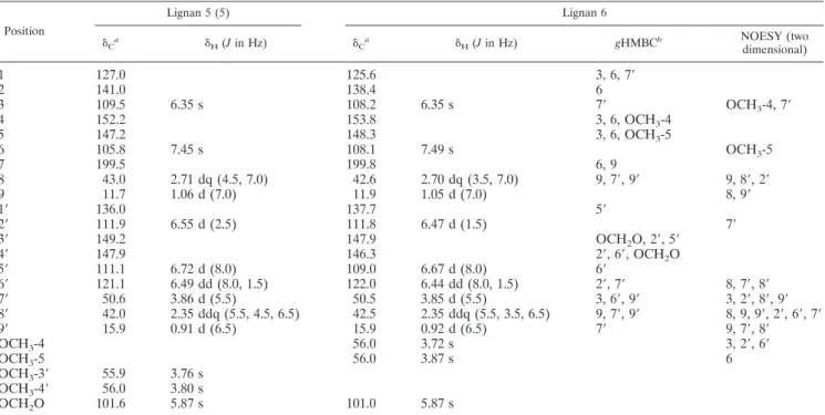
experimen-TABLE 1. NMR spectroscopic data (500 MHz, CDCl
3) for lignans 5 and 6
Position
Lignan 5 (5) Lignan 6
␦Ca ␦H(Jin Hz) ␦Ca ␦H(Jin Hz) gHMBCb NOESY (twodimensional)
1
127.0
125.6
3, 6, 7
⬘
2
141.0
138.4
6
3
109.5
6.35 s
108.2
6.35 s
7
⬘
OCH
3-4, 7
⬘
4
152.2
153.8
3, 6, OCH
3-4
5
147.2
148.3
3, 6, OCH
3-5
6
105.8
7.45 s
108.1
7.49 s
OCH
3-5
7
199.5
199.8
6, 9
8
43.0
2.71 dq (4.5, 7.0)
42.6
2.70 dq (3.5, 7.0)
9, 7
⬘
, 9
⬘
9, 8
⬘
, 2
⬘
9
11.7
1.06 d (7.0)
11.9
1.05 d (7.0)
8, 9
⬘
1
⬘
136.0
137.7
5
⬘
2
⬘
111.9
6.55 d (2.5)
111.8
6.47 d (1.5)
7
⬘
3
⬘
149.2
147.9
OCH
2O, 2
⬘
, 5
⬘
4
⬘
147.9
146.3
2
⬘
, 6
⬘
, OCH
2O
5
⬘
111.1
6.72 d (8.0)
109.0
6.67 d (8.0)
6
⬘
6
⬘
121.1
6.49 dd (8.0, 1.5)
122.0
6.44 dd (8.0, 1.5)
2
⬘
, 7
⬘
8, 7
⬘
, 8
⬘
7
⬘
50.6
3.86 d (5.5)
50.5
3.85 d (5.5)
3, 6
⬘
, 9
⬘
3, 2
⬘
, 8
⬘
, 9
⬘
8
⬘
42.0
2.35 ddq (5.5, 4.5, 6.5)
42.5
2.35 ddq (5.5, 3.5, 6.5)
9, 7
⬘
, 9
⬘
8, 9, 9
⬘
, 2
⬘
, 6
⬘
, 7
⬘
9
⬘
15.9
0.91 d (6.5)
15.9
0.92 d (6.5)
7
⬘
9, 7
⬘
, 8
⬘
OCH
3-4
56.0
3.72 s
3, 2
⬘
, 6
⬘
OCH
3-5
56.0
3.87 s
6
OCH
3-3
⬘
55.9
3.76 s
OCH
3-4
⬘
56.0
3.80 s
OCH
2O
101.6
5.87 s
101.0
5.87 s
aThe13C NMR data were assigned with the assistance of DEPT,gHMQC (optimized for 140 Hz), andgHMBC experiments.
bgHMBC correlations (optimized for 7 Hz) are from the proton(s) stated to the indicated carbon.
TABLE 2. IC
50s and IC
90s of lignans, alone or in mixtures, tested against
P. falciparum
isolate BHz26/86
Lignan(s) Compound IC50(M)a IC90(M)a
1
(7
⬘
R
,8
S
,8
⬘
R
)-4,5-Dimethoxy-3
⬘
,4
⬘
-methylenodioxy-2,7
⬘
-cyclolignan-7-one
0.26
⫾
0.08
3.35
⫾
0.12
2
(7
⬘
R
,8
S
,8
⬘
R
)-3
⬘
,4,4
⬘
,5-Tetramethoxy-2,7
⬘
-cyclolignan-7-one
0.32
⫾
0.11
4.60
⫾
0.30
3
(7
⬘
R
,8
R
,8
⬘
S
)-3
⬘
,4,4
⬘
,5-Tetramethoxy-2,7
⬘
-cyclolignan-7-one
0.20
⫾
0.09
3.00
⫾
0.15
4
(7
⬘
R
,8
S
,8
⬘
S
)-3
⬘
,4,4
⬘
,5-Tetramethoxy-2,7
⬘
-cyclolignan-7-one
0.63
⫾
0.20
2.61
⫾
0.06
5
(7
⬘
R
,8
S
,8
⬘
S
)-3
⬘
,4
⬘
-Dimethoxy-4,5-methylenodioxy-2,7
⬘
-cyclolignan-7-one
8.00
⫾
0.65
19.70
⫾
0.42
6
(7
⬘
R
,8
S
,8
⬘
S
)-4,5-Dimethoxy-3
⬘
,4
⬘
-methylenodioxy-2,7
⬘
-cyclolignan-7-one
⬎
140.00
⬎
140.00
3
⫹
4
Combination (3:8)
2.80
⫾
0.34
9.13
⫾
0.30
2
⫹
3
⫹
4
Combination (3:1:2)
6.00
⫾
0.50
18.20
⫾
0.40
1
⫹
5
⫹
6
Combination (11:3:2)
1.90
⫾
0.09
8.40
⫾
0.15
Chloroquine
b0.19
⫾
0.02
0.70
⫾
0.13
aValues are means⫾standard deviations in triplicate. bAntimalarial reference drug.
on January 16, 2014 by UNESP - Universidade Estadual Paulista
http://aac.asm.org/
tal conditions at the maximal dose tested (140.0
M
⫽
50
g/ml). Mixtures of these lignans, which were not previously
subjected to semipreparative HPLC (3 and 4; 2, 3, and 4; and
1, 5, and 6), also showed some activity and exhibited significant
IC
50(from 1.9 to 6.0
M) and IC
90(from 8.4 to 18.2
M)
values (Table 2).
The cytotoxicities of the active lignans evaluated in vitro
were considered low, since the mean minimum lethal dose that
killed 30% of the cells (450
g/ml) was at least 5,000 times
higher than the mean IC
50value obtained for them.
Compound 6 has not yet been described in the literature. It
was isolated from the active fraction 1, 5, and 6 (11:3:2) by
semipreparative HPLC. The
1H and
13C NMR, UV, IR, and
ESI-MS data for lignan 6 were similar to those reported for
lignan 5 (5).
DISCUSSION
Lignoids with different structural types (up to 60) have been
previously isolated from the family Aristolochiaceae (4–6, 8,
16, 19). The biosynthesis, functions, and pharmacological and
physiological effects of lignans have been studied, and these
compounds have been shown to possess a wide range of
bio-logical activities (15, 18, 23). Lignans have been used as lead
compounds for the development of new drugs, mainly due to
their low cytotoxicity and their antiangiogenic, antiviral,
anti-leishmanial, antifungal, hypolipidemic, and antirheumatic
ac-tivities (2). Here, we show that they also have an
antiplasmo-dial activity, as well as rather low cytotoxicity, as tested for one
cell line so far.
Compound 6 was suggested to be an aryltetralone lignan,
since it showed quasi-molecular ions at
m/z
355 [M
⫹
H]
⫹,
which were consistent with the molecular formula C
21H
22O
5,
and its IR,
1H, and
13C NMR spectra were very similar to those
of lignan 5 (5). A detailed analysis of
1H and
13C NMR,
1H-
1H
COSY, DEPT,
g
HMQC, and
g
HMBC experiments enabled
the precise assignment of all hydrogens and carbons in the
basic structure of lignan 6 (Table 1).
1H-
1H COSY and
1H
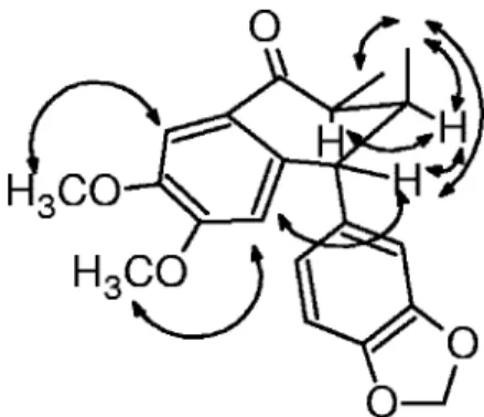
selective-irradiation NMR experiments with lignan 6 allowed
us to establish the same conformations and relative
configura-tion for the B ring as in lignan 5 (Fig. 2) (5). Therefore, the
main difference between lignans 6 and 5 is due to the
inter-change of substituents on the A and C rings. This deduction
was further confirmed by NOESY experiments (Table 1 and
Fig. 2). Moreover, the similarity between the CD curves of
these lignans allowed us to determine the same absolute
con-figuration for stereocenters on the B ring (5, 6). Thus, the
absolute configuration 7
⬘
R
,8
S
,8
⬘
S
was determined for lignan 6.
Based on an analysis of the structure-activity relationships
for these lignans, we could infer that the activity was affected
by the configurations of the stereocenters on the B ring and by
the substituents (methylenedioxy or dimethoxy groups) on the
A and C rings. The best activity was achieved for lignan with
dimethoxy substituents on the A ring and with the substituents
on the B ring (CH
3-9 and CH
3-9
⬘
and veratryl) in a
tran
s-
trans
orientation (lignan 3).
Aryltetralone lignans from
H. reniformis
showed
antimalar-ial activities and low toxicity on hepatic cells, and the three
most active lignans (1 to 3) showed IC
50values of
ⱕ
0.32
M.
Although mixtures of the lignans were at least 10 times less
active than lignan 3 and the standard chloroquine, their IC
50values were still low, i.e., in the micromolar range. However, a
lower antimalarial activity than one would expect for fractions
comprising mixtures of these lignans was observed. Whether
this reflects an antagonist effect is unclear, and further work
must be undertaken to elucidate this finding. These lignans are
worthy of further investigation, including chemical
transforma-tions, to optimize the activity and to study structure-activity
relationships of this class of antiplasmodial compounds. As
toxicity is a very important parameter for a suitable lead
can-didate in the development of antimalarial drugs, it also has to
be further investigated for the active lignans using other cell
lines, as well as animal models.
ACKNOWLEDGMENTS
We thank the Fundac¸a˜o de Amparo a` Pesquisa do Estado de Sa˜o
Paulo (FAPESP) and Fundac¸a˜o de Amparo a` Pesquisa de Minas
Gerais (FAPEMIG) for financial support and Conselho Nacional de
Desenvolvimento Cientı´fico (CNPq) for fellowships for A.U.K.,
T.D.S., and V.F.D.A.N.
We also thank Condorcet Aranha and Lindolpho Cappellari Jr. for
plant identification.
REFERENCES
1.Andrade-Neto, V. F., M. G. L. Branda˜o, F. Q. Oliveira, V. W. D. Casali, B. Njaine, M. G. Zalis, L. A. Oliveira, and A. U. Krettli.2004. Antimalarial activity ofBidens pilosaL. (Asteraceae) ethanol extracts from wild plants collected in various localities of plants cultivated in humus soil. Phytother. Res.18:634–639.
2.Apers, S., A. Vlietinck, and L. Pieters.2003. Lignans and neolignans as lead compounds. Phytochem. Rev.2:201–217.
3.Carvalho, L. H., M. G. L. Branda˜o, D. Santos-Filho, J. L. C. Lopes, and A. U. Krettli.1991. Antimalarial activity of crude extracts from Brazilian plants. Studied in vivo inPlasmodium berghei-infected mice and in vitro against
Plasmodium falciparumin culture. Braz. J. Med. Biol. Res.24:1113–1123. 4.da Silva, T., A. U. Krettli, V. F. de Andrade-Neto, and L. M. X. Lopes.2005.
Lignans, and particularly aryltetralone lignans, extracts containing them, processes for obtaining the extracts and the lignans, and use of the lignans or the extracts in pharmaceutical compositions for treating or preventing ma-laria. Rev. Prop. Ind.1795:1774–1786.
5.da Silva, T., and L. M. X. Lopes.2004. Aryltetralone lignans and 7,8-seco-lignans fromHolostylis reniformis. Phytochemistry65:751–759.
6.da Silva, T., and L. M. X. Lopes.2006. Aryltetralol and aryltetralone from
Holostylis reniformis. Phytochemistry67:929–937.
7.Denizot, F., and R. Lang.1986. Rapid colorimetric assay for cell growth and survival. Modifications to the tetrazolium dye procedure giving improved sensitivity and reliability. J. Immunol. Methods89:271–277.
8.de Pascoli, I. C., I. R. Nascimento, and L. M. X. Lopes.2006. Configurational analysis of cubebins and bicubebin fromAristolochia lagesianaand Aristolo-chia pubescens. Phytochemistry67:735–742.
9.Guerin, P. J., P. Olliaro, F. Nosten, P. Druilhe, R. Laxminarayan, F. Binka, W. L. Kilama, N. Ford, and N. J. White.2002. Malaria: current status of
FIG. 2. Selected nuclear Overhauser enhancement interactions
and conformation for lignan 6.
V
OL. 51, 2007
ACTIVITY OF ARYLTETRALONE LIGNANS FROM
H. RENIFORMIS
2349
on January 16, 2014 by UNESP - Universidade Estadual Paulista
http://aac.asm.org/
control, diagnosis, treatment, and a proposed agenda for research and de-velopment. Lancet Infect. Dis.2:564–573.
10.Hoehne, F. C.1942. Aristolochiaceas, p. 1–265.InF. C. Hoehne (ed.), Flora Brası´lica, vol. 17. Graphicars, Sa˜o Paulo, Brazil.
11.Huber, W., and J. C. Koella.1993. A comparison of three methods of estimating EC50 in studies of drug resistance of malaria parasites. Acta Trop.55:257–261.
12.Kayser, O., A. F. Kiderlen, and S. L. Croft.2003. Natural products as antiparasitic drugs. Parasitol. Res.90:S55–S62.
13.Krettli, A. U., V. F. Andrade-Neto, M. G. L. Branda˜o, and W. M. Ferrari. 2001. The search for new antimalarial drugs from plants used to treat fever and malaria or plants randomly selected: a review. Mem. Inst. Oswaldo Cruz 96:1033–1042.
14.Lambros, C., and J. P. Vanderberg.1979. Synchronization ofPlasmodium falciparumerythrocytic stages in culture. J. Parasitol.65:418–420. 15.Lewis, N. G., and L. B. Davin.1999. Lignans: biosynthesis and function, p.
639–712.InD. H. R. Barton, K. Nakanishi, and O. Meth-Cohn (ed.), Com-prehensive natural products chemistry, vol 1. Elsevier, London, United King-dom.
16.Lopes, L. M. X., I. R. Nascimento, and T. da Silva.2001. Phytochemistry of the Aristolochiaceae family, p. 19–108.InR. M. M. Mohan (ed.), Research advances in phytochemistry, vol. 2. Global Research Network, Kerala, India. 17.Madureira, A. M., A. P. Martins, M. Gomes, J. Paiva, M. J. U. Ferreira, A. P. Cunha, and V. E. Rosa´rio.2002. Antimalarial activity of medicinal plants used in traditional medicine in S. Tome´ and Prı´ncipe Island. J. Ethnophar-macol.81:23–29.
18.Nascimento, I. R., A. T. Murata, S. A. Bortoli, and L. M. X. Lopes.2004. Insecticidal activity of chemical constituents from Aristolochia pubescens
againstAnticarsia gemmatalislarvae. Pest Manage. Sci.60:413–416. 19.Nascimento, I. R., and L. M. X. Lopes.1999. 2,3-Dihydrobenzofuran
neoli-gnans fromAristolochia pubescens. Phytochemistry52:345–350. (Erratum, 53:621.)
20.Peters, W., L. B. Stewart, and B. L. Robinson.2003. The chemotherapy of rodent malaria. LXI. Drug combinations to impede the selection of drug resistance, part 4: the potential role of 8-aminoquinolines. Ann. Trop. Med. Parasitol.97:221–236.
21.Ridley, R. G.2002. Medical need, scientific opportunity and the drive for antimalarial drugs. Nature415:686–693.
22.Schwikkard, S., and F. R. Van Heerden.2002. Antimalarial activity of plant metabolites. Nat. Prod. Rep.19:675–692.
23.Skytte, D. M., S. F. Nielsen, M. Chen, L. Zhai, C. E. Olsen, and S. B. Christensen.2006. Antimalarial and antiplasmodial activities of norneoli-gnans. Syntheses and SAR. J. Med. Chem.49:436–440.
24.Snow, R. W., C. A. Guerra, A. M. Noor, H. Y. Myint, and S. I. Hay.2005. The global distribution of clinical episodes ofPlasmodium falciparummalaria. Nature434:214–217.
25.Trager, W. M., and J. B. Jensen.1976. Human malaria parasites in contin-uous culture. Science193:673–675.
26.Willcox, M. L., and G. Bodeker.2004. Traditional herbal medicines for malaria. Br. Med. J.329:1156–1159.
27.Wright, C. W.2005. Plant derived antimalarial agents: new leads and chal-lenges. Phytochem. Rev.4:55–61.