801
Original article doi: 10.12980/jclm.3.2015j5-114 ©2015 by the Journal of Coastal Life Medicine. All rights reserved.
Alterations of blood serum parameters in patients with chronic hematogenous osteomyelitis
Sadrudin Magomedov, Larisa Polishchuk*
Laboratory of Biochemistry, the Institute of Traumatology and Orthopedics, National Academy of Medical sciences of Ukraine, Kiev, Ukraine Journal of Coastal Life Medicine2015; 3(10): 801-804
Journal of Coastal Life Medicine
*Corresponding author: Larisa Polishchuk, Laboratory of Biochemistry, the Institute of Traumatology and Orthopedics, National Academy of Medical sciences of Ukraine, Kiev, Ukraine.
E-mail: [email protected]
Foundation Project: Supported by Institute of Traumatology and Orthopedics of the National Academy of Medical Sciences of Ukraine (Grant No. 0103U001318).
1. Introduction
Osteomyelitis is an infectious disease that affects bones and surrounding tissues. Based on the ways of microbial penetration in the bone, it evolves hematogenous osteomyelitis and non-hematogenous (posttraumatic). The development of inflammation in bone tissue may be preceded by bone injury and reduce (due to overworking, infectious disease, hypovitaminosis, etc.) total organism’s resistance[1].
Currently, despite the use of new high-performance technologies, the problem of osteomyelitis treatment remains unsolved. Thereby, a further study and clarification of the existing theory of osteomyelitis pathogenesis, and the search for new treatment approaches, diagnosis and disease prevention represent theoretical and practical interest. Medical rehabilitation of orthopedic and trauma patients with chronic osteomyelitis is an extremely important issue. The evidence
is a steady increase in the number of patients with this pathology, elevated rate of complications and failures, as well as patients with permanent disabilities[2].
The significant variations in chemical and cellular composition of internal environment, levels of renal excretion of electrolytes and products of catabolism, changes of hormonal and immunological status have been revealed in patients with osteomyelitis[3,4]. Furthermore, this pathology is caused by prolonged incapacity and patient with high frequency of disability[5].
Alterations of collagen metabolism and glycosaminoglycans, which comprise organic basis of bone matrix, are the most informative biochemical parameters of inflammatory pathology and regeneration of bone tissue[6].
Collagenase activity is increased in tumors and inflammatory processes such as rheumatoid arthritis, osteomyelitis, osteogenesis imperfecta, Paget’s disease, a metabolic disorder in which collagen is accompanied by breach of collagenase activity[7-10].
Glycosaminoglycans comprise intercellular substance of connective tissue, and are contained in bones, synovial fluid, vitreous body and cornea. Together with the fibers of collagen and elastin, proteoglycans form a connective tissue matrix (base substance). One of the representative glycosaminoglycans is heparin, which has A RT I C L E I N F O A B S T R AC T
Objective:To examine metabolic disorders of major components of organic basis of bone tissue in patients with chronic hematogenous osteomyelitis and response to surgical treatment. Methods: The cubital vein puncture was conducted to take blood for analysis in patients with chronic hematogenous osteomyelitis. The activity of collagenase and hyaluronidase, elastin, elastase and total content of glycosaminoglycans were measured in blood serum.
Results: The study revealed an enhancement of catabolic phase of metabolism of the main components in bone organic matrix during the relapse of inflammation. It was evidenced by indicators reflecting the synthetic and catabolic phases of the main components of the connective tissue collagen and glycosaminoglycans. The effective therapeutic treatments led to the reduction and normalization of studied compounds.
Conclusions: The initial development of hematogenous osteomyelitis happens in a background of metabolic disorders of the main components of organic matrix of bone tissue, and normalizes upon effective therapy.
Article history: Received 13 Jul 2015
Received in revised form 23 Jul 2015 Accepted 20 Aug 2015
Available online 28 Sep 2015
Keywords:
Hematogenous osteomyelitis Glycosaminoglycans Collagen
Sadrudin Magomedov and Larisa Polishchuk/Journal of Coastal Life Medicine 2015; 3(10): 801-804
802
anticoagulant activity and is localized in intercellular substance of liver, lung, heart and artery walls. Proteoglycans covering surface of the cells, play an important role in ion exchange, immune reactions, and tissue differentiation. Genetic disorders caused by disturbed decay of glycosaminoglycans lead to the development of a large group of inherited metabolic diseases including mucopolysaccharidosis[1]. The solubility of collagen and elastin decreases with aging, and increases the amount of cross-links in proteins, reduces the content of tissues proteoglycans and glycosaminoglycans. Overall decrease in cellular elements of connective tissue is common. Metabolic disorders of connective tissue play an important role in the development of many acquired diseases. Thus, the excess of collagen synthesis is observed in the fibrosing processes in lungs, liver, and regeneration disorders during wound healing (keloids). Various metabolic disorders underlie diffuse connective tissue diseases[11-13].
Thus, the study of glycosaminoglycans and collagenase activity, enzymes involved in protein metabolism, in serum of patients with chronic hematogenous osteomyelitis, will provide an opportunity to determine the degree of metabolic processes disruption in organic basis of bone tissue.
The purpose of research is to examine metabolic disorders of major components of organic basis of bone tissue in patients with chronic hematogenous osteomyelitis.
2. Materials and methods
A total of 36 patients in the age of 18–50 years with chronic hematogenous osteomyelitis were examined. The cubital vein puncture was conducted to take blood for analysis in patients before meal during the morning hours 8:00–9:00.
Terms of observations were as follows: 1) before treatment (sequestrectomy surgery), 2) 14–15 days after the start of treatment (sequestrectomy surgery) and 3) 35–40 days after the start of
treatment.
Sequestrectomy was a surgical treatment that included four points: 1) removal of necrotic tissue from osteomyelitis pockets, sequestration, pus and granulation; 2) processing of sclerotized wall of sequestered cavity till the emergence of viable areas of bone; 3) opening of medullary canal above and below the center of destruction; 4) treatment of residual bone cavity followed by its plastic.
The following biochemical parameters were measured in blood serum: the activity of collagenase and hyaluronidase, elastin, elastase, and total content of glycosaminoglycans. Similar parameters measured in blood serum of healthy donors in the age of 18–50 years
were taken for control.
The biochemical parameters, listed above, were determined according to methods: activity of collagenase was determined by the method of Lindy et al.[14], glycosaminoglycans in blood serum were determined by orcin method of Klyatskin and Lifshitz[15], and activity of hyaluronidase was determined by the method of Matysiak
et al.[16]. The content of elastin was determined by the method of Anwar[17] and elastase activity was determined by the method of Murashova and Osadchuk[18].
Statistical analysis and graphical display of the results were made using Microsoft Excel 2010. The data were presented as mean ± SD. The biochemical data were analyzed by student’s t-test. The difference was considered as significant at P≤0.05.
3. Results
Figures 1–5 show changes in concentration of the studied
parameters in blood serum of patients with osteomyelitis in comparison with values in the control group. The concentration of the studied compounds in the control group was accepted as 100% and changes were recalculated correspondingly.
250
200
150
100
50
0
% Collagenase acti
vity
Control goup Before treatment 14–15 days 35–40 days Figure 1. Collagenase activity in blood serum of patients with chronic hematogenous osteomyelitis.
Figure 2. The concentration of glycosaminoglycans in blood serum of
patients with chronic hematogenous osteomyelitis. 600
500 400 300 200 100 0
% Glycosaminoglycans
Control goup Before treatment 14–15 days 35–40 days
Figure 3. The activity of hyaluronidase in blood serum of patients with chronic hematogenous osteomyelitis.
350 300 250 200 150 100 50 0
% Hyaluronidase acti
vity
Sadrudin Magomedov and Larisa Polishchuk/Journal of Coastal Life Medicine 2015; 3(10): 801-804
803
Figure 4. The content of elastin in blood serum of patients with chronic
hematogenous osteomyelitis. 160
140 120 100 80 60 40 20 0
% Elastin
Control goup Before treatment 14–15 days 35–40 days
Figure 5. Elastase activity in the blood of patients with chronic hematogenous osteomyelitis.
250 200 150 100 50 0
%
E
la
st
as
e
ac
ti
v
it
y
Control goup Before treatment 14–15 days 35–40 days
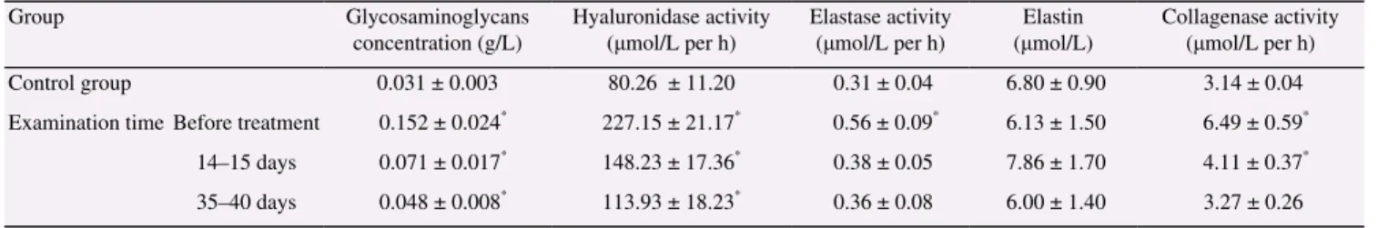
The analysis of enzymes involved in catabolism of collagen had shown that collagenase activity in patients with chronic hematogenous osteomyelitis in the period of disease relapse reached (6.49 ± 0.59) μmol/L per h [normal (3.14 ± 0.04) μmol/L per h], or 207% relative to the norm. On Day 14–15 after the start of treatment, enzyme activity was reduced to (4.11 ± 0.37) μmol/L per h, and on Day 35–40 to (3.27 ± 0.26) μmol/L per h (131% and 104%, respectively) (Table 1 and Figure 1).
Glycosaminoglycans, which were one of the key components of organic matrix of tissue, played an important role in formation
of connective tissue and its function. Hyaluronidase had a great influence on glycosaminoglycans catabolism. It had been
found that, along with the increase in activity of hyaluronidase, concentration of glycosaminoglycans in blood serum increased. The
concentration of glycosaminoglycans in blood serum of healthy people was (0.031 ± 0.003) g/L, whereas its level in serum of
patients with hematogenous osteomyelitis during disease relapse increased more than 4 times reaching (0.152 ± 0.024) g/L, or 492%
(Table 1 and Figure 2).
A f t e r 1 4 – 1 5 d a y s o f t r e a t m e n t , c o n c e n t r a t i o n o f glycosaminoglycans reduced to 228%, and on 35–40 days it reached 155% [(0.048 ± 0.008) g/L] approaching the level typical for healthy
individuals. Data obtained in the study of blood serum in patients with chronic hematogenous osteomyelitis were compared with
activity of hyaluronidase and concentration of glycosaminoglycans. It had revealed direct correlation between parameters in all terms of
patient examinations (Table 1 and Figure 3).
However, data obtained in the study of elastin metabolites in
blood serum of patients with chronic hematogenous osteomyelitis, showed tendency to reduction of concentration during inflammation relapse. Thus, if in healthy people it was equal to (6.80 ± 0.90) μmol/L, in blood of patients before treatment it comprised (6.13 ± 1.50) μmol/L, or 91% relatively to control. On Day 14–15 after
treatment, concentrations of elastin metabolite increased to (7.86 ± 1.70) μmol/L, or 116% comparing with controls. On Day 35–40 this parameter reduced to (6.00 ± 1.40) μmol/L, or 88% (Table 1 and Figure 4).
We suggested that decrease in concentration of elastin metabolites on Day 35–40 was associated with inhibition of elastin synthesis by its high concentration on Day 14–15 after the surgical
treatment. The same as it took place at high concentrations of glycosaminoglycans and collagen due to activation of retrograde
principle. The reduction may also be associated with increased rate of incorporation of elastin metabolites into newly formed fibers of
bone connective tissue.
In our study of elastase, an enzyme involved in elastin catabolism
during relapse of inflammatory focus, before surgical treatment had high activity of elastase reaching (0.56 ± 0.09) μmol/L per h, whereas in control group it comprised (0.31 ± 0.04) μmol/L per
h. It increased to 181%, or 1.8 times comparing to healthy people. On Day 14–15 after surgery and therapy enzyme activity reduced to (0.38 ± 0.05) μmol/L per h, or 123%. This decline continued until Day 35–40 but did not reach the values in the blood of control
group (Table 1 and Figure 5). Table 1
Biochemical parameters of blood serum in patients with chronic hematogenous osteomyelitis.
Group Glycosaminoglycans
concentration (g/L)
Hyaluronidase activity
(μmol/L per h) Elastase activity (μmol/L per h) (μmol/L) Elastin Collagenase activity(μmol/L per h)
Control group 0.031 ± 0.003 80.26 ± 11.20 0.31 ± 0.04 6.80 ± 0.90 3.14 ± 0.04
Examination time Before treatment 0.152 ± 0.024* 227.15 ± 21.17* 0.56 ± 0.09* 6.13 ± 1.50 6.49 ± 0.59*
14–15 days 0.071 ± 0.017* 148.23 ± 17.36* 0.38 ± 0.05 7.86 ± 1.70 4.11 ± 0.37*
35–40 days 0.048 ± 0.008* 113.93 ± 18.23* 0.36 ± 0.08 6.00 ± 1.40 3.27 ± 0.26
*
Sadrudin Magomedov and Larisa Polishchuk/Journal of Coastal Life Medicine 2015; 3(10): 801-804
804
4. Discussion
Our data show intensification of catabolic phase of metabolism in the key components of bone organic matrix in blood of patients with chronic hematogenous osteomyelitis during relapse of inflammation. It is demonstrated by parameters of synthetic and catabolic phases of the major components of connective tissue (collagen and glycosaminoglycans).
The activity of collagenase, an enzyme participating in catabolic phase of collagen metabolism - the major protein of bone tissue, exceeds control parameter in 2 folds before treatment, on Day 14–15 reduces to 131% comparing with control, and on Day 35–40 reaches normal values [(3.27 ± 0.26) μmol/L per h].
The increase of glycosaminoglycans content and activation of hyaluronidase should be noted as well. It suggests about the enhancement of catabolic phase of metabolism of bone organic matrix. Upon Day 14–15, the effective therapy stabilizes metabolic phases in all components of organic matrix of bone tissue, and on Day 35–40 parameters get closer to norm, but do not reach it. Increased elastase activity during amplification of inflammatory process in patients can be explained by the fact that this enzyme is involved in catabolism of elastin, which is collapsing at inflammation.
Our data suggest that the initial development of hematogenous osteomyelitis happens in a background of metabolic disorders in the main components of organic matrix of bone tissue, and normalizes upon effective therapy.
These biochemical parameters may also be the criteria for relapse of disease process and effectiveness of patient treatment.
Conflict of interest statement
We declare that we have no conflict of interest.
Acknowledgments
We are grateful to Professor Mykola Grytsay, M.D., Head of the Department, and Andrrii Hordii, traumatologist-orthopedist at the Department of Osteopurulent Surgery at the Institute of Traumatology and Orthopedics of the National Academy of Medical Sciences of Ukraine for providing blood samples, technical support and discussion. This paper was supported by Institute of Traumatology and Orthopedics of the National Academy of Medical Sciences of Ukraine (Grant No. 0103U001318).
References
[1] Braunlin EA, Harmatz PR, Scarpa M, Furlanetto B, Kampmann C, Loehr JP, et al. Cardiac disease in patients with mucopolysaccharidosis: presentation, diagnosis and management. J Inherit Metab Dis 2011;
34(6): 1183-97.
[2] Bhavan KP, Marschall J, Olsen MA, Fraser VJ, Wright NM, Warren DK. The epidemiology of hematogenous vertebral osteomyelitis: a cohort study in a tertiary care hospital. BMC Infect Dis 2010; 10: 158. [3] Wideman RF, Prisby RD. Bone circulatory disturbances in the
development of spontaneous bacterial chondronecrosis with osteomyelitis: a translational model for the pathogenesis of femoral head necrosis. Front Endocrinol (Lausanne) 2013; 3: 183.
[4] Ferguson PJ, Sandu M. Current understanding of the pathogenesis and management of chronic recurrent multifocal osteomyelitis. Curr Rheumatol Rep 2012; 14(2): 130-41.
[5] Caliendo AM, Gilbert DN, Ginocchio CC, Hanson KE, May L, Quinn TC, et al. Better tests, better care: improved diagnostics for infectious diseases. Clin Infect Dis 2013; 57(Suppl 3): S139-70.
[6] Tiemann AH, Hofmann GO. Principles of the therapy of bone infections in adult extremities: are there any new developments? Strategies Trauma Limb Reconstr 2009; 4(2): 57-64.
[7] Sagai M, Bocci V. Mechanisms of action involved in ozone therapy: is healing induced via a mild oxidative stress? Med Gas Res 2011; 1: 29. [8] Müller G. Microvesicles/exosomes as potential novel biomarkers of
metabolic diseases. Diabetes Metab Syndr Obes 2012; 5: 247-82. [9] Miller JD, Carter E, Hatch DC, Zhubrak M, Giovinco NA, Armstrong
DG. Use of collagenase ointment in conjunction with negative pressure wound therapy in the care of diabetic wounds: a case series of six patients. Diabet Foot Ankle 2015; 6: 24999.
[10] Ternovoĭ KS, Magomedov SM, Perfilova TN, Gritsaĭ NP. [Biochemical criteria for assessing the effectiveness of treating chronic osteomyelitis patients]. Vrach Delo 1987; (3): 74-7. Russian.
[11] Spellberg B, Lipsky BA. Systemic antibiotic therapy for chronic osteomyelitis in adults. Clin Infect Dis 2012; 54(3): 393-407.
[12] Owens CD, Rybicki FJ, Wake N, Schanzer A, Mitsouras D, Gerhard-Herman MD, et al. Early remodeling of lower extremity vein grafts: inflammation influences biomechanical adaptation. J Vasc Surg 2008; 47(6): 1235-42.
[13] Langevin HM, Bouffard NA, Fox JR, Palmer BM, Wu J, Iatridis JC, et al. Fibroblast cytoskeletal remodeling contributes to connective tissue tension. J Cell Physiol 2011; 226(5): 1166-75.
[14] Lindy S, Halme J, Turto H, Rokkanen P, Vainio K, Wegelius O. Collagenolytic activity in rheumatoid synovial tissue. Clin Chim Acta 1973; 47(2): 153-7.
[15] Kliatskiĭ SA, Lifshitz RI. [Analysis of glycosaminoglycans in the blood of patients by the orcinol method]. Lab Delo 1989; (10): 51-3. Russian. [16] Nagaraju S, Girish KS, Pan Y, Easely KA, Kemparaju K. Estimation of
serum hyaluronidase activity overcoming the turbidity interference. Clin Lab Sci 2011; 24(3): 172-7.
[17] Anwar RA. Comparison of elastins from various sources. Can J Biochem 1966; 44: 725-34.