Simplified Method for Rapid Purification
of Soluble Histones
Nives Ivić, Bernhard Groschup, Silvija Bilokapić,* Mario Halić#
Gene Center Munich and Department of Biochemistry, Ludwig-Maximilians-Universität München, 81377 Munich, Germany * Corresponding author’s e-mail address: [email protected]
# Corresponding author’s e-mail address: [email protected]
RECEIVED: December 15, 2015 REVISED: March 08, 2016 ACCEPTED: March 11, 2016
THIS PAPER IS DEDICATED TO THE LOVING MEMORY OF IVANA WEYGAND-ĐURAŠEVIĆ (1952–2014)
Abstract: Functional and structural studies of histone-chaperone complexes, nucleosome modifications, their interactions with remodelers and regulatory proteins rely on obtaining recombinant histones from bacteria. In the present study, we show that co-expression of Xenopus laevis histone pairs leads to production of soluble H2AH2B heterodimer and (H3H4)2 heterotetramer. The soluble histone complexes are purified by simple chromatographic techniques. Obtained H2AH2B dimer and H3H4 tetramer are proficient in histone chaperone binding and histone octamer and nucleosome formation. Our optimized protocol enables rapid purification of multiple soluble histone variants with a remarkable high yield and simplifies histone octamer preparation. We expect that this simple approach will contribute to the histone chaperone and chromatin research.
Keywords: histone, nucleosome, chromatin, protein co-expression.
INTRODUCTION
HE genome of eukaryotic cells has to be packed within a small nucleus, but at the same time accessible to dif-ferent macromolecular machineries required for DNA replication, transcription, recombination, damage and repair.[1] To accomplish this, all Eukaryotes pack their DNA into a highly organized and dynamic complex called chro-matin. The basic repeating unit of the chromatin is the nu-cleosome.[2] The nucleosome structure condenses the DNA and serves to shelter and keep the DNA from harm, but at the same time possesses the flexibility to allow numerous DNA and histone modifications, as well as the DNA infor-mation to be read.
The structure of the nucleosome core particle (NCP) has been determined to near atomic resolution.[3] Each nu-cleosome is built from two copies of four different histone proteins (H2A, H2B, H3 and H4) forming an octamer and an approximately 146 bp of DNA that is wrapped around the histone core. An additional 10–90 bp of linker DNA is com-plexed with a linker histone (H1 or H5) that is located at the point where DNA enters and exits the nucleosome.[4] DNA
binds the octamer through extensive nonspecific interac-tions of the sugar-phosphate backbone. Wrapping of DNA around the histone octamer results in a tenfold reduction of the DNA length and forms what is known as ‘beads on a string’, the 10-nm chromatin fiber.[5]
Croat. Chem. Acta 2016, 89(2), 153–162 DOI: 10.5562/cca2806 play a crucial role in diverse biological processes as gene
expression, chromosome packing, DNA damage repair.[7] Histones are highly conserved proteins in all Eukaryotes, underlining their crucial role in packing of the genomic ma-terial and inheritance of epigenetic information.
The term "Histone chaperone" was first used in 1978 by R. Laskey to describe the nucleoplasmin role in the pre-vention of histone-DNA aggregation during nucleosome as-sembly.[8] Histone chaperones are a broad group of structurally unrelated acidic proteins that bind histones, protect them from nonspecific interactions and aggrega-tion, assist in their folding, trafficking and nucleosome as-sembly and disasas-sembly.[9] Histone chaperones are divided into H2AH2B or H3H4 chaperones depending on their pref-erential histone binding.
There is a considerable interest in understanding his-tone-chaperone interactions, the chromatin structure and the mechanisms by which the chromatin is assembled and regulated. Biochemical and biophysical chromatin studies depend on the availability of large amounts of highly puri-fied components. Before bacterial expression of histone proteins has been established in Richmond lab,[10] these proteins used to be isolated from native sources.[11] Alt-hough high amount of histones could be obtained after chromatin isolation from HeLa cells or calf thymus, the pro-cedures were time-consuming and the purified histones were partially degraded by cellular proteases. An additional heterogeneity was caused by posttranslational modifica-tions of histones or by different histone isoforms.
A drawback of bacterial expression is that the indi-vidual histones end up forming insoluble inclusion bodies. Inclusion bodies are isolated from the whole cell extracts in several washing steps with and without detergent. His-tones are solubilized from the purified inclusion bodies us-ing denaturants as urea or guanidine hydrochloride, further purified on ion exchange and size exclusion chromatog-raphy.[12,13] This approach proved to be very successful and resulted in the crystal structure of the nucleosome core particle.[3] However, the procedure is laborious and time demanding. Several different approaches have been estab-lished in the recent years that tried to overcome these is-sues in the histone preparation. The most straightforward modification of the original protocol is "one pot refolding" method.[14] In this method all four X. laevis histones are ex-pressed individually into inclusion bodies, mixed together and denatured in guanidine hydrochloride. After this solu-bilisation step, histone octamer is reconstituted by dialysis against sodium chloride and further purified using affinity and size exclusion chromatography.
In the ‘‘rapid histone purification’’ inclusion bodies isolation is also omitted and the whole cell extracts with the individually expressed histones are directly solubilized us-ing denaturus-ing conditions.15 Histones are further purified
on ion exchange chromatographies under denaturing con-ditions. Unfolded histones are mixed together and octamer is reconstituted by dialysis against sodium chloride. This protocol has been successfully applied to Drosophila and human H2A, H2B, H3, H4 histones.[15]
An alternative method where histones were co-ex-pressed was also described. It was shown that co-expres-sion of histones H2A and H2B (from S. cerevisiae, chicken and Drosophila) resulted in the production of a soluble his-tone dimer. Drosophila H2A and H2B was further purified using different chromatography steps. In order to purify a soluble Drosophila (H3H4)2 complex, the histones had to be co-expressed with Asf1 histone chaperone and further purified from the chaperone using additional chromatog-raphy steps.[16]
Finally, co-expression of all 4 Xenopus histones from a single polycistronic vector was recently described.[17] Co-expression resulted in the production of the soluble histone octamer within the cell. The histone octamer was purified using affinity and size exclusion chromatography.
All described approaches are trying to reduce time and labour needed for histone and octamer preparation. For structural studies histone proteins from X. laevis and H. sapiens are most commonly used. These studies include de-ciphering the interactions within NCP itself3 or with its bind-ing proteins,[18] as well as studying histone-chaperone mechanisms of recognition.[19] Structural studies require large amounts of highly purified components. Preparation of independent histones from overexpressing bacterial cells is laborious and lengthy. Moreover, significant loss of material is experienced in each purification step as compo-nents remain attached to the column.[13] Although the col-umn can be cleaned with sodium hydroxide, the procedure shortens the column lifetime especially as the procedure has to be repeated for each histone protein.
In this study we were interested in finding conditions for obtaining high amounts of starting material combined with a small number of steps required for purification of the soluble components to minimize the subsequent losses. Our method provides flexibility in the downstream steps as histones can be used either for NCP reconstitution or for chaperone binding.
EXPERIMENTAL
Plasmid Construction
DOI: 10.5562/cca2806 Croat. Chem. Acta 2016, 89(2), 153–162 In addition, the pre-existing S-tag at the H2A N-terminus is
also present. The primers used for iPCR are listed in Table 1. In the next iPCR step we replaced the thrombin cleavage site with a human rhinovirus 3C (HRV 3C) protease recogni-tion site (Table 1).
The plasmid containing all four histones and HRV 3C protease cleavage site in front of 6xHis-tag was created. The primers used for PCR are listed in Table 1. DNA contain-ing H2A and H2B histone pair was PCR amplified, as well as plasmid DNA containing H3 and H4 histones. Two PCR prod-ucts were blunt-end ligated after T4 PNK reaction to add phosphates. The resulting vector was transformed into Escherichia coli BL21 (DE3) Rosetta strain. Genes for Asf1 and Nap1 proteins were amplified from Schizosaccharomy-ces pombe genomic DNA and cloned into an in-house mod-ified version of the pETDuet-1 bacterial expression plasmid (Novagen) to yield an N-terminally 6xHis-SUMO-tagged protein followed by a HRV 3C protease recognition site. The resulting vector was transformed into Escherichia coli BL21 (DE3) RIL strain (Stratagene).
Inverse PCR
PCR reactions were set up in a total volume of 25 µl. Prod-ucts were purified using the NucleoSpin Gel and PCR Clean-up kit (Macherey-Nagel). 6.5 µl of purified PCR product was
incubated in 1× T4 DNA ligase buffer with 5 U T4 PNK for 1 hour at 37 °C. The product was circularized using 200 U T4 DNA ligase for 1 h at room temperature. Template plasmid was digested by adding 10 U DpnI for 1 hour at 37 °C. The total volume of the reaction was 20 µL and 5 µL was used to transform XL21-blue E. coli cells.
Protein expression and purification
Transformed cells were grown in LB medium containing 0.4 % (w/v) glucose and the appropriate antibiotics. The culture was grown at 37 °C to an optical density at 600 nm of 0.6 before it was shifted to 18 °C for 30 min and induced by the addition of 0.2 mmol dm−3 isopropylthioga-lactopyranoside. Cells were harvested by centrifugation after an overnight induction at 18 °C. Pelleted cells were resuspended in the lysis buffer. The lysis buffer for histone purifications contained 50 mmol dm−3 sodium-phosphate, pH 8.0, 2 mol dm−3 NaCl, 20 mmol dm−3 imidazole, 3 mmol dm−3 β-mercaptoethanol, 1 mmol dm−3 phenylmethane-sulfonyl fluoride. When purifying histone chaperones, lysis buffer with 500 mmol dm−3 NaCl was used. Cells were lysed using a French press and clarified by centrifugation at 17000 g for 20 min. The cleared supernatant was incubated with Ni Sepharose 6 Fast Flow resin (GE Healthcare) for 30 min. After binding, the resin
Table 1. List of primers used for cloning A List of primers used for iPCR
OLIGONUCLEOTIDES SEQUENCE
plasmid with H2AH2B genes thrombin recognition site
435R TTACTTGGCGCTGGTG
435F GATCCGGCTGCTAACA
plasmid with H2AH2B genes HRV 3C protease recognition site
438R GAACAGAACTTCCAGGGTACCGTGGTGGT
438F CAGGGGCCCGGATCCATGTCAGGAAGAGGC
plasmid with H3H4 genes thrombin recognition site
436R1 CTAGAGGGGAATTGTTATC
436F GTCGACAATAATTTTGTTT
plasmid with H3H4 genes HRV 3C protease recognition site
439R1 GAACAGAACTTCCAGGCCGCTGCTACCACCG
439F CAGGGGCCCGGATCCCACCACCACCACCACCAC
B List of primers used for blunt end cloning
plasmid amplification
436R1 CTAGAGGGGAATTGTTATC
436F GTCGACAATAATTTTGTTT
insert amplification
437R TAAAGTTAAACAAAATTATTGTCGACTTACTTGGCGCTGG
Croat. Chem. Acta 2016, 89(2), 153–162 DOI: 10.5562/cca2806 was washed three times with 10 bed volumes of lysis
buffer in batch and loaded onto a disposable column (Pierce). The column was washed with 4 bed volumes of washing buffer (50 mmol dm−3 sodium-phosphate, pH 8.0, 2 mol dm−3 NaCl, 40 mmol dm−3 imidazole, 3 mmol dm−3 β-mercaptoethanol, 1 mmol dm−3 phenylmethanesulfonyl fluoride). The bound protein was eluted with 6 bed volumes of elution buffer (50 mmol dm−3 sodium-phosphate, pH 8.0, 2 mol dm−3 NaCl, 300 mmol dm−3 imidazole, 3 mmol dm−3 β-mercaptoethanol, 1 mol dm−3 phenylmethanesulfonyl fluoride). The histone proteins were dialyzed overnight at 4 °C against 25 mmol dm−3 Tris/HCl, pH 7.5, 2 mol dm−3 NaCl, 1 mmol dm−3 EDTA and 1 mmol dm−3 DTT. To obtain the histone octamer from co-expressed and co-purified H2AH2B and H3H4 histone pairs, we used 1.2-1.4 fold excess of H2AH2B dimer. This prevented formation of the free (H3H4)2 tetramer and the histone hexamer that are difficult to separate from the octamer in size exclusion chromatography. The sample was set up for overnight dialysis against 25 mmol dm−3 Tris/HCl, pH 7.5, 2 mol dm−3 NaCl, 1 mmol dm−3 EDTA and 1 mmol dm−3 DTT. Histone chaperones were dialyzed against 20 mmol dm−3 HEPES/KOH, pH 7.5, 150 mmol dm−3 NaCl, 1 mmol dm−3 EDTA and 1 mmol dm−3 DTT.
The tags, when required, were proteolytically re-moved by adding HRV 3C protease in 100:1 mass ratio during dialysis. The digestion was analysed by 17 % SDS-PAGE.
Size Exclusion Chromatography
Histone proteins were concentrated after dialysis and loaded onto a size-exclusion Superdex 200 column 10/30 (GE Healthcare) equilibrated in dialysis buffer (25 mmol dm−3 Tris/HCl, pH 7.5, 2 mol dm−3 NaCl, 1 mmol dm−3 DTT). The elution profile and the SDS-PAGE analysis of H2AH2B histones confirmed dimerization, and tetramerization for co-purified H3H4. Histone octamers obtained by co-expres-sion had the same running behaviour on size excluco-expres-sion chromatography as histone octamers obtained by mixing individually purified soluble H2AH2B histone dimer and (H3H4)2 histone tetramer. For long-term storage the his-tone octamer peak fractions were pooled and dialyzed against 25 mmol dm−3 Tris/HCl pH 7.5, 2 mol dm−3 NaCl, 1 mmol dm−3 DTT and 50 % glycerol (v/v).
Nucleosome Reconstitution
DNA for nucleosome reconstitution was PCR amplified from a plasmid containing the strong positioning Widom 601 DNA sequence.[21] After DNA purification, the salt concen-tration in the sample was adjusted to 2 mol dm−3 NaCl. The histone octamers and DNA were mixed in the molar ratio 1:1. Each reaction was placed into a dialysis button made from the lid of an Eppendorf tube. All dialysis buttons were
placed inside a dialysis bag, filled with ≈30 ml of initial buffer (25 mmol dm−3 Tris/HCl pH7.5, 2 mol dm−3 NaCl, 1 mmol dm−3 EDTA, 1 mmol dm−3 DTT). The dialysis bag was immersed into a 500 ml of buffer containing 1 mol dm−3 NaCl (25 mmol dm−3 Tris/HCl pH7.5, 1 mol dm−3 NaCl, 1 mmol dm−3 EDTA and 1 mmol dm−3 DTT) and dialysed over-night at +4 ºC. The next day the 1 mol dm−3 NaCl buffer was replaced with buffer containing 0.1 mol dm−3 NaCl (25 mmol dm−3 Tris/HCl pH7.5, 100 mmol dm−3 NaCl, 1 mmol dm−3 EDTA and 1 mmol dm−3 DTT). The sample was dialysed into low salt buffer for 6–8 hours. The results of the nucle-osome reconstitution by 'double bag' dialysis were ana-lysed on 6% native polyacrylamide gels run in 0.2x TBE at +4 ºC.
Pull-down Assay
10 μg of His6-H2AH2B and H3H4-His6 was mixed with Nap1 and Asf1, respectively, in a 1:1 stoichiometric ratio to a final volume of 200 μl solution containing binding buffer (20 mmol dm−3 imidazole, 150 mmol dm−3 NaCl and 50 mmol dm−3 sodium-phosphate pH 8.0). After 15 min incubation at room temperature, 180 μl were mixed with equilibrated Ni-NTA resin (60 μl of 50 % bead suspension) and incubated for another 30 min at 25 ºC with continuous shaking. To re-move the unbound proteins the mixture was centrifuged for 3 min at 4000 rpm and washed 3 times with 200 μl of binding buffer. Bound proteins were then eluted in 35 μl elution buffer (400 mmol dm−3 imidazole, 500 mmol dm−3 NaCl and 50 mmol dm−3 sodium-phosphate pH 8.0). 10 μl of input and 15 μl of the elution fraction were analysed by 17 % SDS-PAGE.
Gel Mobility Shift Assay
5 μg of each protein sample (His6-H2AH2B, Nap1, Nap1+His6-H2AH2B, Nap1+H3H4-His6, H3H4-His6, Asf1, Asf1+H3H4-His6 and Asf1+His6-H2AH2B) was mixed with buffer (15 mmol dm−3 HEPES pH 7.5, 150 mmol dm−3 NaCl and 1 mmol dm−3 DTT) to a final volume of 10 μl and incu-bated for 30 min at room temperature. Then 3 μl of loading dye containing 10% glycerol was added and the samples were loaded on 6% native polyacrylamide gel. The gel was run in 1x TBE at +4 ºC (120 V).
Thermal Disassembly of the Nucleosome
DOI: 10.5562/cca2806 Croat. Chem. Acta 2016, 89(2), 153–162
RESULTS
Characterization of Histone Complexes
Produced by Co-expression
Recently, the polycistronic vector for co-expression of X. laevis H2A, H2B, H3, H4 histones was constructed.[17] We reasoned that simultaneous expression of all four histones in Escherichia coli could put a burden on the protein syn-thesis, lowering thus the amounts of the purified complex. An additional drawback of this method was using thrombin protease to remove the affinity tags from purified histones. The lack of a convenient procedure for expression and pu-rification of recombinant thrombin is a noteworthy disad-vantage. Thrombin is typically purified from bovine plasma.[22] Therefore it is often contaminated with other co-purifying and contaminating proteases that can cause cleavage at non-specific sites. The exposed and flexible his-tone tails are very prone to this non-specific cleavage. In addition, the purification of thrombin from a natural source is the reason no affinity tags are added to thrombin to fa-cilitate its removal following the digestion of a fusion pro-tein. In comparison, HRV 3C is a highly specific protease and the recombinant protein with 6xHis and MBP tags has been successfully purified after heterologous expression in E. coli.[23] Therefore, high amounts of tagged HRV 3C can be easily prepared in the laboratories contributing additionally to the advantages of this protease.
Based on a previous study with Drosophila histones where it was possible to obtain soluble histone pairs
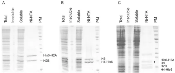
(H2AH2B dimer, (H3H4)2 tetramer after co-expression with the chaperone),[16] we created two polycistronic vectors, each for expression of X. laevis single histone pair. We mod-ified an original plasmid for co-expression of all four X. laevis histones using iPCR.[17] The modified plasmids had a single T7 promoter driving production of a polycistronic mRNA and an original His6-tag at the N-terminus of H2A and the C-terminus of H4. The S-tag in front of H2A His6-tag was also kept. In a second iPCR reaction we replaced the thrombin with an HRV 3C recognition site for conven-ient tag removal (Table 1, see Experimental procedures for more details). Co-expression of Xenopus H2A with H2B leads to the production of soluble histones, suggesting that they correctly associate and fold to form heterodimers (Fig-ure 1A). In contrast to Drosophila histones,[16] co-expres-sion of Xenopus H3 and H4 resulted in soluble histone pairs, which was easily purified using affinity chromatography (Figure 1B). Interestingly, co-expressed histone pairs were so soluble that it was impossible to detect any trace of his-tones in the urea denatured insoluble fraction (Figure 1).
The complexes were purified by Ni-NTA affinity chro-matography under native conditions. Due to a high per-centage of positively charged residues, histones strongly bind nucleic acids. Therefore, histones may co-purify with nucleic acids from E. coli. To remove contaminating nucleic acids from histones purified under denaturing conditions an anion exchange chromatography is routinely em-ployed.[13] We observed that under native conditions, his-tone pairs are free of contaminating nucleic acids from the
Figure 1. SDS–PAGE analysis of purification of co-expressed histones.
(A) Co-expression and Ni-NTA purification of X. laevis His6-tagged H2AH2B construct. (B) Co-expression and Ni-NTA purification of X. laevis His6-tagged H3H4 construct.
(C) Co-expression and Ni-NTA purification of X. laevis His6-tagged H2AH2BH3H4 construct. Partial N-terminal degradation of H2A is labelled with a black cross on the gel.
Croat. Chem. Acta 2016, 89(2), 153–162 DOI: 10.5562/cca2806
Figure 2. Gel filtration analysis of co-expressed and co-purified histones.
(A) Gel filtration profile of the H2AH2B dimer and the H3H4 tetramer. The selected elution fractions, labelled with black bar, were analysed by SDS-PAGE.
(B) Histone octamers were assembled from the H2AH2B dimer and H3H4 tetramer. Size exclusion chromatography of the assembled octamers shows that the affinity tags did not influence octamer formation. The selected elution fractions, labelled with a black bar, were analysed by SDS-PAGE.
DOI: 10.5562/cca2806 Croat. Chem. Acta 2016, 89(2), 153–162 cell if the buffers used for their purification contain 2 mol
dm−3 salt. Moreover, we noticed that even short exposure of the sample to low salt conditions during cell lysis affected the amount and quality of the obtained H3H4 complex. The sample would partition into the insoluble fraction and the remaining soluble fraction when analysed on size exclusion chromatography contained tetramer and higher oligomeric species. To avoid this, we always used French press for cell extract preparation using buffer containing 2 mol dm−3 salt. The size exclusion chromatography of histone pairs is consistent with formation of heterodimeric H2AH2B and heterotetrameric (H3H4)2 (Figure 2A). The yield of purified H2AH2B ranges 45-50 mg dm−3 cell culture, while the yield of purified (H3H4)2 is 15-20 mg dm−3 of induced cell cul-ture. The quality of our co-expressed and co-purified his-tone pairs was tested in a pull-down assay (Figure 3A). The affinity tag was left on each histone pair. H2AH2B dimer was tested for binding to nucleosome assembly protein 1 (Nap1).[24] The interactions between the His6-tagged H3H4 histone pair and untagged Asf1 chaperone were analysed employing Ni-NTA affinity resin.[25] Additionally, the inter-actions between histones and chaperones were analysed by gel shift assay on 6 % native PAGE gels (Figure 3B). Pre-viously it was shown that Nap1 binds H3H4, in addition to the cognate H2AH2B histones.[26] In agreement with the published data, we obtained Nap1:H2AH2B and Nap1:H3H4 complexes in our gel shift assay (Figure 3B). Nap1 makes significant number of interactions with the main chain of the bound histones.[24] In contrary, Asf1 teractions with H3H4 histones are more specific and in-volve side chain recognition.[25] Therefore, in our gel shift assay we obtained the complex between Asf1 and H3H4, but not with H2AH2B histone pair. Both assays showed for-mation of specific histone:chaperone complexes, confirm-ing proper recognition and good quality of the heterodimeric H2AH2B and heterotetrameric (H3H4)2 complexes.
Characterization of Histone Octamer
Assembled from Co-expressed
Histone Pairs
We assembled the polycistronic vector for co-expression of X. laevis H2A, H2B, H3, H4 histones from vectors for expres-sion of each histone pair (Table 1, see Experimental for more details). All three samples were induced and purified in parallel from 1 dm3 of bacterial culture (Figure 1). Inter-estingly, we noticed that co-expression of all four histones is significantly lower when compared to separate expres-sion of each pair (Figure 1C). In addition, histones were de-tected also in the insoluble fraction. Therefore, individual co-expression of each histone pair allows not only more possibilities for further experiments, but also increases the amounts of the obtained protein sample.
After purifying soluble histone pairs by Ni-NTA affin-ity chromatography, the histone octamer was formed by salt dialysis. Using our method we did not observe any his-tone precipitation. When the hishis-tone octamer was refolded from inclusion bodies purified histones or by the 'one pot refolding' method, heavy precipitation was observed and removed by centrifugation.[13,14] Separation of the histone octamer and excess of the H2AH2B dimer is better than separation of the octamer and an excess of (H3H4)2 te-tramer. Adding H2AH2B dimer in a slight excess over (H3H4)2 prevents formation of free tetramers or histone hexamers, enabling thus separation of histone octamers from smaller, by-side assembly products. In our histone oc-tamer assembly reactions we always mix a 1.2–1.4 molar excess of H2AH2B dimer over (H3H4)2 tetramer. During the size exclusion chromatography, two major elution peaks corresponding to histone octamer and H2AH2B dimer were found (Figure 2B).
The quality of the histone octamer assembled from co-expressed and co-purified histone pairs was assessed by their ability to be reconstituted into nucleosome core par-ticles (NCPs) (Figure 3C). We also tested if leaving the affin-ity tags would interfere with the reconstitution into the nucleosome (Figure 2C). The histone octamer samples were mixed in a 1:1 ratio with 146-bp long strong nucleosome-positioning 601 DNA and dissolved in buffer containing 2 mol dm−3 sodium chloride. The nucleosomes were reconsti-tuted by the 'double bag' dialysis method, as described in the experimental procedures. The formation of NCPs was analysed using a gel shift assay (Figure 3C). The structure of the NCP revealed that histone tails flexibly protrude from the structural core.3 Therefore it is to be expected that the affinity tags would not interfere with the nucleosome re-constitution. Our native PAGE gel results clearly show that the NCP is formed using histones with or without an addi-tional tag on N- or C-terminus (Figure 3C). Keeping affinity tags in NCPs could be beneficial for studying binding of dif-ferent proteins to the nucleosomes by pull-down assays.
Finally, we performed thermal disassembly of the re-constituted nucleosomes (Figure 3D). In this assay we com-pared thermodynamic properties of the nucleosomes reconstituted using histones purified with our simplified method with the conventional denaturing protocol.[12] The assay shows the same stability properties for both nucleo-somes and that the affinity tags used for purification of the soluble histones do not affect the NCP stability.
DISCUSSION
Croat. Chem. Acta 2016, 89(2), 153–162 DOI: 10.5562/cca2806
Figure 3. In vitro assembly of a histone chaperone:histone complex.
(A) His pull-down assays using His6-H2AH2B, H3H4-His6 and histone chaperones Nap1 and Asf1. The proteins were visualized by Simply Blue SafeStain (Novex).
(B) Native polyacrylamide gel electrophoresis (PAGE) of histones, histone chaperones Nap1 and Asf1 and their complexes. H2AH2B dimer and H3H4 tetramer cannot enter the native gel due to their positive charge. A histone chaperone:histone complex migrates slower compared to the histone chaperone alone. The proteins were visualized by Simply Blue SafeStain (Novex).
(C) Native polyacrylamide gel electrophoresis (PAGE) demonstrates that the nucleosome core particle assembly does not depend on the presence of the affinity tags used for histone purification. The protein and DNA samples were visualized by silver staining.
(D) Native PAGE analysis of nucleosomes incubated at the indicated temperature for 30 min. Nucleosomes were prepared using our simplified method (left side of the gel) and, for comparison, using conventional denaturing protocol (on the right side of the gel). The affinity tags used for purification of the soluble histones were kept in NCP.
DOI: 10.5562/cca2806 Croat. Chem. Acta 2016, 89(2), 153–162 that histones form heterotypic complexes; H2AH2B form
heterodimers and H3H4 form heterotetramers. Therefore, there is no special need for purification of the individual tones. When compared to preparation of the individual his-tone protein, co-expression and co-purification of soluble histone complexes avoids solubilisation of the inclusion bodies and losses associated with proteins refolding. In ad-dition, working with soluble histones eliminates the need for using urea in the buffers that can induce carbamylation of the histone proteins upon reaction with natural degra-dation products of urea. Using the described protocol, H2AH2B and H3H4 complexes purifications in parallel is straightforward, so that all four histones are available within a day. Pure histone complexes can be stored at −80 °C after Ni-NTA purification and dialysis in size exclusion buffer. The affinity tag removal, if required, is done during the dialysis step. Cleaved tags and HRV 3C protease can be removed by quick incubation with limiting amounts of Ni-NTA resin in a batch mode. Otherwise, if a higher purity of the proteins is needed, histone complexes can be addition-ally polished over size exclusion chromatography.
Soluble H2AH2B dimers and (H3H4)2 tetramers formed complexes with their cognate chaperones (Figure 3A, 3B). The chaperones have been purified under native conditions. Thus, denaturation and subsequent refolding steps could also be omitted for histone-chaperone complex formation.
Purified soluble histone pairs readily formed histone octamer independently of the freezing step. Similarly, re-moval of the affinity tags did not influence octamer or NCP formation (Figure 2B, Figure 3C). We find it convenient to store histone dimers and tetramers, as it gives more free-dom for subsequent experimental work. Otherwise, his-tone octamers can also be stored at −80 °C. This requires previous dialysis in buffer with 50 % glycerol, as already de-scribed.[13] Co-expressed histone complexes readily formed histone octamers, and these octamers were proficient in NCP reconstitution (Figure 3C). Our approach reduces the number of column purification steps and provides high amounts of pure and soluble histones. Our overall proce-dure is very fast and highly efficient so that multiple sam-ples can be processed in parallel. In addition, numerous modifications of each histone gene, like removal of the S-tag from H2A N-terminus, adding FLAG or HA-S-tag, removal of N-terminal tails, introducing mutations or histone isoforms, can be done in a day using iPCR.
We hope that our simplified and rapid approach will help researchers to obtain high amounts of high quality nu-cleosomes in a short period of time.
Acknowledgment. We thank Jung-Hyun Min for the polycistronic vector containing histone genes. We thank Ilaria Ugolini and Cornelia Brönner for critically reading the
manuscript. This work was supported by the core funding of Gene Center, BioSysNet, and ERC-smallRNAhet-309584. The research leading to these results has received funding from the European Union Seventh Framework Programme (FP7 2007-2013) under grant agreement n° 291823 Marie Curie FP7-PEOPLE-2011-COFUND (The new International Fellow-ship Mobility Programme for Experienced Researchers in Croatia - NEWFELPRO). This article has been prepared as a part of a project “Importins-H1 complex" which has received funding through NEWFELPRO project under grant agree-ment n° 26.
REFERENCES
[1] P. Tessarz, T. Kouzarides, Nat. Rev. Mol. Cell. Biol. 2014, 15, 703.
[2] R. K. McGinty, S. Tan, Chem. Rev. 2015, 115, 2255. [3] K. Luger, A. W. Mäder, R. K. Richmond, D. F. Sargent,
T. J. Richmond, Nature 1997, 389, 251.
[4] B. R. Zhou, J. Jiang, H. Feng, R. Ghirlando, T. S. Xiao, Y. Bai, Mol. Cell 2015, 59, 628.
[5] G. Felsenfeld, M. Groudine, Nature 2003, 421, 448. [6] P. Tessarz, T. Kouzarides, Nat. Rev. Mol. Cell Biol.
2014, 15, 703.
[7] G. E. Zentner, S. Henikoff, Nat. Struct. Mol. Biol. 2013, 20, 259.
[8] R. A. Laskey, B. M. Honda, A. D. Mills, J. T. Finch, Na-ture 1978, 275, 416.
[9] M. Hondele, A. G. Ladurner, Curr. Opin. Struct. Biol. 2011, 21, 698.
[10] K. Luger, T. J. Rechsteiner, A. J. Flaus, M. M. Waye, T. J. Richmond, J. Mol. Biol. 1997, 26, 301.
[11] E. L. Böhm, W. N. Strickland, M. Strickland, B. H. Thwaits, D. R. van der Westhuizen, C. von Holt, FEBS Lett. 1973, 34, 217.
[12] K. Luger, T. J. Rechsteiner, T. J. Richmond, Methods Enzymol. 1999, 304, 3.
[13] K. Luger, T. J. Rechsteiner, T. J. Richmond, Methods Mol. Biol. 1999, 119, 1.
[14] Y. T. Lee, G. Gibbons, S. Y. Lee, Z. Nikolovska-Coleska, Y. Dou, Protein Expr. Purif. 2015, 110, 89.
[15] H. Klinker, C. Haas, N. Harrer, P. B. Becker, F. Mueller-Planitz, PLoS One 2014, 9, e104029. [16] M. Anderson, J. H. Huh, T. Ngo, A. Lee, G. Hernandez,
J. Pang, J. Perkins, R. N. Dutnall, Protein Expression Purif. 2010, 72, 194.
[17] Y. Shim, M. R. Duan, X. Chen, M. J. Smerdon, J. H. Min, Anal. Biochem. 2012, 427, 190.
[18] R. K. McGinty, R. C. Henrici, S. Tan, Nature 2014, 514, 591. [19] C. M. English, M. W. Adkins, J. J. Carson, M. E.
Churchill, J. K. Tyler, Cell 2006, 127, 495.
Croat. Chem. Acta 2016, 89(2), 153–162 DOI: 10.5562/cca2806 [21] P. T. Lowary, J. Widom, J. Mol. Biol. 1998, 276,19.
[22] Z. Ding, Y. Xu, Prep. Biochem. 1995, 25, 21.
[23] P. A. Walker, L. E. Leong, P. W. Ng, S. H. Tan, S. Waller, D. Murphy, A. G. Porter, Biotechnology 1994, 12, 601. [24] S. D'Arcy, K. W. Martin, T. Panchenko, X. Chen, S. Bergeron, L. A. Stargell, B. E. Black, K. Luger, Mol.
Cell. 2013, 51, 662.
[25] R. Natsume, M. Eitoku, Y. Akai, N. Sano, M. Horikoshi and T. Senda, Nature 2007, 446, 338.