0103 - 5053 $6.00+0.00
A
r
ti
c
le
* e-mail: [email protected]
Fatty Acids and Terpenoids from
Trigonia fasciculata
Jair Mafezoli a,b, Regina Helena A. Santosc, Maria Teresa P. Gambardelac and Edilberto R. Silveira*,a
a
Departamento de Química Orgânica e Inorgânica, Centro de Ciências, Universidade Federal do Ceará, CP 12.200, 60021-940 Fortaleza - CE, Brazil
b
Centro de Ciências da Saúde, Universidade de Fortaleza, CP 1258, 60811-450 Fortaleza - CE, Brazil
c
Departamento de Química e Física Molecular, Instituto de Física e Química de São Carlos, 13560-250 São Carlos - SP, Brazil
A fração lipídica do extrato hexânico das raízes de Trigonia fasciculata foi analisada por CG/EM sob a forma de seus ésteres metílicos, identificando-se dezessete ácidos graxos, sendo o ácido oléico (38,8%) o principal componente. A análise por CG/EM da fração menos polar da parte insaponificável do extrato hexânico das raízes permitiu identificar quinze sesquiterpenos não oxigenados, sendo o α-santaleno (28,4%) o componente majoritário. Cromatografia em gel de sílica da fração mais polar permitiu o isolamento de dois triterpenos conhecidos, ácido betulínico e friedelina, e o diterpeno inédito 7-(2-hidroxiacetil)-10-hidroxitetradecaidro-1-metileno-4b,7,10a-trimetilfenantreno (6α -hidroxi-15-oxo-allodevadarool). Todas as substâncias isoladas e identificadas são inéditas para a espécie. A determinação estrutural foi realizada por derivatização química, comparação com dados da literatura, análise espectral, incluíndo RMN 2D (COSY, HETCOR e COLOC), e cristalografia de raios X.
The fatty portion of the hexane extract from roots of Trigonia fasciculata has been determined by GC/MS analysis of the methyl ester mixture. Seventeen fatty acids were identified and oleic acid (38.8%) was the major component. The GC/MS analysis of the less polar fraction of the non-saponifiable part of the root hexane extract allowed the identification of fifteen sesquiterpenes and α-santalene (28.4%) was the major component. Chromatography over silica gel of the more polar fraction allowed the isolation of two known compounds: betulinic acid and friedelin, and a novel diterpene 7-(2-hydroxy-acetyl)-10-hydroxy-tetradecahydro-1-methylene-4b,7,10a-trimethyl-phenantrene (6α-hydroxy-15-oxo-allodevadarool), all unknown for the species. Structure determination was accomplished by chemical derivatization, comparison to literature data and spectral analysis, including 2D NMR (COSY, HETCOR, COLOC) and X-ray crystallography.
Keywords: Trigonia fasciculata,Trigoniaceae, terpenoids, 6α-hydroxy-15-oxo-allodevadarool, fatty acids
Introduction
In our search for Bredemeyera species of plants in the Northeast of Brazil because of its pharmacological properties,1,2 we have collected another liana like plant
later identified as Trigonia fasciculata Griseb.
(Trigoniaceae).
According to Barroso,3 the Trigoniaceae family consists
of two genera comprising 33 species. Only the Trigonia genus occurs in Brazil. A literature survey in the Chemical and Biological Abstracts revealed no phytochemical study of T. fasciculata, a shrub well dispersed through Bahia
and Minas Gerais States. Thus, it was chosen for phytochemical analysis.
Results and Discussion
portion (TFRH(1)/NS) after usual work up. Methylation of an aliquot of TFRH(1)/FA with BF3/MeOH 14% solution followed by GC/MS analysis of the methyl ester mixture allowed the identification of the fatty acids listed in the Experimental Section. Silica gel chromatography of TFRH(1)/NS yielded a yellowish fraction by elution with hexane. 1H and 13C NMR analysis indicated its terpenoid
character. GC/MS analysis of this fraction, followed by comparison of the mass spectra obtained for each compound with a computer library data bank,4 and literature data5 in
addition to Kovats’ Indices comparison6 allowed the
identification of the sesquiterpenes listed in the Experimental Section.
Successive column chromatography over silica gel of the more polar fraction obtained by ethyl acetate elution from TFRH followed by rotational planar chromatography yielded
1, a colorless solid, homogenous by TLC. The BB 13C NMR
spectrum of 1 showed 20 spectral lines. Comparison of the BB spectrum with the DEPT 135o and DEPT 90o spectra
revealed the presence of 3 methines, 9 methylenes, 3 methyls and consequently 5 non-hydrogenated carbons. A closer analysis of the 13C NMR data allowed the identification of a
ketone carbonyl (δ 215.2, C-15), a sp2 methylene (δ 102.9,
C-18) and a non-hydrogenated sp2 carbon (δ 157.4, C-4),
very characteristic of an exocyclic double bond, an
oxygenated methylene (δ 63.8, C-16) and an oxygenated methine (δ 74.5, C-6). All other signals indicated non-functionalized, saturated carbons. The FT-IR spectrum confirmed the ketone moiety (1723 cm-1), its carbinolic
character (3300 and 1020 cm-1) and the exocyclic double
bond [3100 (ν=C-H), 1642 (νC=C) and 900 cm-1 (δ
=C-H)]. The
EI-Mass spectrum showed the molecular ion [M]+ at m/z 320
compatible with the molecular formula C20H32O3 corres-ponding to a tricyclic structure if a carbonyl and a carbon-carbon bond have already been characterized. The 1H NMR
spectrum clearly showed 3 angular methyl groups as singlets at δ 0.73, 1.06 and 1.21, a carbinolic methine at δ 3.99 (1H, dd, J 9.9 and 4.9 Hz, H-6), a carbinolic methylene at δ 4.36 (2H, s, H-16), and two sp2 hydrogens at δ 4.64 (1H, br s) and δ
4.70 (1H, br s).
Direct one bond attached hydrogen-carbon correlations
were established by 1H, 13C-COSY (HETCOR) NMR
experiment. Thus, the methyls at δ 0.73, 1.06 and 1.21 correlate with carbons at δ 12.0, 15.7 and 20.5, respectively. The carbinolic carbons at δ 74.5 and 63.8 correlate with hydrogens at δ 3.99 and 4.36, respectively. The hydrogens at δ 4.64 and 4.70 correlate both with the carbon at δ 102.9. All observed direct C, H correlations are depicted in Table 1. The more downfield character of the carbinolic methylene protons relative to the carbinolic methine
Table 1. 1H and 13C NMR data assignment for 1, 1a and 1b
1 1 a 1b
HETCOR COLOC
#C δC δH 2J
C-H
3J
C-H δC δH δC δH
1 20.6 1.20 & 1.60 20.5 21.0
2 28.7 - 27.8 29.9
3 33.3 1.40-1.50 H-18 33.2 35.3
4 157.4 - H-19 156.0 157.4
5 46.8 - H-19 46.2 47.5
H-18
6 74.5 3.99 H-19 77.0 5.06 74.5 4.26
7 33.2 1.50 & 2.10 30.6 33.9
8 38.8 - 38.6 39.8
9 37.0 - H-20 36.8 37.2
1 0 55.7 0.95 56.0 56.2
1 1 34.0 1.50 H-20 34.1 36.2
1 2 27.9 - H-17 28.5 29.2
1 3 45.4 - H-17 45.2 37.5
1 4 34.7 - H-17 34.4 36.2
1 5 215.2 - H-17 207.8 81.5 3.95
1 6 63.8 4.36 64.5 4.82 63.0 3.72
4.13
1 7 20.5 1.21 20.5 1.20 20.0 1.32
1 8 102.9 4.70 102.9 4.50 104.8 5.40
4.64 4.30 4.89
1 9 15.7 1.06 16.6 1.14 16.1 1.11
2 0 12.0 0.73 12.0 0.74 12.3 0.76
C=O (Ac) 170.9
C=O (Ac) 170.4
CH3 (Ac) 21.3 2.13
protons is indicative of its neighboring position to an anisotropic deshielding group such as the carbonyl or the carbon-carbon double bond. Acetylation of 1 yielded a colorless solid, designated 1a, [M]+ m/z 404 (C
24H36O5).
The 1H NMR spectrum of 1a, showed two extra methyl
groups at δ 2.13 and 1.98 (3H each, s), confirming the
dihydroxy character of 1a. It also showed a more
deshielded carbinolic methine (δ 5.06) with respect to the carbinolic methylene (δ 4.82) in an opposite relationship relative to the non-acetylated compound. These features are indicative of the presence of an α-hydroxy-acetyl moiety in 1a. The upfield shift of the carbonyl (δ 207.8, for 1a) due to the loss of the chelation through the five member ring hydrogen bonding is also in agreement with the ketone carbonyl bearing the carbinolic hydrogen. Reduction of 1 with NaBH4/ethanol yielded a colorless solid, designated 1b, [M]+ m/z 322 (C
20H34O3). The BB 13C
NMR spectrum of 1b showed a signal at δ 81.5 related to a carbinolic methine carbon and the absence of the signal at
δ 215.2 which confirmed the presence of the ketone
carbonyl in 1. A search in the literature for tricyclic diterpene skeletons that could accommodate three angular methyls, an exocyclic double bond and an α -hydroxy-acetyl moieties, yielded three possible diterpene general structure types: cassane, cleistanthane and erythroxylane.7-9
The long-range heteronuclear correlation spectrum (COLOC) of 1 showed a correlation between the angular methyl at δ 1.21 with the carbonyl carbon at δ 215.2 (3J
H-17,
C-15). The same angular methyl also correlated with two methylenes at δ 34.7 (3J
H-17,C-14) and δ 27.9 ( 3J
H-17,C-12)
and the quaternary carbon at δ 45.4 (2J
H-17,C-13). The
COLOC spectrum also showed correlations of the angular methyl at δ 1.06 to the non-hydrogenated sp2 carbon at δ
157.4 (3J
H-19,C-4), the quaternary carbon at δ 46.8 ( 2J
H-19,
C-5), and the carbinolic methine carbon at δ 74.5 (3J H-19,
C-6). Moreover, the exocyclic double-bond hydrogens at
δ 4.70 and 4.64 correlated to a methylene at δ 33.3 (3J H-18,
C-3) and to the same quaternary carbon at δ 46.8 (3J H-18,
C-5). Thus, 1 was suggested to be the 6α -hydroxy-15-oxo-allodevadarool. This carbon skeletal-pattern, also designated by dolabrane, has been observed for compounds from Erythroxylum species.10-13 The junction of the A/B
ring is trans, since the 13C NMR spectra did not show any
signal referent to a methyl group at approximately δ 32.0.14
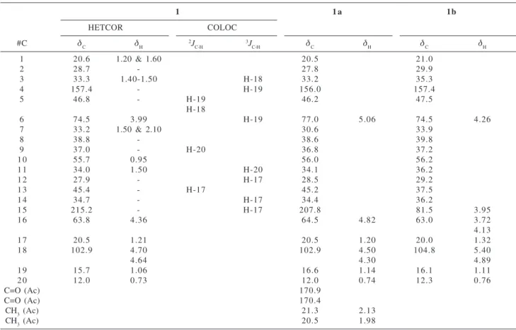
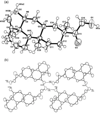
Finally, the X-ray analysis of a crystal obtained from MeOH confirmed the structure of 1 as 7-(2-hydroxy-acetyl)-10- hydroxy-tetradecahydro-1-methylene-4b,7,10a-trimethyl-phenantrene. Figure 1a shows an ORTEP three-dimensional structural drawing of the relative stereochemistry of 1, whereas Figure 1b highlights the intermolecular hydrogen bonding among the molecules, further stabilizing the molecular structure arrangement. The absolute stereochemistry shown in Figure 1 is arbitrary.
Experimental
General
mps.: uncorr.; IR: KBr pellets; 1H NMR: 200 and 400
MHz; 13C NMR: 50 and 100 MHz, Bruker AC-200 or Varian
Unit Plus, respectively; GC/MS Low resolution EIMS: (70 eV) Hewlett-Packard HP-5995-A.
Figure 1. Three-dimensional structural drawing of the relative
X-ray analysis
A single crystal of 1 was mounted on an Enraf-Nonius CAD-4 diffractometer equipped with graphite mono-chromated MoKα radiation. Unit cell parameters and intensity data were obtained at room temp. The structure was solved by SIR and refined by full matrix least squares. Figure 1a is an ORTEP view of the molecule, with hydrogens on idealized position and Figure 1b shows the intermolecular hydrogen bonds between O(1)-O(2)* (do-o= 2.679(4)Å, * -x,y,z), and O(2)-O(1)# (do-o=2.682(5)Å, # 2-x, 1/2+y, 2-z).
Plant material
Trigonia fasciculata was collected in Jacobina-BA, Brazil and identified by Dr. Afrânio G. Fernandes and Dr. Edson de Paula Nunes (Botanists, Universidade Federal do Ceará). A voucher specimen (#20366) representing the collection has been deposited at Herbário Prisco Bezerra, Departamento de Biologia, Universidade Federal do Ceará, Brazil.
Extraction and isolation of constituents
After removal of the aerial part, the roots (4.0 Kg) were dried, ground and extracted exhaustively with hexane to yield, after solvent evaporation, 26.2 g of a yellowish viscous extract designated TFRH. After standing at room temperature, a precipitate was formed, which yielded 1.6 g of a yellowish solid upon filtration and was designated TFRH-p. Two-hundred mg of TFRH-p was chroma-tographed over silica gel to yield 57.6 mg of TFRH-1, as a colorless solid, and 33.4 mg of another colorless solid designated TFRH-2. TFRH-1 mp. 260.2-261.2 oC, showed
all spectrometric (MS, IR, 1H and 13C NMR) data identical
to friedelin (lit.15,16 263.0-263.5 oC). TFRH-2, mp.
273.3-276.4 oC, was identified as betulinic acid (lit.7
275.0-278.0 oC) after spectral (MS, IR, 1H and 13C NMR)
comparison to literature data.7
Fatty acids and sesquiterpenes
Coarse chromatography over silica gel of the mother liquor TFRH (24.5 g) yielded a fraction by elution with hexane (6.9 g). An aliquot (3.5 g) was hydrolyzed with KOH/EtOH 1:1 solution yielding 0.56 g of fatty acids and 2.18 g of unsaponifiable material after usual work-up. 100 mg of the fatty acids were methylated with BF3/MeOH 14% and the methyl ester mixture obtained after work-up was analyzed by GC/MS allowing the identification of
methyl esters of lauric (C12:O, 2.00%), miristic (C14:O, 2.63%), pentadecanoic (C15:O, 0.41%), pentadecadienoic (C15:2, 0.96%), 14-methylpentadecanoic (C16:O, 0.39%), palmitic (C16:O, 13.87%), palmitoleic (C16:1, 0.50%), margaritic (C17:O, 0.51%), 14-methyl-hexadecanoic (C17:O, 0.55%), stearic (C18:O, 4.20%), oleic (C18:1, 38.84%), linoleic (C18:2, 19.54%), arachydic (C20:O, 0.96%), gondic (C20:1, 1.58%), behenic (C22:O, 2.87%), tricosanoic (C23:O, 0.57%) and lignoceric acid (C24:O, 1.58%). The unsapo-nifiable portion (2.15 g) was chromatographed over silica gel to yield a clear viscous fraction, by elution with hexane, whose 1H NMR spectrum showed its terpenoid features.
GC/MS analysis, utilizing the method developed by Alencar,4 allowed the identification of the sesquiterpenes:
d-elemene (4.68%), cyclosativene (1.20%), α-copaene (1.51%), β-elemene (11.64%), α-santalene (28.40%), α -bergamotene (7.71%), epi-β-santalene (2.66%), α -humulene (2.14%), cis-β-farnesene (6.74%), β-santalene (3.78%), γ-muurolene (2.22%), β-selinene (1.37%), β -bisabolene (9.20%), γ-cadinene (8.14%), and δ-cadinene (3.64%).
6α-Hydroxy-15-oxo-allodevadarool (1). The most polar fraction obtained from column chromatography of TFRH after elution with EtOAc, 120 mg, was submitted to
rotational planar chromatography (CHROMATOTRON
) with a mixture of hexane/EtOAc 3:2 to yield 31.2 mg of 1
as colorless crystals from MeOH. The next closest fraction, also eluted with EtOAc (1.6 g), was submitted to successive CC over silica gel to yield 120 mg more of 1, mp. 140-144 oC, [α]
D
25= +4.73o (c. 0.5, CHCl
3) FT-IR νmax/cm -1: 3301,
3100, 2934, 2852, 1723, 1642, 1089, 1018, 890 (KBr). EI-MS m/z (rel. int.): 320 ([M]+, 14), 302 (13, M- H
20), 289
(28, M - CH2OH), 271 (6, 302 - CH2OH), 261 (89, M - O=C-CH2OH), 243 (73, 271 - CO or 261 - H2O), 109 (100). 1H
NMR (200 MHz, CDCl3): Table 1 13C NMR (50 MHz,
CDCl3): Table 1.
6α,16-Acetoxy-15-oxo-allodevadarool (1a) . 32 mg of
1 was acetylated by usual procedure with pyridine/Ac2O to yield 24 mg of the acetylated material 1a after usual work-up and filtration of the reaction product over a small layer of silica gel. 1a, a yellowish amorphous solid, mp. 114.0-116.0 oC.
EI-MS m/z (rel. int.): 404 ([M]+,26), 362 (33, M+
-CH2CO), 347 (11, 362- CH3), 344 (50, M+- CH
3CO2H), 329
(36, 344- Me), 303 (58, 362- CH2OHCO), 261 (71, 303-CH2CO), 243 (100, 261- H2O). Supplementary material under author request. FT-IR νmax/cm
-1: 3093, 2932-2858,
1750, 1720, 1642, 1248, 889 (KBr). 1H NMR (200 MHz,
CDCl3): Table 1 13C NMR (50 MHz, CDCl
3): Table 1.
work-up the reaction mixture was chromatographed over a small layer of silica gel to yield 29 mg of an white amorphous solid 1b, mp. 184.5-187.5 oC.
EI-MS m/z (rel. int.): 322 ([M]+,32), 307 (26, M
-CH3),304 (14, M+- H
2O), 291 (27, M - CH2OH), 273 (18,
291- H2O), 109 (100). Supplementary material under author request. FT-IR νmax/cm
-1: 3386, 3106, 2932, 2858, 1642,
1095, 1068, 892 (KBr). 1H NMR (200 MHz, CDCl 3): Table
1. 13C NMR (50 MHz, CDCl
3): Table 1.
Supplementary Information
Crystallographic data (excluding structure factors) for the structure 1 in this paper have been deposited with the Cambridge Crystallographic Data Centre as supplementary publication on CCDC 176082. Copies of the data can be obtained, free of charge, on application to CCDC, 12 Union Road, Cambridge CB2 1EZ, UK, (fax: +44 1223 336033 or e-mail: [email protected]).
Acknowledgements
The authors wish to acknowledge Dr. Afrânio G. Fernandes and Dr. Edson de Paula Nunes (Botanists, UFC-CE) for plant identification; Dr. R. Braz-Filho (UENF-RJ) for his helpful discussion about MS data; CNPq for the fellowships and CAPES/PADCT/FINEP/PRONEX/ FUNCAP for financial support.
References
1. Rao, V.S.N.; Menezes, A.M.S.; Viana, G.S.B.; Gadelha, M.G.T.; Silveira, E.R.; Fitoterapia1990, LXI, 9.
2. Silveira, E.R.; Falcão, M.J.C.; Menezes, A.Jr.; Kingston, D.G.I.; Glass, T.E.; Phytochemistry1995, 39, 1433.
3. Barroso, G.M.; Sistemática de Angiosperma do Brasil, Imprensa Universitária-Universidade Federal de Viçosa: Viçosa, 1986, vol. 2.
4. Alencar, J.W.; Craveiro, A.A.; Matos, F.J.A.; Machado, M.I.L.;
Quim. Nova1990, 13, 282.
5. Adams, R. P.; Identification of Essential Oil Components by Gas Chromatography/Mass Spectrometry; Academic Press: San Diego, 1995.
6. Alencar, J.W.; Craveiro, A.A.; Matos, F.J.A.; J. Nat. Prod.
1984, 47, 890.
7. Buckingham, J. B. ed.; Dictionary of Natural Products, Chapman and Hall: London, 1994, v. 1.
8. Leal, R.S.; Lima, M.A.S.; Silveira, E.R.; J. Braz. Chem. Soc.
2003, 14, 120.
9. Silva, G.C.; Patitucci, M.L.; Pinto, A.C.; Menezes, N.L.; Quim. Nova2001, 24, 619.
10. Soman, R.; Dev, S.; Misra, R.; Pandey, R.C.; Tetrahedron Lett.
1964, 3767.
11. Soman, R.; Dev, S.; Indian J. Chem.1983, 22B, 984. 12. Connolly, J.D.; McCrindle, R.; Murray, R.D.H.; Renfrew, A.J.;
Overton, K.H.; Melera, A.; J. Chem. Soc. (C)1966, 268. 13. Ansell, S.M.; Pegel, K.H.; Taylor, D.A.H.; Phytochemistry
1993, 32, 945.
14. Kijjoa, A.; Polonia, M.A.; Pinto, M.M.M.; Kitiratakarn, T.; Gedris, T.E.; Herz, W.; Phytochemistry1994, 37, 197. 15. Windholz, M.; The Merck Index, 10th ed., Merck & Co.:New
Jersey, 1983.
16. Beierbeck, H.; Saunders, J.K.; Apsimon, J.W.; Can. J. Chem.
1977, 55, 2813.
Received: January 6, 2002 Published on the web: May 8, 2003