Malaysian Orthopaedic Journal 2015 Vol 9 No 3 Taran S, et al
75 ABSTRACT
Upper cervical chordoma (UCC) is rare condition and poses unique challenges to surgeons. Even though transoral approach is commonly employed, a minimally invasive technique has not been established. We report a 44-year old Malay lady who presented with a 1 month history of insidious onset of progressive neck pain without neurological symptoms. She was diagnosed to have an axial (C2) chordoma. Intralesional resection of the tumour was performed transorally using the Destandau endoscopic system (Storz, Germany). Satisfactory intralesional excision of the tumour was achieved. She had a posterior fixation of C1-C4 prior to that. Her symptoms improved postoperatively and there were no complications noted. She underwent adjuvant radiotherapy to minimize local recurrence. Endoscopic excision of UCC via the transoral approach is a safe option as it provides an excellent magnified view and ease of resection while minimizing the operative morbidity.
Key Words:
Cervical spine, endoscopic resection, transoral, chordoma
INTRODUCTION
Chordoma is a rare primary malignant tumour of notochordal origin. It is slow growing and locally invasive, however, may metastasize to other organs. The current treatment of choice for chordomas of the mobile spine and sacrum is en-bloc excision with wide margins and postoperative external-beam radiation therapy1-3. Only 6% of chordomas are localized at the cervical spine3. The resection of an upper cervical chordoma poses unique challenges to surgeons, hence, making wide resection surgery very challenging. Currently, transoral approach is the most widely performed technique to approach this area and has been accepted as the standard procedure among spine surgeons. To our knowledge, there is no report on the usage of Destandau endoscopic technique to excise an axial chordoma using the transoral approach.
CASE REPORT
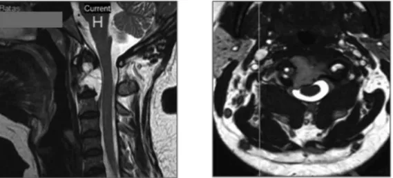
We report a 44-year old Malay lady who presented to us with a 1 month history of insidious onset of progressive severe neck pain without neurological symptoms, dysphagia or evidence of metastasis. Her physical examination revealed hypertonia, hyperreflexia and a positive Hoffman’s sign for both upper limbs. Her erythrocyte sedimentation rate (ESR) on presentation was 100 mm/hr. Magnetic resonance imaging (MRI) revealed a lesion of the axis (2nd cervical vertebra C2) with extension superiorly along the dens and postero-inferiorly, as well as indentation of the spinal cord. The radiological features were suggestive of a chordoma (Figure 1).
She underwent a 2-stage surgical procedure. The first stage surgery was a posterior C1-C4 instrumented fusion (B. Braun Surgical system), while the second stage surgery was a transoral endoscopic excision of the axis, which was performed 3 months later.
With regards to the operative technique for the second stage surgery, the patient was positioned in the supine position and orotracheal intubation was utilized for general anaesthesia, as shown in Figure 3. A Boyle-Davis mouth gag was employed to maintain the mouth open throughout the surgery (Figure 2) and the uvula was retracted using a soft rubber catheter trans-nasally. A needle and image intensifier was used to confirm the level of the anterior tubercle of the atlas, before a 3cm midline vertical incision was made on the posterior oropharyngeal mucosa and muscle (Figure 3A). Stay sutures were used to retract the incision edges. The anterior longitudinal ligament was exposed subperiosteally and the longus coli was mobilized laterally (Figure 3B). Next, the Destandau endoscopic system with the mobile Endospine® operating tube (Storz, Germany) was incorporated. A high-speed burr was introduced through the working portal of the operating tube and was used to remove the anterior cortex of the atlas and axis (Figure 3C). Intralesional excision of the tumour was performed entirely
Endoscopic Transoral Resection of an Axial Chordoma: A
Case Report
Taran S,MMed Orth, Yusof AH,MMed Orth, Yusof MI,MMed Orth
Department of Orthopaedics, Universiti Sains Malaysia, Kubang Kerian, Kelantan
Date of submission: July 2015 Date of acceptance: October 2015
Corresponding Author: Taran Singh Pall Singh, Department of Orthopaedics, Universiti Sains Malaysia, 16150 Kubang Kerian, Kelantan, Malaysia
Email: [email protected]
Malaysian Orthopaedic Journal 2015 Vol 9 No 3 Taran S, et al
76
under endoscopic image guidance, using the burr and a bone punch, to achieve satisfactory anterior decompression (Figure 3D). The incision was closed using interrupted absorbable sutures. The blood loss was 350 ml and the duration of surgery was 9 hours. There were no immediate or delayed neurovascular complications, CSF leak or infection following the surgery. A 3-day prophylactic antibiotic course of intravenous cefuroxime and metronidazole was given in view of the long surgical duration, the usage of the oropharyngeal approach and the presence implants. The patient was able to swallow clear fluids by postoperative day 3. Histopathological examination of the bone and tissue samples taken intra-operatively confirmed the diagnosis of chordoma (Figure 4).
She underwent radiotherapy to the area of C1 – C3 nine months after the second stage surgery (45 Gys in 25 fractions over 5 weeks), in a different center due to logistic reasons. The patient is still under our follow up and 3 years post surgery, she is free from recurrence, metastasis and has no Fig. 1: Pre-operative sagittal and axial views demonstrating the extent of the tumour on the cervical MRI.
Fig. 2: The mouth was maintained open using the Boyle-Davis mouth gag to enable the incorporation of the Destandau endoscopic system.
Fig. 4: Photomicrograph demonstrating the chordoma cells with eccentrically located oval nuclei and vacuolated cytoplasm (physaliphorous cells) forming sheets that are embedded in mucoid stroma (H&E: 20X).
Endoscopic Transoral Resection of an Axial Chordoma: A Case Report
77 REFERENCES
1. Walcott BP, Nahed BV, Mohyeldin A, Coumans J-V, Kahle KT, Ferreira MJ. Chordoma: current concepts, management, and
future directions. Lancet Oncol. 2012; 13(2): e69-76.
2. Hsu W, Kosztowski TA, Zaidi HA, Gokaslan ZL, Wolinsky J-P. Image-guided, endoscopic, transcervical resection of cervical
chordoma: technical note. J Neurosurg Spine. 2010; 12(4): 431-5.
3. Delfini R, Marruzzo D, Tarantino R, Marotta N, Landi A. Multilevel oblique corpectomies as an effective surgical option to treat
cervical chordoma in a young girl. World J Clin Cases.2014; 2(3): 57-61.
4. Barrenechea IJ, Perin NI, Triana A, Lesser J, Costantino P, Sen C. Surgical management of chordomas of the cervical spine. J
Neurosurg Spine.2007; 6(5): 398-406.
5. Depreitere B, Vander Poorten V, Vranckx J, Pans S, Menten J, Goitein G. Management of Cervical Spine Chordomas. Case
Report. World Neurosurg. 2012; 77(1): 215.
significant neurological deficit. Her screening was done using serial CT scans and physical examination.
DISCUSSION
Endoscopic excision of upper cervical chordoma using the Destandau endoscopic system through the transoral approach is an innovative and safe technique. Applying our experience of lumbar decompressive surgery using this system, we successfully performed excision of an upper cervical tumour.
In this case, the operative time was lengthy because it was our first time performing such a procedure. In addition, the tumour margin was wide and extra caution was needed. Nevertheless, the benefit of low blood loss and a magnified view throughout surgery is truly advantageous, especially in reducing complications such as cerebrospinal fluid leakage in the event of a dural injury.
Our decision to only perform intra-lesional excision to achieve satisfactory decompression was governed by the fact that a true en-bloc resection is not possible in most cases4, 5. In the experience of Barrenechea et al., 2007, piecemeal intra-lesional excision of cervical chordoma remains an effective technique when combined with post-operative radiotherapy4. The importance of adjuvant radiotherapy has also been stressed upon in literature, irrespective of the extent of the excision2, 3, 5.