CASE REPORT
Idiopathic radial artery aneurysm: case report
Aneurisma idiopático de artéria radial: relato de caso
Luiz Ernani Meira Jr.1, halis Marcelo Gouvêa1, hiago Jardim de Macedo2
Introduction
he upper limbs are considered less vulnerable to arte-rial diseases, especially those of aneurysmatic nature. Due to their low incidence, such aneurysms are diagnostic and therapeutic challenges. In this area, the arteries most fre-quently afected by the aneurysmatic process are, in decreas-ing order: subclavian, brachial, ulnar and radial arteries1,2.
Radial artery aneurysms are mostly pseudoaneurysms associated with iatrogenic conditions resulting from inva-sive procedures for endovascular diagnosis and/or therapy, invasive monitoring of mean arterial pressure and repeated punctures in the intensive care unit or in drug users. Blunt traumas are another cause of radial artery pseudoaneurysms, especially those related to fracture with vascular injury, as well as penetrating traumas caused by cold weapon3.
In contrast, the main cause of true aneurysms in ar-teries of the upper limbs, below the axillary artery and specifically the radial artery, is repeated blunt trauma, which is followed by idiopathic causes, atherosclerosis,
metabolic and congenital diseases or associated with disorders such as neurofibromatosis and vasculitis such as Buerger’s disease, Kawasaki’s disease, Bechet’s disease and polyarteritis nodosa4. Aneurysmatic formations have
also been reported, associated with arteriovenous fistu-las created for hemodialysis, poststenotic dilatations, one case related to hemophilia A and one case related to severe anemia4-9.
he purpose of this study is to present the case re-port of a three-year-old child with true idiopathic radial aneurysm.
Case description
A healthy three-year-old male patient, no history of comorbidities. According to the mother, pre-natal period without occurrences, normal delivery, proper vaccina-tion and no history of admissions, traumas or surgical interventions.
Abstract
Radial artery aneurysms are extremely rare. Post-traumatic pseudoaneurysms are the vast majority. True radial artery aneurysms can be idiopathic, congenital, poststenotic, or associated with some pathologies, such as vasculitis and conjunctive tissue diseases. We report a case of an idiopathic aneurysm of the radial artery in a three-year-old child who was submitted to surgical resection after a complete diagnostic approach.
Keywords: aneurysm; radial artery; child.
Resumo
Os aneurismas da artéria radial são extremamente raros. Em sua maioria, consistem de pseudoaneurismas pós-traumáticos. Os aneurismas da artéria radial verdadeiros podem ser idiopáticos, congênitos, pós-estenóticos ou associados a patologias, tais como vasculites e doenças do tecido conjuntivo. Foi relatado um caso de aneurisma idiopático de artéria radial em uma criança de três anos, que, após completa investigação diagnóstica complementar, foi submetida à ressecção cirúrgica.
Palavras-chave: aneurisma; artéria radial; criança.
Study carried out at the Vascular Surgery Service of Hospital Regional Dr. João Penido from Fundação Hospitalar do Estado de Minas Gerais (FHEMIG) – Juiz de Fora (MG), Brazil. 1 Vascular and Endovascular Surgeon at the Hospital Regional João Penido from FHEMIG – Juiz de Fora (MG), Brazil.
2 Attending Medicine at Universidade Presidente Antônio Carlos (UNIPAC) – Juiz de Fora (MG), Brazil.
Financial support: none.
Idiopathic radial aneurysm - Meira Jr. L.E. et al. J Vasc Bras 2011, Vol. 10, Nº 4
316
Around two months ago, a pulsatile nodular injury was observed, in middle third, anterior face of the right forearm. he arterial echo Doppler exam was performed in the right upper limb, and radial artery aneurism was identiied (Figure 1).
he patient was sent to the Vascular Surgery Service of Hospital Regional Dr. João Penido, Fundação Hospitalar do Estado de Minas Gerais (FHEMIG), in the city of Juiz de Fora, Minas Gerais, in May 2009. he patient complained of pain on palpation and reported mild intermittent swell-ing in the right hand and did not present any ischemic sign, neurological alteration or local or systemic infectious sign. In addition, the patient did not have history of trauma or invasive procedure in the right upper limb. he Allen’s test result was bilaterally negative.
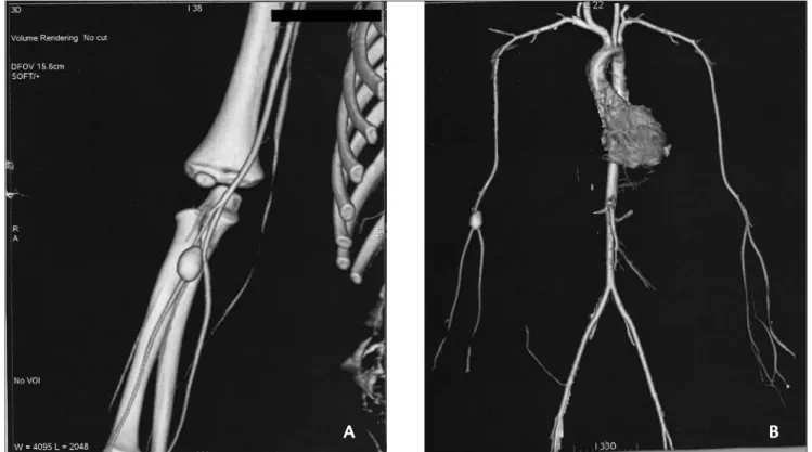
The diagnosis of vasculitis or metabolic diseases was ruled out, based on clinical and laboratorial exams. The patient was sent to imaging propedeutics, and the follow-ing exams were performed: abdominal ultrasound, lower extremity ultrasound, transthoracic echocardiogram, multislice helical angiotomography of upper limbs, aor-tic arch and great vases, from thoracic and abdominal aorta to femoral vessels. No other aneurysmatic lesions were identified besides the focal dilatation of proximal right radial artery, with the largest diameter of 1.1 cm and 1.5 cm length. The aneurysm started around 2.0 cm from the origin of radial artery, which had 0.33 cm proximal diameter, distal to the dilatation (Figures 2a and 2b).
he patient was then submitted to aneurysm resection. he selected procedure was radial artery ligature, due to a satisfactory relux from the distal stump, reduced arterial caliber and the resected segment length (around 3 cm). he surgical specimen was sent for anatomopathological and culture exam. he histological exam showed arterial ves-sel wall with all layers and negative culture for infectious agents (Figures 3 and 4).
he procedure included a 30-day follow-up and the pa-tient presented good post-operative progress, with no isch-emic or neurological deicit.
Discussion
he true radial artery aneurysm is a diagnostic and therapeutic challenge. he diagnosis is deined through complaint and physical examination. he main manifesta-tion is local pain, followed by symptoms and signs of distal ischemia, secondary to episodes of thrombosis or distal microembolization. Symptoms resulting from the com-pression process are also described, such as hand swell-ing and neurological symptoms, such as paresthesias5,9.
Rupture is not described in any published series. Aneurysms can be asymptomatic and are presented only as a pulsatile mass, with fremitus and blow in the physi-cal exam. he diferential diagnosis includes synovial cyst, ganglions, lipomas and neuromas. In a case of radial ar-tery aneurysm, complete anamnesis and rigorous physical
Idiopathic radial aneurysm - Meira Jr. L.E. et al. J Vasc Bras 2011, Vol. 10, Nº 4 317
Figure 2. (a) Multislice helical pan-computed angiotomography. (b) Helical angiotomography showing the right radial artery aneurysm.
A
B
Figure 4. Resected true radial artery aneurysm.
Figure 3. Dissection and repair of true radial artery aneurysm.
examination are essential. Ater traumatic or iatrogenic events are ruled out, the rare causes of true aneurysms should be investigated. For such purpose, laboratorial and imaging exams should be performed, as well as investiga-tions for collagenosis, vasculitis and metabolic diseases, before considering the lesion is congenital or idiopathic. Imaging propedeutics includes echo Doppler exam, ab-dominal ultrasound, echocardiogram, angiotomography, angioresonance and arteriography3,4,10.
Idiopathic radial aneurysm - Meira Jr. L.E. et al. J Vasc Bras 2011, Vol. 10, Nº 4
318
aneurysm should be submitted to pan-computed angioto-mography before any treatment is started10.
he treatment of radial artery aneurysms is still con-troversial; it is dependent on the etiology, location, pres-ence of thrombi, associated symptoms and mainly the cir-culation status collaterally and distally to the lesion. he surgical options range from proximal and distal ligature of the vessel combined with the aneurysmatic sac resec-tion to revascularizaresec-tion procedures, with termino-termi-nal primary anastomosis or bypass with venous grat10-12.
here is no deinition regarding the clinical follow-up without any surgical treatment; therefore, due to the risk of thrombotic and microembolic complications that can lead to distal ischemia, the surgical correction of aneu-rysm is always indicated.
References
1. De Luccia N. Aneurismas nos Membros Superiores. In: Leão PP, Kaufman P (org.). Aneurismas Arteriais. São Paulo: Fundo Editorial Bik; 1998. v.1.
2. Brito CJ, Azevedo Jr AC, Silva RM. Aneurismas dos membros su-periores. In: Brito CJ. Cirurgia Vascular: Cirurgia Endovascular, Angiologia. São Paulo: Revinter; 2008; p. 605-8.
3. Santos ACB, Oliveira FM, Oliveira JG, Bolanho E, Roberti T, Mathias UUM, et al. Aneurisma idiopático de artéria radial na região da ta-baqueira anatômica: relato de caso. J Vasc Bras. 2008;7(4):380-3.
4. Yaghoubian A, Virgílio C. Noniatrogenic Aneurysm of Distal Radial Artery: A case report. Ann Vasc Surg. 2006;20:784-6.
5. Walton NP, Choudhary F. Idiophatic radial artery aneurysm in ana-tomical snuf box. Acta Orthop Belg. 2002;68(3):292-4.
6. Filis K, Arhontovassilis F, heodorou D, heodossiades G, Manouras A. True radial artery aneurysm in a mild haemophilia A patient. Haemophilia. 2007;13(4):440-2.
7. Grey AC, Vallelly SR. Spontaneous false aneurysm of the radial ar-tery in neuroibromatosis. Clin Radiol. 1999;54(3):185-6.
8. Singh S, Riaz M, Wilmshurst AD, Small JO. Radial artery aneurysm in a case of neuroibromatosis. Brit J Plast Surg. 1998;51:564-5.
9. Hattori N, Furuta Y, he radial artery aneurysm within the ana-tomical snuf box. J Vasc Surg. 2004;13:597-601.
10. Pagès ON, Alicchio F, Keren B, Diallo S, Lefebvre F, Valla JS, et al Management of brachial artery aneurisms in infants. Pediatr Surg Int. 2008;24:509-13.
11. Gray RJ, Stone WM, Fowl RJ, Cherry KJ, Bower TC. Management of true aneurysms distal to the axillary artery. J Vasc Surg. 1998;28(4):606-10.
12. Miura S, Kigawa I, Miyari T, Fukuda, S. A surgically treated case of true radial aneurysm in the anatomical snuf box. J Vasc Surg. 2004;13:687-90.
Correspondence
Luiz Ernani Meira Jr. Rua Walter Linhares Frota Machado, 541 – Ibituruna
CEP: 39400-000 – Montes Claros (MG), Brazil E-mail: [email protected]
Author’s contributions