Reproductive and Respiratory Syndrome Virus by
Multiplex Real-Time RT-PCR
Kerstin Wernike1, Bernd Hoffmann1, Malte Dauber2, Elke Lange2, Horst Schirrmeier1, Martin Beer1* 1Institute of Diagnostic Virology, Friedrich-Loeffler-Institut, Greifswald-Insel Riems, Germany,2Department of Experimental Animal Facilities and Biorisk Management, Friedrich-Loeffler-Institut, Greifswald-Insel Riems, Germany
Abstract
Porcine reproductive and respiratory syndrome (PRRS) causes economic losses in the pig industry worldwide, and PRRS viruses (PRRSV) are classified into the two distinct genotypes ‘‘North American (NA, type 2)’’ and ‘‘European (EU, type 1)’’. In 2006, a highly pathogenic NA strain of PRRSV (HP-PRRSV), characterized by high fever as well as high morbidity and mortality, emerged in swine farms in China. Therefore, a real-time reverse transcription polymerase chain reaction (RT-qPCR) assay specific for HP-PRRSV was developed and combined with type 1- and type 2-specific RT-qPCR systems. Furthermore, an internal control, based on a heterologous RNA, was successfully introduced. This final multiplex PRRSV RT-qPCR, detecting and typing PRRSV, had an analytical sensitivity of less than 200 copies perml for the type 1-assay and 20 copies perml for the type 2- and HP assays and a high diagnostic sensitivity. A panel of reference strains and field isolates was reliably detected and samples from an animal trial with a Chinese HP-PRRS strain were used for test validation. The new multiplex PRRSV RT-qPCR system allows for the first time the highly sensitive detection and rapid differentiation of PRRSV of both genotypes as well as the direct detection of HP-PRRSV.
Citation:Wernike K, Hoffmann B, Dauber M, Lange E, Schirrmeier H, et al. (2012) Detection and Typing of Highly Pathogenic Porcine Reproductive and Respiratory Syndrome Virus by Multiplex Real-Time RT-PCR. PLoS ONE 7(6): e38251. doi:10.1371/journal.pone.0038251
Editor:Todd Davis, Centers for Disease Control and Prevention, United States of America
ReceivedDecember 27, 2011;AcceptedMay 4, 2012;PublishedJune 29, 2012
Copyright:ß2012 Wernike et al. This is an open-access article distributed under the terms of the Creative Commons Attribution License, which permits unrestricted use, distribution, and reproduction in any medium, provided the original author and source are credited.
Funding:The study was funded by SAB, Sa¨chsische Aufbaubank - Fo¨rderbank. The funders had no role in study design, data collection and analysis, decision to publish, or preparation of the manuscript.
Competing Interests:The authros have read the journal’s policy and have the following conflicts: MB is a PLoS ONE Editorial Board member (Academic Editor). The study was funded by the SAB, Sa¨chsische Aufbaubank - Fo¨rderbank, as a collaboration project with the company Labor Diagnostik Leipzig. KW was funded by the SAB. This does not alter the authors’ adherence to all the PLoS ONE policies on sharing data and materials.
* E-mail: martin.beer@fli.bund.de
Introduction
Porcine reproductive and respiratory syndrome (PRRS), one of the economically most important diseases of swine worldwide, is characterized by reproductive failure in pregnant sows and respiratory disease in piglets [1]. The causative agent is the antigenically, genetically and clinically heterogeneous PRRS virus (PRRSV) [2–4], an enveloped positive-strand RNA virus that belongs to the order Nidovirales, family Arteriviridae [5]. PRRSV isolates are generally classified into genotype 1 (EU, type 1) and genotype 2 (NA, type 2) [6].
In 2006, a highly pathogenic strain of type 2-PRRSV (HP), causing high fever and severe morbidity and mortality in pigs of all ages, emerged in swine farms all over China [7]. HP-PRRSV, characterized by a unique discontinuous deletion of 30 amino acids (aa) in the non-structural protein 2 (Nsp 2) [7], affected a large number of pigs and causes enormous economic losses [8,9]. Apart from China, HP-PRRSV has already been detected in other Asian countries such as Vietnam, where it caused a serious epidemic [9,10]. Therefore it is essential to monitor the spread of these highly pathogenic PRRSV strains. For the detection of HP-PRRSV several real-time reverse transcription polymerase chain reaction (RT-qPCR) assays have been developed [8,11].
In this study a HP-PRRSV specific RT-qPCR assay was developed and combined with systems specific for type 1 and 2. An
internal control (IC) using heterologous RNA [12] was successfully included to verify efficient RNA extraction and uninhibited amplification. Furthermore, an animal trial was conducted with a Chinese HP-PRRSV strain in pigs and the multiplex assay was further validated with samples from the experimental infection.
Materials and Methods
Viruses and Diagnostic Samples
The PRRSV strains used in this study (Table 1) were provided by the virus collection of the Friedrich-Loeffler-Institut, Isle of Riems, Germany. The Eastern European isolate ‘‘Lena’’ [13] was kindly provided by U. Karniychuk (Ghent University, Belgium).
RNA Extraction
Primers, Probes and Real-time PCR
Published sequence information of highly virulent, Chinese-type, isolates of PRRSV was used for the selection of primers and probes. Based on sequence alignments, primers and probes were selected with Beacon Designer 7.01 (PREMIER Biosoft In-ternational, Palo Alto, CA, USA). Primers and probes of the type 1 and 2 assays have been described previously [14]. In contrast to the type 2 assay by Kleiboeker et al. (2005), which uses two forward primers, a single modified forward primer was applied in this study. Furthermore the type 1 system was adapted using the available sequence information about European strains. Primers and probe of the EGFP internal control assay have been published
by Hoffmann et al. [12]. Sequences of all primers and probes are shown in Table 2.
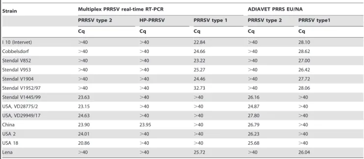
The multiplex PRRSV RT-qPCR was carried out using the RNA UltraSenseTM One-Step Quantitative RT-PCR System (Invitrogen, Carlsbad, CA, USA). The multiplex assay was optimized using a total reaction volume of 25ml. For a single reaction, 8.75ml RNase-free water, 5.0ml RNA UltraSenseTM5X Reaction Mix, 1.25ml RNA UltraSenseTM Enzyme Mix, 1.0ml type 1-PRRSV-specific FAM-labelled primer-probe mix (20 pmol/ml PRRSV-EU-2.1F, 20 pmol/ml PRRSV-EU-2.1R, 6.25 pmol/ml PRRSV-EU-2.1FAM), 1.0ml type 2-PRRSV-spe-cific Texas Red-labelled primer-probe mix (10 pmol/ml PRRSV-Table 1.Virus strains used in this study.
Strain Multiplex PRRSV real-time RT-PCR ADIAVET PRRS EU/NA
PRRSV type 2 HP-PRRSV PRRSV type 1 PRRSV type 2 PRRSV type1
Cq Cq Cq Cq Cq
I 10 (Intervet) .40 .40 22.84 .40 28.10
Cobbelsdorf .40 .40 24.66 .40 28.62
Stendal V852 .40 .40 23.22 .40 27.00
Stendal V953 .40 .40 25.27 .40 26.42
Stendal V1904 .40 .40 24.46 .40 27.72
Stendal V1952/97 .40 .40 32.73 .40 28.06
Stendal V1445/99 23.63 .40 .40 26.16 .40
USA, VD28775/2 23.15 .40 .40 24.87 .40
USA, VD29949/17 24.63 .40 .40 27.80 .40
China 23.90 23.95 .40 26.79 .40
USA 2 24.01 .40 .40 26.23 .40
USA 18 20.86 .40 .40 25.68 .40
Lena .40 .40 25.72 .40 26.04
doi:10.1371/journal.pone.0038251.t001
Table 2.Sequences of primers and probes used in the study.
Name Sequence 59-39 Genome position* Reference
PRRSV-US-1dF ATRATGRGCTGGCATTC 15257–15273 Kleiboeker et al., 2005, modified
PRRSV-US-1R ACACGGTCGCCCTAATTG 15353–15370
PRRSV-US-1TEX ACACGGTCGCCCTAATTG 15307–15330
PRRSV-HP-1F CCGCGTAGAACTGTGACAAC 2914–2933 this study
PRRSV-HP-1R TCCAGGATGCCCATGTTCTG 3035–3016
PRRSV-HP-1Cy5aS Cy5-ACGCACCAGGATGAGCCTCTGGAT-BHQ3 2941–2964
PRRSV-EU-2.1F GCACCACCTCACCCRRAC 14792–14809 Kleiboeker et al., 2005, modified
PRRSV-EU-2.1R CAGTTCCTGCRCCYTGAT 14851–14868
PRRSV-EU-2.1FAM FAM-CCTCTGYYTGCAATCGATCCAGAC-BHQ1 14819–14842
EGFP-11-F CAGCCACAACGTCTATATCATG 537–558 Hoffmann et al., 2006
EGFP-10-R CTTGTACAGCTCGTCCATGC 813–794
EGFP-HEX HEX-AGCACCCAGTCCGCCCTGAGCA-BHQ1 703–724
*genome position according to.
type 1-PRRSV: type 1 prototype strain Lelystad (accession number M96262). type 2-PRRSV: type 2 prototype strain VR-2332 (accession number U87392). HP-PRRSV: Porcine respiratory and reproductive syndrome virus strain JXwn06. (accession number EF641008).
US-1dF, 10 pmol/ml PRRSV-US-1R, 2.5 pmol/ml PRRSV-US-1TEX), 1.0ml HP-PRRSV-specific Cy5-labelled primer-probe mix (10 pmol/ml PRRSV-HP-1F, 10 pmol/ml PRRSV-HP-1R, 2.5 pmol/ml PRRSV-HP-1Cy5aS) and 2ml IC-specific HEX-labelled primer-probe mix (2.5 pmol/ml EGFP-11-F, 2.5 pmol/ml EGFP-10R, 1.25 pmol/ml EGFP-HEX) were combined in a
mas-ter mix. Finally, 5ml RNA template were added and the
RT-qPCR was carried out in a Bio-Rad CFX 96 Real-Time Detection System (Bio-Rad, Hercules, CA, USA) using the following thermal profile: reverse transcription at 50uC for 15 min, initial PCR activation step at 95uC for 2 min; 45 cycles of denaturation at 95uC for 15 s, annealing at 56uC for 20 s, and extension at 72uC for 30 s. All samples were tested in duplicate.
The commercially available ADIAVETH PRRS EU/NA Kit (ADIAGENE, Saint-Brieuc, France) for PRRSV detection and concurrent genotype differentiation was used according to the manufacturer’s recommendations.
Positive Standard RNA
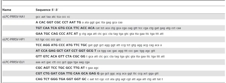
For each assay an oligonucleotide (PRRSV-EU1, oLPC-PRRSV-NA1, oLPC-PRRSV-HP1) was designed containing a T7-promoter and the respective primer and probe binding sequences connected by short spacer sequences (Table 3).
The oligonucleotides were transcribed in vitro with the SP6/T7 Transcription Kit (Roche Diagnostics Deutschland GmBH, Mannheim, Germany) according to the manufacturer’s instruc-tions. The T7-transcribed positive controls were digested with DNaseI and purified using the RNeasy Kit (Qiagen GmbH, Hilden, Germany). The RNA concentration was determined by spectrophotometry and the exact number of RNA molecules was calculated.
Animal Experiment with HP-PRRSV
On day 0 of the study, three serologically PRRSV negative swine (6–8 weeks of age) were inoculated intranasally with 105.8 Table 3.Sequences of positive standards used in this study, primer and probe binding regions are depicted in capitals.
Name Sequence 59-39
oLPC-PRRSV-NA1 gcc aat taa atc tca ccc cc
A CAC GGT CGC CCT AAT TGa ata ggt gac tta gag gca caa
TGT CAA TCA GTG CCA TTC ACC ACAcat tct aca ctg gca cga cag gtt tcc cga ctg gat gag atg cct caa
GAA TGC CAG CCC ATC ATg ctg aga att ctc gcc cta tag tga gtc gta tta gaa ttc tga ttt att oLPC-PRRSV-HP1 tct tgc ccc ccc gcc
TCC AGG ATG CCC ATG TTC TGCgat ggt gct agg ggt att ccg tct gtg agg acg cag aca a
AT CCA GAG GCT CAT CCT GGT GCG Tca tgg cac gac agg ttt ccc gac tgg agc gtt
GTT GTC ACA GTT CTA CGC GGt gca att ctc gcc cta tag tga gtc gta tta gaa ttc tga ttt att oLPC-PRRSV-EU1 aaa act gac ctt ccc gct gga tga aag cga
CGC AGT TCC TGC GCC TTG ATt gaa agc
CGT CTG GAT CGA TTG CAA GCA GAG Gga gct ggc acg aca ggt ttc ccg act gga gtt
CAG TCT GGG TGA GGT GGT GCc aat tct cgc cct ata gtg agt cgt att aga att ctg att tat t
doi:10.1371/journal.pone.0038251.t003
Figure 1. Analytical sensitivity of the PRRSV-multiplex-assay based on 10-fold dilution series of positive standard RNA.Regression curve was estimated by Bio-Rad CFX ManagerTMSoftware (Bio-Rad, Hercules, CA, USA).
50% tissue culture infective doses (TCID50) of the HP-PRRSV strain ‘‘China’’ in 10 ml. On day 3 post infection two further pigs were placed into the same room. During the entire study, blood samples were taken at regular intervals, rectal body temperatures were taken daily, and the pigs were monitored for clinical signs. On days 0, 3, 5, 7 and 10 after infection the quantity of white blood cells (WBC) was measured. Total RNA from serum was extracted and analysed with the multiplex PRRSV RT-qPCR. All animals were euthanized 14 days post infection. The animal experiment was reviewed by a state ethics commission and has been approved by the competent authority (State Office for
Agriculture, Food Safety and Fisheries Mecklenburg-Vorpom-mern, ref. LALLF M-V/TSD/7221.3-2.1-024/08.
Results
Sensitivity
The analytical sensitivity of the multiplex PRRSV RT-qPCR was determined with a series of 10-fold dilutions of the positive standards. The type 2 and HP-assays showed an amplification efficiency of more than 98% with a linear dynamic range from 106 copies down to 20 copies perml (Fig. 1). For the type 1-assay, the Figure 2. Chequerboard titration of a type 1- and a HP-PRRSV strain.Cq-values (z-axis) of the type 2- (2a) and HP- (2b) specific assays while co-amplification of different concentrations of the type 1 strain (y-axis) are shown. 2c depicts results of the type 1-specific assay when co-amplifying varying concentrations of HP-specific sequences (x-axis). Cq-values of the internal control RT-qPCR are shown in figure 2d. Images were designed supported by SigmaPlotHsoftware.
linear dynamic range reached down to 200 copies per ml with a PCR efficiency similar to the type 2-assay (Fig. 1).
The sensitivity of each PRRSV assay in the presence of the other genotype was tested in a checkerboard titration where the EU strain ‘‘Stendal V953’’ was titrated against the HP strain ‘‘China’’. Internal control RNA was added to the RT-qPCR master mix. Since the HP-strains belong to type 2, amplification was observed in both RT-qPCR systems, PRRSV-HP and type 2-PRRSV. The sensitivity of the type 2 and HP-assay was hardly affected even by the co-amplification of high amounts of a type 1-PRRSV (Fig. 2a, 2b). Both assays showed comparable quantifi-cation cycle (Cq; [15]) values. The 1027dilution of the HP-strain scored positive in three samples out of six in the type2-assay, and four out of six in the HP system. In the samples which were tested negative in one or both assays, the concentration of the type 1-strain, also present in the reaction, ranged from 1022 to 1027. Independent of the HP- and type 2 strain amplification, type 1 PRRSV was reliably detected even in low concentrations (Fig. 2c). In addition, the IC-assay was obviously not affected by the co-amplification of PRRSV (Fig. 2d).
The multiplex RT-qPCR was also compared to the ADIA-VETHPRRS EU/NA Kit. Both systems achieved equal results for diverse virus strains (Table 1).
The diagnostic sensitivity was evaluated by testing different sample types, including serum and lung samples (Table 4). All samples showed comparable results when tested with the novel multiplex PRRSV RT-qPCR and the ADIAVETH PRRS EU/ NA Kit. The multiplex PRRSV RT-qPCR assay failed to detect the IC in one case, the ADIAVETHPRRS EU/NA kit with two samples.
Specificity
The sequences of primers and probes included in the PRRSV multiplex RT-qPCR were compared to published sequence information (NCBI GenBank) with a special focus on porcine viruses. There was no indication of possible cross-reactions.
The diagnostic specificity of the multiplex RT-qPCR assay was validated using 132 serum samples from animals previously tested negative for PRRSV by a commercial RT-qPCR kit. All scored clearly negative in the type 1 and 2, as well as PRRSV-HP-specific assay within the multiplex approach with positive results in the IC-specific RT-PCR.
When testing the virus strains (Table 1) as well as the diagnostic samples (Table 4), the multiplex PRRSV RT-qPCR and ADIAVETH PRRS EU/NA Kit correctly determined the genotype for all samples.
Table 4.Analyses of diagnostic sample from PRRSV infected swine with the multiplex RT-qPCR and with the ADIAVETHPRRS EU/ NA Kit.
sample material Multiplex PRRSV real-time RT-PCR ADIAVET PRRS EU/NA
PRRSV type 2 HP-PRRSV PRRSV type 1 IC PRRSV type 2 PRRSV type 1 IC
Cq Cq Cq Cq Cq Cq Cq
serum .40 .40 33.31 26.35 .40 36.10 30.79
.40 .40 35.87 27.02 .40 33.13 29.04
.40 .40 24.89 24.82 .40 28.42 30.37
.40 .40 24.45 28.14 .40 29.08 29.64
.40 .40 23.06 28.32 .40 24.14 29.74
.40 .40 22.49 27.27 .40 28.90 27.55
.40 .40 29.78 26.85 .40 35.97 27.71
.40 .40 24.21 27.87 .40 30.53 28.63
.40 .40 29.55 26.21 .40 33.50 29.86
.40 .40 29.75 25.60 .40 32.20 29.61
.40 .40 29.36 25.65 .40 32.99 31.31
.40 .40 32.63 28.15 .40 36.69 38.68
34.79 .40 33.06 28.00 35.80 33.13 .40
30.68 .40 .40 26.88 32.65 .40 36.64
.40 .40 36.35 27.34 .40 31.44 .40
.40 .40 32.52 28.92 .40 33.42 .40
lung 21.09 .40 .40 27.89 27.80 .40 28.03
22.19 .40 .40 28.69 27.80 .40 22.93
26.75 .40 .40 28.04 32.76 .40 25.68
26.61 .40 30.35 28.52 34.28 30.46 25.20
.40 .40 28.59 27.55 .40 28.48 24.56
29.32 .40 .40 27.94 30.73 .40 24.35
23.80 24.89 .40 26.10 .40 27.16 23.54
29.49 30.15 .40 29.64 .40 34.25 22.71
22.30 23.13 .40 .40 .40 25.89 22.98
Reproducibility, Repeatability
Intra-assay reproducibility of the developed multiplex real-time RT-PCR was assessed with four replicates each of a 10-fold dilution series of RNA from PRRSV strains ‘‘USA VD 28775/20, ‘‘Stendal V953’’ and ‘‘China’’, respectively.
The inter-assay repeatability was determined for 10-fold dilution series on four separate days. Mean values and standard deviations of the PRRSV specific assays are shown in Fig. 3. The IC-specific PCR showed a mean Cq value of 33.86 with a standard deviation of 0.72.
Animal Experiment with HP-PRRSV
The body temperature of the infected pigs reached 40uC at day 3 post infection (dpi) and 41–42uC at 8 dpi. The fever then lasted until the end of the study. In-contact animals reached 40uC 3 days after first exposure and more than 41uC after 8 days. Monitoring of clinical signs revealed symptoms like red to purple dermal discoloration and purple and blue ears 5 days after infection or 7 days after first exposure, respectively. The discolorations persisted until the end of the study. Additionally, one of the infected and one of the in-contact animals showed diarrhoea and nasal discharge. Coughing was observed in one of the infected animals, while sneezing was seen in one of the in-contact animals.
Serum samples of infected and in-contact animals were investigated using the multiplex PRRSV RT-qPCR (Fig. 4). Samples from day 0 of the study scored negative for all animals. On 3 dpi the infected animals showed positive results, while the in-contact pigs remained negative. On 5 dpi PRRSV-RNA could be detected in samples of all animals.
In serum samples of the infected animals, Cq values declined until 7 dpi, and subsequently increased till 14 dpi. The in-contact animals showed decreasing Cq values up to day 10, followed by increasing values also until the end of the study. All samples showed equal results in the type 2 as well as the HP-specific system (Figure 4). However, one weak positive sample scored negative with the type 2-specific assay, whereas the HP-specific assay gave a positive result.
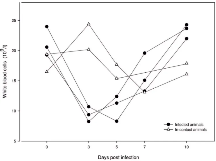
The WBC count of infected animals dropped from 0 dpi until 3 dpi, and subsequently increased again. In-contact animals showed a decreasing number of WBC from day 3 until day 7 (Figure 5).
Discussion
In 2006, a highly pathogenic disease emerged in swine farms in China. The morbidity rate was 50–100% and the mortality rate was 20–100% [16]. In the affected swine herds, high and continuous fever, anorexia, red discolorations in the bodies and blue ears were observed, followed by diarrhoea and other clinical signs in the late phase of the disease. A highly pathogenic PRRSV type with a 30-aa deletion within the Nsp2 encoding genome region could be demonstrated as the causative agent. Under experimental conditions, the virus could induce clinical symptoms identical to those observed in the field [7,17–19]. However, none of these trials used in-contact control animals to study virus transmission.
In our animal experiment the typical clinical symptoms of high and continuous fever and dermal discolorations could be observed in infected as well as in contact animals. The in-contact animals showed clinical symptoms 3 days later than the infected swine. As the control animals were placed in contact to the infected swine on day 3 of the study, a very rapid infection has to be assumed. This is reinforced by the decrease of WBC two days after the first exposure. Samples from the animal trial were used to validate the Figure 3. Reproducibility and repeatability of the multiplex
PRRSV assay.The Cq-values (y-axis) of 16 replicates of the type 2- (3a), PRRSV-HP- (3b) and type 1- (3c) assays using 10-fold dilution series of adequate virus strains (x-axis: log 10 initial quantity) are shown. Mean values are indicated alongside of each box plot, standard deviations are depicted in parentheses. Box plots were designed supported by R software [38].
newly developed PRRSV multiplex assays, allowing a differentia-tion of type 1 and 2 as well as a direct characterizadifferentia-tion of a PRRSV-strain as the HP-type. This is possible, since the Nsp2-encoding region within the 15 kb genome of PRRSV [20] shows a remarkable genetic variation [21,22]. Although the 30-aa deletion in Nsp-2 coding region of HP-PRRSV is not related to its virulence [23], the deletion can be used as a marker to distinguish the highly pathogenic Chinese virus from other NA-PRRSV strains.
For the detection of HP-PRRSV several RT-qPCR assays have been developed [8,11]. None of these assays, however, can detect other type 2- or even type 1-strains. Furthermore, RT-PCR assays for the detection and differentiation of both genotypes have been described [14,24,25], but no HP-PRRSV detection system was included in these assays.
Type 1 PRRSV isolates have been recently introduced into North American swine herds infected with type 2 strains [2]. Natural dual infection of swine with strains of both genotypes has been documented in Europe [26]. Even in two diagnostic samples used in this study, the type 1 and 2specific assays scored positive, emphasizing the necessity for simultaneous detection and differ-entiation of both genotype strains.
By testing dilution series of appropriate positive standards, it was demonstrated that the novel multiplex PRRSV RT-qPCR system is a highly sensitive approach for the detection and differentiation
of type 1 and 2 PRRSV and allows also the direct characterization of HP-specific genome sequences. Checkerboard titration con-firmed that the sensitivity of the respective assays was not affected by the co-amplification of any other target included in the multiplex assay. In the lowest detectable dilution of HP-PRRSV either the HP- or the type 2 assay or both scored positive, independently of the concentration of the type 1-strain RNA in the same sample. Consequently, the presence of HP-PRRSV genome in weak type 2-positive, and HP-negative, samples cannot be fully excluded.
When testing diverse virus strains and diagnostic samples including serum and lung samples, the new multiplex RT-qPCR showed a high diagnostic sensitivity. However, the number of used strains is limited consequently not representing the full range of the diversity of both genotypes. Especially the Eastern European type 1 isolates are highly diverse [27,28]; one of those strains (‘‘Lena’’) was tested by the multiplex RT-qPCR and correctly detected. Sequence information of Eastern Europe isolates, in particular ORF 7 sequences, which covers the primers and probes used in this study, are scarce. As a consequence, the applicability of the multiplex PCR for those strains in not completely proven. Nevertheless, the known sequences of primers and probes enable a rapid adaption to new sequence information and emerging strains, which is an important feature in PCR diagnostics of viruses exhibiting a large genetic diversity, such as PRRSV [29–32]. Figure 4. Analyses of serum samples from PRRSV-infected and in-contact pigs with the multiplex RT-qPCR.Graphs showing the same symbol belong to an identical animal, filled symbols depict results of the type 2-specific RT-qPCR unfilled of the HP assay. One to three are infected, four and five in-contact animals. Figure was designed supported by SigmaPlotHsoftware.
In addition to the PRRSV type 1, 2 and HP-specific assays, an internal control PCR based on heterologous RNA sequences was introduced. To verify efficient RNA extraction and uninhibited amplification, IC based on housekeeping genes [33–36] or heterologous internal controls [12,37] are used. Depending on the quality of the clinical sample, even housekeeping genes have unknown and variable mRNA concentrations, which could negatively influence the amplification and detection of the virus-derived nucleic acid. As the concentration of the heterologous IC RNA is known, sub-optimal RNA extractions or partial inhibitions of the RT-PCR can be detected, therefore avoiding false-negative results. However, a negative result in the IC specific PCR could also be caused by the absence of detectable RNA, but this is not significant when the virus-specific PCR scored positive.
A large number of samples from PRRSV-negative pigs were also tested and scored clearly negative in the PRRSV assays, whereas all of them scored positive in the IC-specific RT-qPCR. Using the checkerboard titration, it could be shown that the IC-specific assay was not affected by the co-amplification of PRRSV. In serum samples obtained from the animal trial, viral genome was first detectable at day 3 after infection or first exposure, demonstrating the rapid infection of the introduced contact animals. Except in a weak positive sample, where the HP-assay
scored positive and the type 2 negative, both detection systems showed comparable Cq-values.
In conclusion, the newly developed 4-colour multiplex RT-qPCR is a sensitive and specific method to confirm the presence of PRRSV genomes in field samples. For the first time, this assay can also detect and distinguish type 1 and 2 PRRSV as well as the Chinese HP-strain in a single qPCR reaction.
Acknowledgments
We thank Christian Korthase and Patrick Zitzow for excellent technical assistance, Jens Brackmann (LUFA Nord-West, Institut fu¨r Tiergesundheit, Oldenburg), Hubert Wonnemann (Landeslabor Schleswig-Holstein, Neu-mu¨nster) and Tilman Ku¨hn (synlab, Leipzig) for providing diagnostic samples and Uladzimir U. Karniychuk (Ghent University) for strain ‘‘Lena’’.
Author Contributions
Conceived and designed the experiments: KW BH MB. Performed the experiments: KW HS MD EL. Analyzed the data: KW BH HS MB. Contributed reagents/materials/analysis tools: KW BH MD HS. Wrote the paper: KW BH MB.
References
1. Rossow KD (1998) Porcine reproductive and respiratory syndrome. Vet Pathol 35: 1–20.
2. Ropp SL, Wees CE, Fang Y, Nelson EA, Rossow KD, et al. (2004) Characterization of emerging European-like porcine reproductive and re-spiratory syndrome virus isolates in the United States. J Virol 78: 3684–3703. 3. Key KF, Haqshenas G, Guenette DK, Swenson SL, Toth TE, et al. (2001)
Genetic variation and phylogenetic analyses of the ORF5 gene of acute porcine reproductive and respiratory syndrome virus isolates. Vet Microbiol 83: 249– 263.
4. Wensvoort G, Terpstra C, Pol JM, ter Laak EA, Bloemraad M, et al. (1991) Mystery swine disease in The Netherlands: the isolation of Lelystad virus. Vet Q 13: 121–130.
5. Cavanagh D (1997) Nidovirales: a new order comprising Coronaviridae and Arteriviridae. Arch Virol 142: 629–633.
6. Nelsen CJ, Murtaugh MP, Faaberg KS (1999) Porcine reproductive and respiratory syndrome virus comparison: divergent evolution on two continents. J Virol 73: 270–280.
7. Tian K, Yu X, Zhao T, Feng Y, Cao Z, et al. (2007) Emergence of fatal PRRSV variants: unparalleled outbreaks of atypical PRRS in China and molecular dissection of the unique hallmark. PLoS One 2: e526.
8. Xiao XL, Wu H, Yu YG, Cheng BZ, Yang XQ, et al. (2008) Rapid detection of a highly virulent Chinese-type isolate of Porcine Reproductive and Respiratory Syndrome Virus by real-time reverse transcriptase PCR. J Virol Methods 149: 49–55.
9. Feng Y, Zhao T, Nguyen T, Inui K, Ma Y, et al. (2008) Porcine respiratory and reproductive syndrome virus variants, Vietnam and China, 2007. Emerg Infect Dis 14: 1774–1776.
10. Normile D (2007) Virology. China, Vietnam grapple with ‘rapidly evolving’ pig virus. Science 317: 1017.
11. Chen NH, Chen XZ, Hu DM, Yu XL, Wang LL, et al. (2009) Rapid differential detection of classical and highly pathogenic North American Porcine Re-productive and Respiratory Syndrome virus in China by a duplex real-time RT-PCR. J Virol Methods 161: 192–198.
12. Hoffmann B, Depner K, Schirrmeier H, Beer M (2006) A universal heterologous internal control system for duplex real-time RT-PCR assays used in a detection system for pestiviruses. J Virol Methods 136: 200–209.
13. Karniychuk UU, Geldhof M, Vanhee M, Doorsselaere JV, Saveleva TA, et al. (2010) Pathogenesis and antigenic characterization of a new East European subtype 3 porcine reproductive and respiratory syndrome virus isolate. BMC Vet Res 6: 30.
14. Kleiboeker SB, Schommer SK, Lee SM, Watkins S, Chittick W, et al. (2005) Simultaneous detection of North American and European porcine reproductive and respiratory syndrome virus using real-time quantitative reverse transcrip-tase-PCR. J Vet Diagn Invest 17: 165–170.
15. Bustin SA, Benes V, Garson JA, Hellemans J, Huggett J, et al. (2009) The MIQE guidelines: minimum information for publication of quantitative real-time PCR experiments. Clin Chem 55: 611–622.
16. Tong GZ, Zhou YJ, Hao XF, Tian ZJ, An TQ, et al. (2007) Highly pathogenic porcine reproductive and respiratory syndrome, China. Emerg Infect Dis 13: 1434–1436.
17. Lv J, Zhang J, Sun Z, Liu W, Yuan S (2008) An infectious cDNA clone of a highly pathogenic porcine reproductive and respiratory syndrome virus variant associated with porcine high fever syndrome. J Gen Virol 89: 2075–2079. 18. Li Y, Wang X, Bo K, Tang B, Yang B, et al. (2007) Emergence of a highly
pathogenic porcine reproductive and respiratory syndrome virus in the Mid-Eastern region of China. Vet J 174: 577–584.
19. Zhou YJ, Hao XF, Tian ZJ, Tong GZ, Yoo D, et al. (2008) Highly virulent porcine reproductive and respiratory syndrome virus emerged in China. Transbound Emerg Dis 55: 152–164.
20. Meulenberg JJ, Hulst MM, de Meijer EJ, Moonen PL, den Besten A, et al. (1993) Lelystad virus, the causative agent of porcine epidemic abortion and respiratory syndrome (PEARS), is related to LDV and EAV. Virology 192: 62– 72.
21. Gao ZQ, Guo X, Yang HC (2004) Genomic characterization of two Chinese isolates of porcine respiratory and reproductive syndrome virus. Arch Virol 149: 1341–1351.
22. Fang Y, Kim DY, Ropp S, Steen P, Christopher-Hennings J, et al. (2004) Heterogeneity in Nsp2 of European-like porcine reproductive and respiratory syndrome viruses isolated in the United States. Virus Res 100: 229–235. 23. Zhou L, Zhang J, Zeng J, Yin S, Li Y, et al. (2009) The 30-amino-acid deletion
in the Nsp2 of highly pathogenic porcine reproductive and respiratory syndrome virus emerging in China is not related to its virulence. J Virol 83: 5156–5167. 24. Egli C, Thur B, Liu L, Hofmann MA (2001) Quantitative TaqMan RT-PCR for
the detection and differentiation of European and North American strains of porcine reproductive and respiratory syndrome virus. J Virol Methods 98: 63– 75.
25. Lurchachaiwong W, Payungporn S, Srisatidnarakul U, Mungkundar C, Theamboonlers A, et al. (2008) Rapid detection and strain identification of porcine reproductive and respiratory syndrome virus (PRRSV) by real-time RT-PCR. Lett Appl Microbiol 46: 55–60.
26. Oleksiewicz MB, Botner A, Madsen KG, Storgaard T (1998) Sensitive detection and typing of porcine reproductive and respiratory syndrome virus by RT-PCR amplification of whole viral genes. Vet Microbiol 64: 7–22.
27. Stadejek T, Oleksiewicz MB, Potapchuk D, Podgorska K (2006) Porcine reproductive and respiratory syndrome virus strains of exceptional diversity in eastern Europe support the definition of new genetic subtypes. J Gen Virol 87: 1835–1841.
28. Stadejek T, Oleksiewicz MB, Scherbakov AV, Timina AM, Krabbe JS, et al. (2008) Definition of subtypes in the European genotype of porcine reproductive and respiratory syndrome virus: nucleocapsid characteristics and geographical distribution in Europe. Arch Virol 153: 1479–1488.
29. Darwich L, Gimeno M, Sibila M, Diaz I, de la Torre E, et al. (2011) Genetic and immunobiological diversities of porcine reproductive and respiratory syndrome genotype I strains. Veterinary Microbiology.
30. Pesch S, Meyer C, Ohlinger VF (2005) New insights into the genetic diversity of European porcine reproductive and respiratory syndrome virus (PRRSV). Veterinary Microbiology 107: 31–48.
31. Thanawongnuwech R, Amonsin A, Tatsanakit A, Damrongwatanapokin S (2004) Genetics and geographical variation of porcine reproductive and respiratory syndrome virus (PRRSV) in Thailand. Veterinary Microbiology 101: 9–21.
32. Shi M, Lam TT, Hon CC, Murtaugh MP, Davies PR, et al. (2010) Phylogeny-based evolutionary, demographical, and geographical dissection of North American type 2 porcine reproductive and respiratory syndrome viruses. J Virol 84: 8700–8711.
33. Oleksiewicz MB, Donaldson AI, Alexandersen S (2001) Development of a novel real-time RT-PCR assay for quantitation of foot-and-mouth disease virus in diverse porcine tissues. J Virol Methods 92: 23–35.
34. Wakeley PR, Johnson N, McElhinney LM, Marston D, Sawyer J, et al. (2005) Development of a real-time, TaqMan reverse transcription-PCR assay for detection and differentiation of lyssavirus genotypes 1, 5, and 6. J Clin Microbiol 43: 2786–2792.
35. Toussaint JF, Sailleau C, Breard E, Zientara S, De Clercq K (2007) Bluetongue virus detection by two real-time RT-qPCRs targeting two different genomic segments. J Virol Methods 140: 115–123.
36. Moniwa M, Clavijo A, Li M, Collignon B, Kitching PR (2007) Performance of a foot-and-mouth disease virus reverse transcription-polymerase chain reaction with amplification controls between three real-time instruments. J Vet Diagn Invest 19: 9–20.
37. Hoffmann B, Beer M, Schelp C, Schirrmeier H, Depner K (2005) Validation of a real-time RT-PCR assay for sensitive and specific detection of classical swine fever. J Virol Methods 130: 36–44.