Diagnosis of Dentigerous Cyst
Antonione Santos Bezerra Pinto,
1André Luiz Ferreira Costa,
2Neiandro dos Santos Galvão,
3Thásia Luiz Dias Ferreira,
2and Sérgio Lúcio Pereira de Castro Lopes
41Department of Morphology, Faculty of Medicine, Federal University of Ceara, Fortaleza, CE, Brazil 2Department of Orthodontics and Radiology, University of S˜ao Paulo City (UNICID), S˜ao Paulo, SP, Brazil 3Piracicaba Dental School, State University of Campinas (UNICAMP), Piracicaba, SP, Brazil
4Department of Diagnosis and Surgery, S˜ao Jos´e dos Campos Dental School, S˜ao Paulo State University (UNESP),
S˜ao Jos´e dos Campos, SP, Brazil
Correspondence should be addressed to Andr´e Luiz Ferreira Costa; [email protected]
Received 1 July 2016; Revised 24 August 2016; Accepted 18 September 2016
Academic Editor: Yuk-Kwan Chen
Copyright © 2016 Antonione Santos Bezerra Pinto et al. his is an open access article distributed under the Creative Commons Attribution License, which permits unrestricted use, distribution, and reproduction in any medium, provided the original work is properly cited.
Odontogenic cysts have a high prevalence in the dental clinic population, with dentigerous cyst being one of the most frequent ones and whose aetiology involves accumulation of luid between the reduced enamel epithelium and the crown of an unerupted tooth. In the diagnostic process of these lesions, one should consider complementary imaging exams such as conventional radiography and computed tomography, which are commonly used for providing anatomical information on the tissues compromised by the lesion, but not on the nature of it. Magnetic resonance imaging (MRI) scans are noninvasive modalities which, due to their unique acquisition characteristics, can provide distinct information on the nature of the lesion. his study reports on a case of dentigerous cyst in the mandible of a 9-year-old patient, documented by means of diferent imaging modalities. MRI played an important role in both diagnosis of the lesion and diferential diagnosis between neoplastic lesions presenting similar imagenological behaviour under other techniques of radiography.
1. Introduction
A considerable number of lesions can compromise the dentomaxillofacial complex. Among these, one can highlight tumours and cysts of odontogenic origin as they are highly prevalent in the dental clinic population. he majority of the gnathic cysts are lined by odontogenic epithelium—the reason by which they are termed odontogenic cysts [1, 2].
Dentigerous cyst is the most common odontogenic cyst originating from the separation of the pericoronal follicle from the unerupted tooth, with prevalence of about 20 percent among all cysts lined with epithelium in the maxillae [3].
In the diagnostic process of these lesions, one should con-sider imaging exams, such as the complementary modalities
due to mainly their easiness of access, acquisition, and use of relevant information for conducting the diagnostic process [4]. Imaging techniques (panoramic, occlusal, and periapical radiographs) allow localisation of lesions, but they are not speciic [5]. In the case of dentigerous cyst, it is necessary a three-dimensional view of the cortical bone and assessment of the cyst content to make a better diagnosis [6, 7].
(a) (b)
(c)
Figure 1: Clinical aspect of the lesion. Facial asymmetry (a) and tumefaction in the right posterior region of the mandible. Front (b) and upper (c) views.
compared to MRI, with exposure to X-ray radiation being the main one [8].
Recent research has shown that MRI provides infor-mation which are beyond the image quality, not exposing the patient to the harmful efect of ionising radiation and thus helping determining a precise diagnosis both spatially and anatomically for conducting the treatment. he deter-mination of the content of the lesions is another advantage consistent with MRI compared to other imaging modalities [6, 9].
he aim of this study is to discuss on several diagnostic imaging modalities by means of a case report of a dentigerous cyst, emphasising the singular role of MRI in the analysis and treatment planning for such a lesion.
2. Case Presentation
A 9-year-old male patient was referred to our clinics for evaluation of the presence of asymptomatic tumefaction, hard on palpation, in the right posterior region of the mandible, resulting in a facial asymmetry. Intraoral exami-nation showed evidence of lingual displacement of tooth #45 as well as presence of root remnants of tooth #46, including
a solid mass lined by mucosa of normal appearance in the region corresponding to the entire alveolar crest extending occlusally (Figure 1).
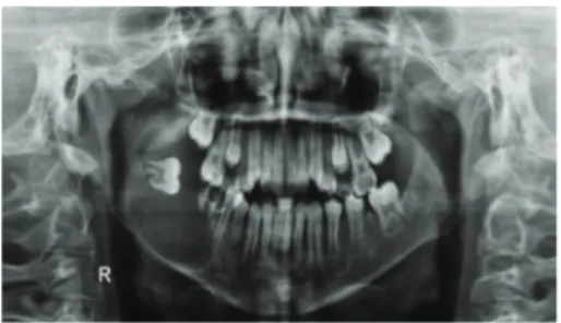
Panoramic radiograph was performed and showed a radiolucid, unilocular image of precisely corticalised limits involving tooth #47, which was in horizontal position and projecting towards the ipsilateral region of the mandibular ramus, with the crown medially positioned. his lesion extended from the apical region of tooth #45 to the corre-sponding mandibular ramus, with a very thin mandibular base encompassing the root remnants of tooth #46 (Fig-ure 2). he radiographic appearance was suggestive of benign odontogenic lesion. Among the diagnostic hypotheses raised, dentigerous cyst, ameloblastoma, and keratocystic odonto-genic tumour were considered.
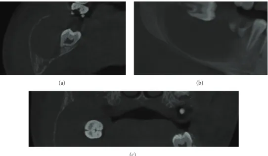
Cone beam computed tomography (GXCB-500, pow-ered by i-CAT) was used to evaluate the lesion
three-dimensionally, operating with ield of view (FOV) of 16×
Figure 2: Panoramic radiograph: radiolucid image of right mandibular body and ramus, with involvement of the included tooth #47.
were also observed. No region suggesting bone in continuity at the borderline areas of the lesion was observed either, which characterised its expansive nature (Figure 3).
he CBCT images were consistent with the diagnostic hypotheses suggested by the panoramic radiograph, which enabled us to conduct a more precise investigation of the lesion in terms of damage to the surrounding osseous struc-tures. However, the clinical, radiographic, and tomographic characteristics were not enough to clarify the real nature of the lesion, thus making the decision-making process diicult for an initial surgical approach.
In order to conduct the diferential diagnostic process and evaluate the internal content of the lesion, MRI was also performed by using a Sigma Tesla machine (General Electric, Milwaukee, USA) with head coil at axial, sagittal, and coronal anatomical planes for T1- and T2-weighted images, which were obtained by using an 8-channel phased array head coil with T1 sequence (TR = 478 ms, isotropic voxel size of
0.72 mm, TE = 16 ms, FOV of 1.0 ×21.0 cm, and slice gap
= 2.0 mm) and T2 sequence (TR = 6.5 ms, isotropic voxel
size of 0.72 mm, TE = 90.0 ms, FOV 21 ×21 cm, and slice
gap = 2.0 mm). hese images have evidenced the previously viewed expansive characteristics. he T1-weighted images showed the internal intermediate signal of the lesion, which are homogeneous characteristics demonstrating the absence of calciication process and fat content. On the other hand, the T2-weighted images showed hypersignal content delimited by areas of signal ranging from intermediate to hyposignal intensity, indicating presence of liquid content (i.e., cystic content) with consequent diferentiation from a solid tumour (Figure 4).
Ater evaluation of the MR images and aspiration punc-tion characterising the cystic content of the lesion, an inci-sional biopsy was performed and a decompression device was installed. he biopsy material was sent for histopathological exam. We have observed on the laminas fragments of cystic capsule partially lined by non-keratinised stratiied epithe-lium. In the focal area, one could note an eosinophilic staining in the epithelium as well as columnar cells in the periphery and cuboidal cells in the basal region. he capsule consisted of dense conjunctive tissue and exhibited multiple cordon-nest-like structures typical of odontogenic epithelium. In some areas, there was a tissue with myxoid appearance and in others it was possible to identify inlammatory iniltrate which was predominantly lymphoplasmocitary, ranging from
radiographic evolution of the case by comparing the images, it was observed that there was a signiicant decrease in the cystic volume and discrete reorientation of the eruption axes of the displaced teeth (Figure 6).
3. Discussion
he radiographic and tomographic aspects, including the clinical characteristics, were found to be relevant as they allowed us to determine the diferential diagnosis of the present case, namely, dentigerous cyst, unicystic ameloblas-toma, or KOT. his happened because these lesions are clinically and radiographically similar [3, 5–7, 9, 10].
In general, these lesions are considered diagnostic hypotheses when one observes a single lesion located in the posterior region of the mandible close to the third molar, with enhanced density at the centre, and well-delimited [5– 7, 9, 10].
For this reason, MRI has played a key role in the diagnostic process by providing new information which enabled us to determine a consistent presumptive diagnosis and consequently a more coherent surgical approach [5, 7, 11]. he T1-weighted image of the lesion showed an inter-mediate signal, thus not being useful for determination of the content of the lesion. Nevertheless, T2-weighted image enabled us to observe an intense brightness inside the lesion, which contributed signiicantly to the interpretation of a probable cystic lesion rather than tumoural. hese MRI data were important for us to consider the dentigerous cyst as the most probable lesion among the options raised in the diferential diagnosis [7, 11].
Such information had notable repercussions on both diagnosis and initial therapeutic approach. In fact, the dentist-surgeon who performed the biopsy had considered the presumptive diagnosis of dentigerous cyst but then considered decompression as the most plausible option of treatment [12].
Regardless whether the case involves a tumoural or cystic lesion, the biopsy would be indicated anyway because the inal diagnosis was based on histopathological examination as recommended in the clinical practice. he histopathological results conirmed the presumptive diagnosis of dentigerous cyst, showing that the MRI data should be considered relevant in the diagnostic process as it allows for diferentiation between cystic and tumoural lesions in terms of content [13– 15].
(a) (b)
(c)
Figure 3: CBCT images showing axial (a), sagittal (b), and coronal (c) slices demonstrating the expansive aspect of the lesion and its internal homogeneous appearance.
(a) (b)
(c)
Figure 4: MR images showing T1 axial view (a), T2 axial view (b), and T2 coronal view (c).
in preparing the inal diagnosis [14], but MRI scans can pro-vide a characterisation of the lesion composition and its alter-ations [7], thus contributing to the type of biopsy procedure to be performed. Some authors have already discussed the relevance of MRI in the dental clinic practice by highlighting its advantages and disadvantages. hese authors apparently agree that the advantages overcome the disadvantages, but it is worth considering that the accessibility to MRI services becomes more diicult in the clinical routine [5, 11, 16].
Considering the real contribution of MRI to the diagnosis and treatment of the present case as well as to other cases reported in the literature, it is clear that the dentist-surgeon
should consider it as an essential modern tool for optimisa-tion of the patient treatment [5, 9, 16].
In conclusion, the beneits of MRI in the diagnostic process were found to be obvious in the present case report. he use of this complementary examination enabled the practitioner to have a safer and more eicient performance, thus optimising the treatment for the patient.
Consent
Figure 5: Microphotographs of the lesion stained with haematoxylin and eosin (H&E); scale bar = 80�m.
(a) (b)
Figure 6: Clinical (a) and radiographic (b) aspects of the compression device following 4 months of installation.
Competing Interests
he authors declare that there is no conlict of interests regarding the publication of this paper.
References
[1] H. Konouchi, J.-I. Asaumi, Y. Yanagi, M. Hisatomi, and K. Kishi, “Adenomatoid odontogenic tumor: correlation of MRI with
histopathological indings,”European Journal of Radiology, vol.
44, no. 1, pp. 19–23, 2002.
[2] G. M. Murray, M. Bhutada, C. C. Peck, I. Phanachet, D. Sae-Lee,
and T. Whittle, “he human lateral pterygoid muscle,”Archives
of Oral Biology, vol. 52, no. 4, pp. 377–380, 2007.
[3] K. Srinivasan, A. Seith, A. Gadodia et al., “Evaluation of the inferior alveolar canal for cysts and tumors of the mandible— comparison of multidetector computed tomography and 3-dimensional volume interpolated breath-hold examination
magnetic resonance sequence with curved multiplanar
refor-matted reconstructions,” Journal of Oral and Maxillofacial
Surgery, vol. 70, no. 10, pp. 2327–2332, 2012.
[4] M. Hara, H. Matsuzaki, N. Katase et al., “Central odontogenic ibroma of the jawbone: 2 case reports describing its imaging
features and an analysis of its DCE-MRI indings,”Oral Surgery,
Oral Medicine, Oral Pathology and Oral Radiology, vol. 113, no. 6, pp. e51–e58, 2012.
[5] F. A. Probst, M. Probst, C. Pautke et al., “Magnetic resonance imaging: a useful tool to distinguish between keratocystic
odontogenic tumours and odontogenic cysts,”British Journal of
Oral and Maxillofacial Surgery, vol. 53, no. 3, pp. 217–222, 2015. [6] K. Srinivasan, A. Seith Bhalla, R. Sharma, A. Kumar, A. Roychoudhury, and O. Bhutia, “Difusion-weighted imaging
in the evaluation of odontogenic cysts and tumours,”British
Journal of Radiology, vol. 85, no. 1018, pp. e864–e870, 2012. [7] M. Minami, T. Kaneda, K. Ozawa et al., “Cystic lesions of
odontogenic keratocysts and ameloblastomas from other cysts,”
American Journal of Roentgenology, vol. 166, no. 4, pp. 943–949, 1996.
[8] M. Cassetta, S. Di Carlo, N. Pranno, A. Stagnitti, V. Pompa, and G. Pompa, “he use of high resolution magnetic reso-nance on 3.0-T system in the diagnosis and surgical planning of intraosseous lesions of the jaws: preliminary results of a
retrospective study,”European Review for Medical and
Pharma-cological Sciences, vol. 16, no. 14, pp. 2021–2028, 2012.
[9] M. Fujita, H. Matsuzaki, Y. Yanagi et al., “Diagnostic value of
MRI for odontogenic tumours,”Dentomaxillofacial Radiology,
vol. 42, no. 5, Article ID 20120265, 2013.
[10] O. Gamba Tde, I. L. Flores, A. B. Pinto, A. L. Costa, M. E. Moraes, and S. L. Lopes, “Keratocystic odontogenic tumor: role of cone beam computed tomography and magnetic resonance
imaging,”General Dentistry, vol. 64, no. 1, pp. 36–39, 2016.
[11] U. N. Yilmaz, F. Yaman, and S. S. Atilgan, “MR T1 and T2
relaxations in cysts and abscesses measured by 1.5 T MRI,”
Dentomaxillofacial Radiology, vol. 41, no. 5, pp. 385–391, 2012. [12] B. C. Kirtaniya, V. Sachdev, A. Singla, and A. K. Sharma,
“Mar-supialization: a conservative approach for treating dentigerous
cyst in children in the mixed dentition,”Journal of Indian Society
of Pedodontics and Preventive Dentistry, vol. 28, no. 3, pp. 203– 208, 2010.
[13] T. Tilakraj, N. Kiran, K. Mukunda, and S. Rao, “Non syndromic unilateral dentigerous cyst in a 4-year-old child: a rare case
report,”Contemporary Clinical Dentistry, vol. 2, no. 4, pp. 398–
401, 2011.
[14] M. Demirkol, B. Ege, S. Yanik, M. H. Aras, and S. Ay, “Clinico-pathological study of jaw cysts in southeast region of Turkey,”
European Journal of Dentistry, vol. 8, no. 1, pp. 107–111, 2014. [15] S. Passi, K. Gauba, A. Agnihotri, and R. Sharma, “Dentigerous
cyst in primary dentition: a case report,” Journal of Indian
Society of Pedodontics and Preventive Dentistry, vol. 26, no. 4, pp. 168–170, 2008.
[16] L. Demiriz, A. F. Misir, and D. I. Gorur, “Dentigerous cyst in
a young child,”European Journal of Dentistry, vol. 9, no. 4, pp.
Submit your manuscripts at
http://www.hindawi.com
Hindawi Publishing Corporation
http://www.hindawi.com Volume 2014
Oral Oncology
Journal ofHindawi Publishing Corporation
http://www.hindawi.com Volume 2014
International Journal of
Biomaterials
Hindawi Publishing Corporation
http://www.hindawi.com Volume 2014
BioMed
Research International
Hindawi Publishing Corporation
http://www.hindawi.com Volume 2014
Oral Implants
Journal ofHindawi Publishing Corporation
http://www.hindawi.com Volume 2014
Anesthesiology Research and Practice
Hindawi Publishing Corporation
http://www.hindawi.com Volume 2014 Radiology
Research and Practice Environmental and
Public Health
Journal of
Hindawi Publishing Corporation
http://www.hindawi.com Volume 2014
Hindawi Publishing Corporation
http://www.hindawi.com Volume 2014
Dental Surgery
Journal ofDrug Delivery
Journal of Hindawi Publishing Corporationhttp://www.hindawi.com Volume 2014
Hindawi Publishing Corporation
http://www.hindawi.com Volume 2014
Oral Diseases
Journal ofHindawi Publishing Corporation
http://www.hindawi.com Volume 2014
Computational and Mathematical Methods in Medicine
Pain
Research and Treatment
Hindawi Publishing Corporation
http://www.hindawi.com Volume 2014
Endocrinology
International Journal ofHindawi Publishing Corporation
http://www.hindawi.com Volume 2014
Hindawi Publishing Corporation
http://www.hindawi.com Volume 2014