Evaluation of urogenital fistulas by magnetic resonance
urography*
Avaliação das fístulas urogenitais por urorressonância magnética
Augusto Elias Mamere1, Rafael Darahem Souza Coelho2, Alexandre Oliveira Cecin1, Leonir Terezinha Feltrin1, Fabiano Rubião Lucchesi2, Marco Antônio Lopes Pinheiro3, Ana Karina Nascimento Borges3, Gustavo Fabene Garcia3, Daniel Seabra4
OBJECTIVE: Vesicovaginal and ureterovaginal fistulas are unusual complications secondary to pelvic sur-gery or pelvic diseases. The therapeutic success in these cases depends on an appropriate preoperative evaluation for diagnosis and visualization of the fistulous tract. The present study is aimed at demonstrating the potential of magnetic resonance urography for the diagnosis of vesicovaginal and ureterovaginal fistulas as well as for defining the fistulous tracts. MATERIALS AND METHODS: Seven female patients clinically diagnosed with vesicovaginal or ureterovaginal fistulas had their medical records, radiological and magnetic resonance images retrospectively reviewed. Magnetic resonance urography included 3D-HASTE sequences with fat saturation. RESULTS: Six patients presented vesicovaginal fistulas and, in one patient, a right-sided ureterovaginal fistula was diagnosed. Magnetic resonance urography allowed the demonstration of the fis-tulous tract in six (85.7%) of the seven patients evaluated in the present study, without the need of bladder catheterization or contrast injection. CONCLUSION: This study demonstrates both the potential and applica-bility of magnetic resonance urography in the evaluation of these types of fistulas.
Keywords: Urinary fistula; Vesicovaginal fistula; Urological diagnostic techniques; Magnetic resonance im-aging; Vaginal fistula – diagnosis.
OBJETIVO: As fístulas vesicovaginais e ureterovaginais são complicações incomuns, secundárias a doenças ou a cirurgias pélvicas. O sucesso terapêutico dessas fístulas depende de adequada avaliação pré-operatória para o diagnóstico e visualização do seu trajeto. Este trabalho tem o objetivo de demonstrar o potencial da urorressonância no diagnóstico das fístulas urogenitais e na visualização dos seus trajetos. MATERIAIS E MÉTODOS: Foram analisados, retrospectivamente, os prontuários médicos e as imagens radiológicas e de urorressonância magnética de sete pacientes do sexo feminino com diagnóstico de fístula urogenital. Para a urorressonância foram realizadas seqüências 3D-HASTE com saturação de gordura. RESULTADOS: Seis pa-cientes apresentavam fístula vesicovaginal e uma paciente tinha diagnóstico de fístula ureterovaginal à direita. Com a utilização da urorressonância magnética, foi possível demonstrar o trajeto da fístula em seis das sete pacientes (85,7%), sem a necessidade de cateterização vesical ou da injeção de contraste. CONCLUSÃO: Este estudo demonstra o potencial e a aplicabilidade da urorressonância na avaliação dessas fístulas. Unitermos: Fístula urinária; Fístula vesicovaginal; Técnicas de diagnóstico urológico; Ressonância magnética; Fístula vaginal – diagnóstico.
Abstract
Resumo
* Study developed in the Departamento de Diagnóstico por Imagem do Hospital de Câncer de Barretos – Fundação Pio XII, Barretos, SP, Brazil.
1. Master, MDs, Radiologists at Hospital de Câncer de Barre-tos – Fundação Pio XII, BarreBarre-tos, SP, Brazil.
2. PhD, MDs, Radiologists at Hospital de Câncer de Barretos – Fundação Pio XII, Barretos, SP, Brazil.
3. Titular Members of Colégio Brasileiro de Radiologia e Diag-nóstico por Imagem (CBR), MDs, Radiologists at Hospital de Câncer de Barretos – Fundação Pio XII, Barretos, SP, Brazil.
4. PhD, MD, Urologist at Hospital de Câncer de Barretos – Fundação Pio XII, Barretos, SP, Brazil.
Mailing address: Dr. Augusto Elias Mamere. Hospital de Cân-cer de Barretos, Fundação Pio XII. Rua Antenor Duarte Vilela, 1331. Barretos, SP, Brazil, 14784-400. E-mail: mamere@uol. com.br
Received January 23, 2007. Accepted after revision July 13, 2007.
INTRODUCTION
Several types of pelvic fistulas second-ary to pelvic diseases or surgeries have al-ready been described: vesicovaginal, vesi-couterine, ureterovaginal, ureteroenteric, enterovaginal and rectovaginal fistulas(1–3).
The close proximity of pelvic organs makes the genitourinary system susceptible to in-jury, so the majority of fistulas occur in the pelvic cavity(1).
Main causes of pelvic fistulas are sur-gical or obstetric procedures, malignant
tu-mors, radiotherapy, pelvic infections, trau-mas and inflammatory intestinal dis-eases(2,4,5). Vesicovaginal and
ureterovagi-nal are some of the most frequent types of fistulas. The most frequent predisposing factor for vesicovaginal fistula is uterine cervix cancer treated with radiotherapy, with an incidence ranging between 1% and 10%(6). Ureterovaginal fistulas occur most
These fistulas subtypes occurring in the female lower urinary tract (vesicovaginal and ureterovaginal) cause social and psy-chological anguish, and frequently repre-sent a therapeutic problem for the sur-geon(7), particularly when they appear
af-ter radiotherapy, associated with involve-ment of the vascular supply and difficulty in cicatrization and regeneration of the ir-radiated tissue(8).
The main symptom of patients with ure-terovaginal or vesicovaginal fistulas sec-ondary to pelvic surgery or disease is a continuous involuntary discharge of urine into the vaginal vault(1,7). Hematuria,
uri-nary infections or perineal dermatitis may be associated(1,2).
The following procedures can be uti-lized in the diagnosis of these fistulas: cys-toscopy, vaginoscopy, computed tomogra-phy and magnetic resonance imaging(1,4).
Magnetic resonance imaging allows the identification of the fistulous tract, as well as the evaluation, by means of sections in different planes, of alterations which even-tually may be present in the adjacent pel-vic structures, allowing an appropriate sur-gical planning. Magnetic resonance urog-raphy images allow a global, non-invasive visualization of the whole urinary tract, without the need for ionizing radiation and administration of contrast agents.
In the medical literature review, few studies were found investigating the utili-zation of magnetic resonance imaging for the diagnosis and evaluation of urogenital fistulas(1–5). In these studies, the patients
have been evaluated only with axial and sagittal sections on conventional se-quences, with no magnetic resonance urog-raphy sequence.
The present study is aimed at demon-strating the potential and applicability of magnetic resonance urography in the diag-nosis of vesicovaginal and ureterovaginal fistulas as well as in the visualization of fistulous tracts.
MATERIALS AND METHODS
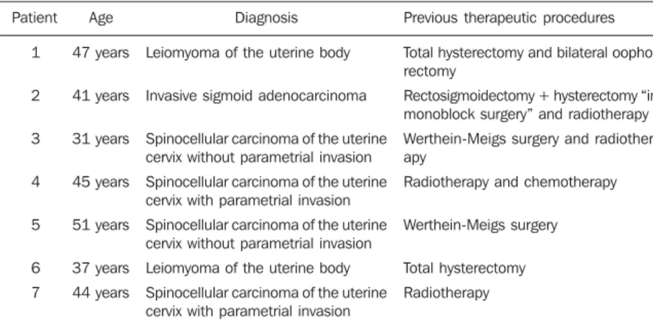
Dossiers, radiological and magnetic resonance urography images of seven fe-male patients diagnosed with vesicovagi-nal or ureterovagivesicovagi-nal fistulas were retro-spectively evaluated. The
clinical-patho-logical profiles of these patients are sum-marized on Table 1.
Magnetic resonance imaging studies were performed in a 1.5 tesla Magnetom Symphony equipment (Siemens®;
Erlan-gen, Germany), with 30 mT/m gradient am-plitude and surface coil. All the patients re-ceived intravenous furosemide (10 mg) 30 minutes prior the examination.
For the magnetic resonance urography studies, three-dimensional half-Fourier acquisition single-shot turbo spin echo (3D-HASTE) T2-weighted sequences with fat saturation were performed with 2800 ms repetition time, 1080 ms echo time, 512 matrix, 2.0 s acquisition time, and block thickness (slab) ranging between 7 cm and 10 cm to include the whole urinary tract and bladder, at different angles in relation to the transverse axis to obtain anterior, lateral (sagittal) and oblique views. The breathhold acquisition time was short (2.0 s) and, consequently, was well toler-ated by the patients. This sequence is the same utilized in magnetic resonance cho-langiography.
In the evaluation of patients 2 and 5, only the magnetic resonance urography se-quence was performed. For the other pa-tients, besides magnetic resonance urogra-phy, conventional axial, sagittal and coro-nal turbo spin echo (TSE) sequences were acquired for evaluation of their pelvic structures.
The study of the patients 4 and 6 also included conventional TSE T1-weighted sequences both before and after intrave-nous contrast agent (gadolinium) adminis-tration.
The cystoscopy reports, excretory urog-raphy and cystogurog-raphy images of the pa-tients also were reviewed and utilized for comparison.
RESULTS
Six patients presented vesicovaginal fis-tulas (patients 1, 2, 4, 5, 6 and 7 – see Table 1); the patient 3 was diagnosed with right-sided ureterovaginal fistula.
The fistulous tract could be visualized on magnetic resonance urography images of five (1, 2, 5, 6 and 7 – see table 1) of the six patients diagnosed with vesicovaginal fistulas. The patients 1 and 6 were submit-ted only to this study (Figure 1), and the patients 2, 5 and 7, besides magnetic reso-nance urography, also were submitted to cystography (Figures 2 and 3).
The vesicovaginal fistulous tract of the patient 4 could not be visualized on the magnetic resonance urography neither on the conventional magnetic resonance im-ages. However, after intravenous paramag-netic contrast injection, the contrast uptake could be detected in the vaginal vault on the delayed sequences after renal excretion, allowing the diagnosis of urogenital fistula, despite the non-visualization of the fistu-lous tract. In this patient, the fistufistu-lous tract could not be visualized also on the cystog-raphy neither on the excretory urogcystog-raphy; the fistula orifice was visualized only dur-ing cystoscopy. Also in the patients 1, 2 and 6, the vesicovaginal fistula orifice was vi-sualized during cystoscopy.
The magnetic resonance urography im-ages of the patient 3 demonstrated a
right-Table 1 Clinical-pathological profile of the patients.
Patient 1 2 3 4 5 6 7 Age 47 years 41 years 31 years 45 years 51 years 37 years 44 years Diagnosis
Leiomyoma of the uterine body
Invasive sigmoid adenocarcinoma
Spinocellular carcinoma of the uterine cervix without parametrial invasion
Spinocellular carcinoma of the uterine cervix with parametrial invasion
Spinocellular carcinoma of the uterine cervix without parametrial invasion
Leiomyoma of the uterine body
Spinocellular carcinoma of the uterine cervix with parametrial invasion
Previous therapeutic procedures
Total hysterectomy and bilateral oopho-rectomy
Rectosigmoidectomy + hysterectomy “in monoblock surgery” and radiotherapy
Werthein-Meigs surgery and radiother-apy
Radiotherapy and chemotherapy
Werthein-Meigs surgery
Total hysterectomy
Figure 2. Imaging studies of the patient 2, a 41-year-old woman, demonstrating vesicovaginal fistula (arrows on A, B and C). On A, magnetic resonance urogram 3D-HASTE; on B, axial, TSE, T2-weighted sequence; on C, cystography. On B a flow artifact is observed within the vaginal vault, generated by the urine passage through the fistula, from the bladder into the vagina.
A B C
sided ureterovaginal fistula (Figure 4). This patient was also submitted to excretory urography, which failed in the diagnosis of the fistula.
Additionally, the patient 4 presented right-sided renal hypotrophy, and the pa-tients 2, 3 and 5, bilateral hydronephrosis. The patient 5 had been previously
submit-ted to bilateral ureteral surgery (uretero-il-eal-vesicoplasty), which also could be ap-propriately demonstrated by magnetic reso-nance urography (Figure 3).
DISCUSSION
Vesicovaginal and ureterovaginal fistu-las are infrequent complications secondary to inflammatory diseases, neoplasms, ra-diotherapy or pelvic surgeries, which cause severe psychosocial problems for affected patients(7). The therapeutic strategies
suc-cess depends on an appropriate preopera-tive evaluation for diagnosis and visualiza-tion of the fistulous tract. Classically, the imaging methods for evaluation of these fistulas include excretory urography, cys-tography and vaginography(1).
In the last years the increasing utiliza-tion of computed tomography urography and magnetic resonance urography for the urinary system evaluation has been re-ported. Besides demonstrating abnormali-ties in the urinary tract, these methods al-low the visualization of adjacent abdomi-nal and pelvic structures on conventioabdomi-nal images. Excretory urography and com-puted tomography urography present the disadvantage of requiring intravenous io-dinated contrast injection and ionizing ra-diation; for this reason, magnetic resonance Figure 1. Magnetic resonance urogram with
3D-HASTE sequence of the patient 6, a 37-year-old woman previously submitted to total hysterectomy, who progressed with symptoms of urogenital fistula, whose image demonstrated the vesicovaginal fis-tulous tract (curved arrow).
imaging is the method of choice for chil-dren, pregnant women and patients with any contraindication to iodinated contrast agents, such as previous allergic reaction, severe cardiopathy, asthma or renal failure(9).
Magnetic resonance urography allows acquisition of images with a diagnostic quality that has been continuously im-proved with the development of sequences technically more sophisticated and with in-creasingly shorter acquisition times(10).
Presently, two techniques can be utilized for this study: non-contrast enhanced, T2-weighted sequences (hydrographic se-quences), or contrast-enhanced (intrave-nous paramagnetic contrast – gadolinium – injection) T1-weighted sequences dem-onstrating contrast excretion(11). Magnetic
resonance urography T2-weighted se-quence has already proved be an excellent technique for investigating a dilated urinary tract, even in the absence of renal excretion (severe renal failure). T1-weighted se-quences with intravenous contrast (gado-linium) injection demonstrate the renal excretory function and the urinary flow through the urinary tract to the bladder(12).
Both MR urography techniques may be combined as necessary(13).
hydronephrosis and obstructive uropa-thies(10,11,14–20).
With the utilization of magnetic reso-nance urography, the fistulous tract could be demonstrated in six of the seven patients evaluated in the present study (85.7%) without the necessity of vesical probing or contrast agent injection.
The vesicovaginal fistulous tracts were appropriately demonstrated by magnetic resonance urography in the patients 2, 5 and 7, and the images presented a perfect correlation with cystographic images. In the patients 1 and 6, the fistulous tracts were also appropriately demonstrated on magnetic resonance urography, and the fis-tulas orifices were visualized on cystos-copy in agreement with the clinical diag-nosis, despite de absence of correlation with cystography.
The fistulous tract of the patient 4 was not demonstrated by magnetic resonance urography, and also could not be visualized on the cystography, probably because of the narrow caliber and low output of the fistula. Therefore, in four of the patients with vesicovaginal fistulas included in the present study there was a total agreement
between the findings on magnetic reso-nance urography and cystography images; and in the other two patients who were not submitted to radiological study, also there was agreement between the magnetic reso-nance urography and the clinical diagnosis of urogenital fistula.
In the single patient diagnosed with ure-terovaginal fistula, the fistulous tract can be perfectly visualized on the magnetic reso-nance urography sequences confirmed by the conventional TSE sequences, although it could not be appropriately visualized at the excretory urography. So, the magnetic resonance images were decisive for the diagnosis in this patient.
Despite the feasibility of urogenital fis-tula diagnosis by conventional magnetic resonance imaging with multiplanar, thick slices sequences, the magnetic resonance urography with 3D-HASTE sequences al-lows the acquisition of images quite simi-lar to those usually seem by clinicians and surgeons on conventional radiographic studies (excretory urography, cystography and pyelography, with a broad, non-inva-sive fast and safe visualization of the whole urinary tract.
Figure 3. Vesicovaginal fistula demonstrated by magnetic resonance urography (arrow on A) and by cystography (arrow on B) in the patient 5, a 51-year-old woman, that appeared after Werthein-Meigs surgery. On C, coronal magnetic resonance urography 3D-HASTE image demonstrating uretero-ileal vesicoplasty with visualization of the anatomoses of the ileum segment with the ureters (straight arrows) and with the bladder (curved arrow) with bilateral hydronephrosis.
A B C
CONCLUSION
Considering that urogenital fistula is an infrequent condition, the number of pa-tients evaluated in the present study is not sufficient to determine the sensitivity, specificity and accuracy of this diagnostic method. Additional controlled studies with a higher number of patients are necessary to determine conclusive results. However, the images obtained in this study demon-strate the potential capacity and applicabil-ity of magnetic resonance urography in the evaluation of urogenital fistulas.
REFERENCES
1. Moon SG, Kim SH, Lee HJ, et al. Pelvic fistulas complicating pelvic surgery or diseases: spectrum of imaging findings. Korean J Radiol. 2001;2:97– 104.
2. Outwater E, Schiebler ML. Pelvic fistulas: find-ings on MR images. AJR Am J Roentgenol. 1993; 160:327–30.
3. Murphy JM, Lee G, Sharma SD, et al. Vesicou-terine fistula: MRI diagnosis. Eur Radiol. 1999; 9:1876–8.
4. Semelka RC, Hricak H, Kim B, et al. Pelvic fis-tulas: appearances on MR images. Abdom Imag-ing. 1997;22:91–5.
5. Blomlie V, Rofstad EK, Tropé C, et al. Critical soft tissues of the female pelvis: serial MR imaging before, during, and after radiation therapy. Radi-ology. 1997;203:391–7.
6. Kuhlman JE, Fishman EK. CT evaluation of en-terovaginal and vesicovaginal fistulas. J Comput Assist Tomogr. 1990;14:390–4.
7. Akman RY, Sargin S, Ozdemir G, et al. Vesico-vaginal and ureteroVesico-vaginal fistulas: a review of 39 cases. Int Urol Nephrol. 1999;31:321–6.
8. Tabakov ID, Slavchev BN. Large post-hysterec-tomy and post-radiation vesicovaginal fistulas: repair by ileocystoplasty. J Urol. 2004;171:272– 4.
9. Kawashima A, Glockner JF, King BF Jr. CT urography and MR urography. Radiol Clin North Am. 2003;41:945–61.
10. Regan F, Bohlman ME, Khazan R, et al. MR urography using HASTE imaging in the assess-ment of ureteric obstruction. AJR Am J Roent-genol. 1996;167:1115–20.
11. Blandino A, Gaeta M, Minutoli F, et al. MR urography of the ureter. AJR Am J Roentgenol. 2002;179:1307–14.
12. Nolte-Ernsting C, Staatz G, Wildberger J, et al. MR-urography and CT-urography: principles, ex-amination techniques, applications. Rofo. 2003; 175:211–22.
13. Nolte-Ernsting CC, Staatz G, Tacke J, et al. MR urography today. Abdom Imaging. 2003;28:191– 209.
14. Khanna PC, Karnik ND, Jankharia BG, et al.
Mag-netic resonance urography (MRU) versus intra-venous urography (IVU) in obstructive uropathy: a prospective study of 30 cases. J Assoc Physi-cians India. 2005;53:527–34.
15. Chahal R, Taylor K, Eardley I, et al. Patients at high risk for upper tract urothelial cancer: evalu-ation of hydronephrosis using high-resolution magnetic resonance urography. J Urol.2005;174: 478–82.
16. Erdogmus B, Bozkurt M, Bakir Z. Diagnostic value of HASTE technique and excretory MR urography in urinary system obstructions. Tani Girisim Radyol. 2004;10:309–15.
17. Karabacakoglu A, Karakose S, Ince O, et al. Di-agnostic value of diuretic-enhanced excretory MR urography in patients with obstructive uropathy. Eur J Radiol. 2004;52:320–7.
18. Magno C, Blandino A, Anastasi G, et al. Lithiasic obstructive uropathy. Hydronephrosis character-ization by magnetic resonance pyelography. Urol Int. 2004;72 Suppl 1:40–2.
19. Shokeir AA, El-Diasty T, Eassa W, et al. Diagno-sis of ureteral obstruction in patients with com-promised renal function: the role of noninvasive imaging modalities. J Urol. 2004;171(6 Pt 1): 2303–6.