Spectrometry. Fast and Reliable Identification from Agar
Plates and Blood Cultures
Laura Ferreira1, Silvia Vega Castan˜o2, Fernando Sa´nchez-Juanes1, Sandra Gonza´lez-Cabrero3, Fabiola Menegotto3, Antonio Ordun˜a-Domingo3, Jose´ Manuel Gonza´lez-Buitrago1,4., Juan Luis Mun˜oz-Bellido2,5*.
1Unidad de Investigacio´n, Hospital Universitario de Salamanca, Salamanca, Spain,2Departamento de Microbiologı´a, Hospital Universitario de Salamanca, Salamanca, Spain,3Departamento de Anatomı´a Patolo´gica, Microbiologı´a, Medicina Preventiva y Salud Pu´blica, Medicina Legal y Forense, Universidad de Valladolid, Valladolid, Spain,
4Departamento de Bioquı´mica y Biologı´a Molecular, Universidad de Salamanca, Salamanca, Spain,5Departamento de Medicina Preventiva, Salud Pu´blica y Microbiologı´a Me´dica, Universidad de Salamanca, Salamanca, Spain
Abstract
Background:MALDI-TOF mass spectrometry (MS) is a reliable method for bacteria identification. Some databases used for this purpose lack reference profiles forBrucellaspecies, which is still an important pathogen in wide areas around the world. We report the creation of profiles for MALDI-TOF Biotyper 2.0 database (Bruker Daltonics, Germany) and their usefulness for identifying brucellae from culture plates and blood cultures.
Methodology/Principal Findings: We created MALDI Biotyper 2.0 profiles for type strains belonging to B. melitensis biotypes 1, 2 and 3;B. abortusbiotypes 1, 2, 5 and 9;B. suis,B. canis,B cetiandB. pinnipedialis. Then, 131 clinical isolates grown on plate cultures were used in triplicate to check identification. Identification at genus level was always correct, although in most cases the three replicates reported different identification at species level. Simulated blood cultures were performed with type strains belonging to the main human pathogenic species (B. melitensis,B. abortus,B. suisandB. canis), and studied by MALDI-TOF MS in triplicate. Identification at genus level was always correct.
Conclusions/Significance:MALDI-TOF MS is reliable forBrucellaidentification to the genus level from culture plates and directly from blood culture bottles.
Citation:Ferreira L, Vega Castan˜o S, Sa´nchez-Juanes F, Gonza´lez-Cabrero S, Menegotto F, et al. (2010) Identification ofBrucellaby MALDI-TOF Mass Spectrometry. Fast and Reliable Identification from Agar Plates and Blood Cultures. PLoS ONE 5(12): e14235. doi:10.1371/journal.pone.0014235
Editor:Edgardo Moreno, Universidad Nacional, Costa Rica
ReceivedJune 23, 2010;AcceptedNovember 15, 2010;PublishedDecember 6, 2010
Copyright:ß2010 Ferreira et al. This is an open-access article distributed under the terms of the Creative Commons Attribution License, which permits unrestricted use, distribution, and reproduction in any medium, provided the original author and source are credited.
Funding:This work was partly supported by a grant from the Consejerı´a de Sanidad, Junta de Castilla y Leo´n (Spain) and by the Instituto de Salud Carlos III (Ministerio de Sanidad, Spain) (Ayuda de Infraestructura). The funders had no role in study design, data collection and analysis, decision to publish, or preparation of the manuscript.
Competing Interests:The authors have declared that no competing interests exist. * E-mail: [email protected]
.These authors contributed equally to this work.
Introduction
Brucellosis is a zoonosis that remains an important public health problem in wide areas, such as the Mediterranean basin, the north of Africa, Mexico, and Central and South America [1–4]. Six species have been described, based on host preferences, metab-olism, culture and antigenic features, including the two most recent species (Brucella pinnipedialisandB. ceti), isolated from seals and cetaceans (dolphins and whales), but most human cases remain to be caused byB. melitensisandB. abortus [5]. However, DNA-DNA hybridization shows a high homology between strains, indicating that current species should be rather considered as subspecies corresponding to evolutionary lineages adapted to specific hosts [6].
Classically, biphasic blood cultures such as the Ruiz-Castan˜eda method were used to isolate brucellae from blood and bone
marrow. Now, most laboratories use continuous-monitoring automated blood culture systems, which can shorten the time to isolation and have been shown to be highly sensitive [7]. Nevertheless, subculture is necessary to identify the microorgan-ism, and brucellae may require 2–3 days to grow on chocolate or blood agar. Rapid automated bacterial identification systems must be interpreted with caution, because brucellae have been misidentified with some of these systems [8]. PCR have shown high sensitivity and specificity, but its use remains infrequent, mainly due to standardization problems [9].
has not been still incorporated to some of the main databases available, because of problems derived from their potential bioterrorist use. This is an important problem for the routine use of MALDI-TOF MS for the direct diagnosis of blood cultures in countries where brucellosis is still frequent.
The aim of our study was to identify and differentiateBrucella
species by MALDI-TOF MS, combining MALDI-TOF MS with dedicated bioinformatics and statistical methods (database search and pattern-matching algorithm). Initial spectra from three type strains ofB. melitensis, five type strains ofB. abortusand one type strain ofB. suis,B. canis,B. cetiandB. pinnipedialiswere used to set up database entries for re-identification of Brucella strains. This database was evaluated with 131 blind-coded Brucella clinical isolates previously identified by conventional methods. We also tested the reliability of this method for identifying brucellae directly from blood cultures, as soon as artificially inoculated blood cultures were reported as positive by a continuous-monitoring automated blood culture system.
Materials and Methods Ethic Statement
Sheep blood was used for some experiments, i.e. simulated blood cultures. Since Sheep blood is obtained, as a conventional laboratory product, from commercial sources (Pronadisa Conda, Madrid, Spain), we did not consider any ethics approval to be necessary for this study.
Microorganisms
The strains used for generating reference spectra were the following: B. melitensisbiotype 1, strain 16M (ATCC 23456); B. melitensisbiotype 2, strain 63/9 (ATCC 23457);B. melitensisbiotype 3, strain ETHER (ATCC 23458);B. abortusbiotype 1, strain 544 (ATCC 23448);B. abortusbiotype 1, strain 45/20 (NCTC 11361);
B. abortus biotype 2, strain 86/8/59 (ATCC 23449); B. abortus
biotype 5, strain B3196 (ATCC 23452);B. abortusbiotype 9, strain C68 (ATCC 23455), B. suis (NCTC 10098), B. canis (NCTC 10854),B. ceti(NCTC 12891) and B. pinnipedialis(NCTC 12890) Microorganisms were plated onto chocolate agar plates (bioMe´r-ieux, France), and incubated at 37uC for 48 hours. Colonies were used for creating Biotyper 2.0 database profiles.
The same isolates were also spread onto blood agar plates (bioMe´rieux, France), under the same conditions, to check the score reported by MALDI-TOF for colonies obtained from different culture media.
One hundred and thirty one human clinical isolates were used as blind coded isolates to check the reliability of the Biotyper 2.0 database, once spectra forB. melitensis,B. abortus,B. canis,B. ceti,B. pinnipedialisandB. suishad been created. The clinical isolates were plated onto chocolate agar plates (bioMe´rieux, France), and incubated at 37uC for 48 hours. Then, colonies were identified by conventional microbiology methods and PCR, according previ-ously described methods [12], and by MALDI-TOF MS.
Colonies samples preparation for MALDI-TOF MS
Cells of a whole colony were transferred from the plate to a 1.5 mL tube (Eppendorf, Germany) with a pipette tip and mixed thoroughly in 300mL of water to resuspend the bacterial cells. Then, 900mL of absolute ethanol was added and the mixture was
centrifuged at 15,500g for 2 min and the supernatant was discarded. The pellet was air-dried at room temperature for 1 hour. Subsequently, 50mL of formic acid (70% v/v) were added
to the pellet and mixed thoroughly by pipetting before the addition of 50mL of acetonitrile to the mixture. The mixture was
centrifuged again at 15,500g for 2 min. One microliter of the supernatant was placed onto a spot of the steel target and air-dried at room temperature. Each sample was overlaid with 1mL of matrix solution (saturated solution of HCCA (alpha-cyano-4-hydroxy cinnamic acid) in organic solvent (50% acetonitrile and 2.5% trifluoroacetic acid) and air-dried.
Simulated blood cultures. The same 3 B. melitensis, 5 B. abortus, 1 B. canisand 1B. suis type strains used for establishing reference spectra, were used to perform simulated blood cultures. Three to four colonies were suspended into sterile water to reach a concentration around 104CFU/mL. Then, 4 mL of brucellae suspension were added to 4 mL of blood, and these 8 mL were inoculated into BACTEC Plus + Aerobic/F bottles (Becton Dickinson, NJ, USA), and incubated at 37uC in a BACTEC 9240 device (Becton Dickinson, NJ, USA), until they were reported as positive.
Blood cultures samples preparation for MALDI-TOF MS
Four mL of the positive blood culture were centrifuged at 2,000g for 30 seconds to remove leucocytes. Supernatant was centrifuged at 15,500gfor 5 minutes to collect bacteria. The pellet was washed once with de-ionized water. Then, the ethanol/formic acid extraction procedure described above was applied.
MALDI-TOF MS
Measurements were performed on an Autoflex III MALDI-TOF/TOF mass spectrometer (Bruker Daltonics, Leipzig, Ger-many) equipped with a 200-Hz smartbeam laser. Spectra were recorded in the linear positive mode at a laser frequency of 200 Hz within a mass range from 2,000 to 20,000 Da. The IS1 voltage was 20 kV, the IS2 voltage was maintained at 18.6 kV, the lens voltage was 6 kV, and the extraction delay time was 40 ns.
For each spectrum, 500 laser shots were collected and analyzed (10650 laser shots from different positions of the target spot). The spectra were calibrated externally using the standard calibrant mixture (Escherichia coliextracts including the additional proteins RNase A and myoglobin, Bruker Daltonics). Calibration masses were as follows: RL36, 4364.3 Da; RS22, 5095.8 Da; RL34, 5380.4 Da; RL33meth, 6254.4 Da, RL32, 6315 Da; RL29, 7273.5 Da; RS19, 10299.1 Da; RNase A, 13682.2 Da; myoglo-bin, 16952.5 Da.
Spectrum generation and data analysis
For reference library construction, 36 independent spectra were recorded for each bacterial isolate (three independent measure-ments at twelve different spots each).
Manual and visual estimation of the mass spectra was performed using Flex Analysis 3.0 (Bruker Daltonics GmbH, Germany) performing smoothing and baseline substraction. Checking existence of flatlines, outliers or single spectra with remarkable peaks differing from the other spectra was done, taking into account that mass deviation within the spectra set should be less than 500 ppm. Finally, 20 spectra were selected, removing questionable spectra from the collection. To create peak lists of the spectra, the BioTyper software was used as described above. The 70 independent peaks of a strain were used for automated ‘‘main spectrum’’ generation with default settings of the BioTyper software. Thereby, for each library entry a reference peak list (main spectrum) which contains information about averaged masses, averaged intensities, and relative abundances in the 20 measurements for all characteristic peaks of a given strain was created, so a main spectrum displayed the most reproducible peaks typical for a certain bacterial strain.
Cluster analysis was performed based on comparison of strain-specific main spectra created as described above. The dendrogram was constructed by the statistical toolbox of Matlab 7.1 (Math-Works Inc., USA) integrated in the MALDI Biotyper 2.0 software. The parameter settings were: ‘Distance Measure = Correlation’ and ‘Linkage = average’. The linkage function is normalized according to the distance between 0 (perfect match) and 1000 (no match).
Results
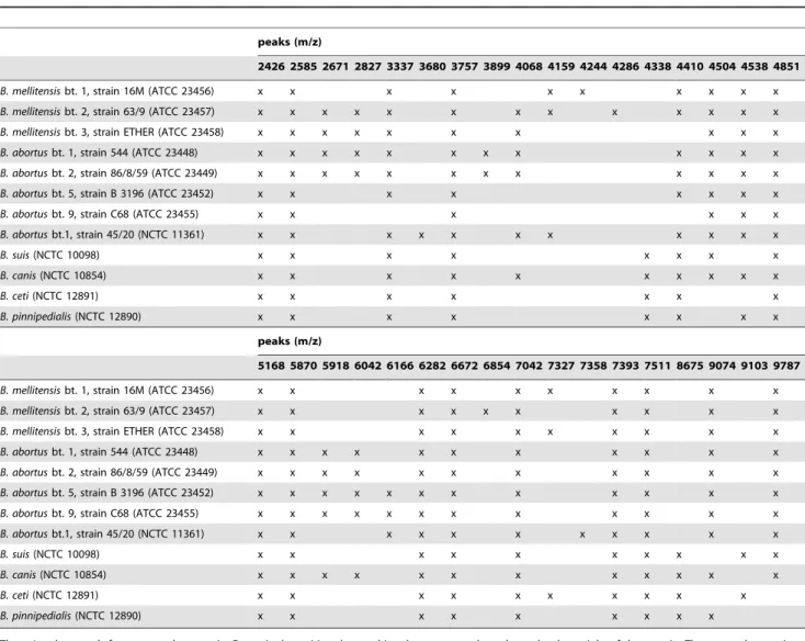
MALDI Biotyper 2.0 database (Bruker Daltonics) used for routine identification of microorganism in clinical microbiology contains 3,476 entries, involving bacteria and fungi groups. Although it contains a wide variety of clinically relevant microorganisms, some important pathogenic microorganisms have not been yet included (e.g.Brucella). This is an important limitation in some countries, where brucellosis is still a relevant infectious problem and it is not infrequent to isolate Brucella species from febrile patients. For this reason, 3B. melitensis, 5B. abortus, 1B. suis, 1B. canis, 1B. cetiand 1B. pinnipedialistype strains were used to generate reference spectra and to extend the MALDI Biotyper database. The visual inspection of these spectra from whole-cell extracts revealed a high similarity among them (Figure 1), with specific peaks commons for all the strains at 2426 Da, 2585 Da, 3757 Da, 4851 Da, 5168 Da, 5870 Da, 6282 Da, 6672 Da, 7042 Da, 7393 Da, and 7511 Da (Table 1). Although some differences can be observed among all the strains, only some strains have discriminating peaks at species level. Thus, peaks at 4244, 4286 and 6854 Da appear only inB. melitensis, but none of these peaks are constant in all the B. melitensisstrains. The same happens with peaks at 3899, 6166 and 7358 Da in B. abortus. According these data, there is a peak profile characteristic of
Brucellaat genus level, but there isn’t at species level.
A cluster analysis with the 12 type strains was performed, using the integrated tools of the MALDI Biotyper 2.0 software package. The resulting dendrogram (Figure 2) shows a close proximity betweenB. ceti and B. pinnipedialis, and betweenB. suisandB. canis, and more proximity between these four species than between any of them andB. melitensisandB. abortus. Otherwise,B. melitensisand
B. abortusbiotypes are extremely close and interrelated, excepting
B. abortusbiotype 1 strain 544 (ATCC 23448).
In addition, we tested the influence of different culture conditions. Therefore the different strains were cultivated on
blood agar and chocolate agar and comparison between identification and scores obtained did not reveal any significant difference between both culture media in any aspect (data not shown).
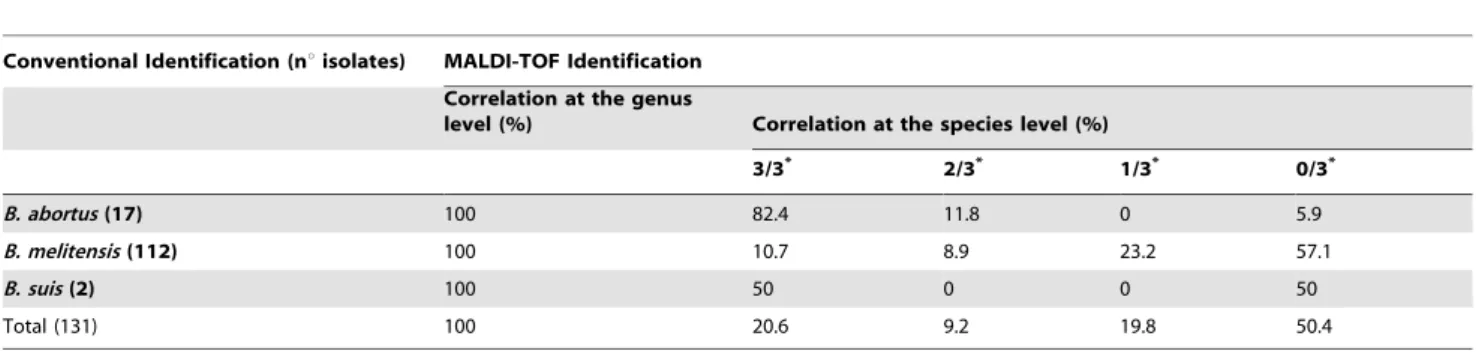
Once established the spectra for the Brucellareference strains, 131 blind-codedBrucellastrains were matched against the MALDI Biotyper 2.0 database in triplicate, to prove its suitability for routine identification and discrimination of Brucellaat the genus and species levels (Table 2). The MALDI-TOF device reported the same identification at species level for the three replicates in 14 out of 17B. abortusisolates (82.4%), but in only 12 out of 112B. melitensisisolates (10.7%) and in 1 out of 2B. suisisolates (50%). Therefore only 20.6% isolates reported the same identification at species level for the three replicates. Nevertheless, in all of them this identification was coincident with conventional and genetic identifications at genus level. In all the clinical isolates tested, the score difference between the ten most probably matched species reported by the MALDI Biotyper software for each isolate was very low and higher than 2 in several of them. As an example, we show in Table 3 the score difference between the ten most probably matched species reported for one B. melitensis clinical isolate. Taking into account that a reliable identification at species level is accepted with scores higher than 2, we can’t discriminate between different Brucella species, since different Brucella species give scores higher than 2 in most clinical isolates.
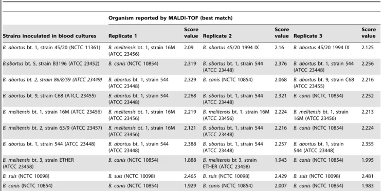
To test the reliability of MALDI-TOF MS for identifying brucellae directly from blood culture, we inoculated blood culture bottles with type strains belonging to the four main human pathogenic species (B. melitensis,B. abortus,B. suisandB. canis). All the blood cultures inoculated were reported as positive by 48 hours of incubation. Three replicates of each positive blood culture were studied using MALDI-TOF MS. All the blood cultures were reported by the MALDI-TOF as Brucella. Never-theless, in several cases the identification did not match with the organism inoculated in the blood culture at the species level, or different identifications were obtained at the species level among the three replicates of the same blood culture (Table 4).
Discussion
Brucellosis remains a serious problem in wide areas around the world. Symptoms are nonspecific (fever, malaise, back pain, profuse night sweating). This lack of specificity of symptoms may delay the diagnosis for weeks. Though mortality rate is currently low, it remains a severe disease, and complications such as epididymo-orchitis, arthritis (especially sacroiliitis), and CNS complications are not infrequent. The current taxonomy of this genus is confusing because traditional, phenotype-based classification, and genetic-based classification are used simultaneously [5]. Genetic taxonomic studies have suggested that the 8 currently accepted species, including the 2 marine mammal-associated species, represent a single species [6,13]. Nevertheless, recent studies show that traditional species, and the marine mammals-associated species, can be differentiated by outer membrane genes polymorphism, insertion sequences and whole-chromosome preparations [5,14].
Human brucellosis has been described associated toB. melitensis, B. abortus, B. suis and B. canis, among the traditional species. Human cases associated to the newer marine mammals-associated species have also been reported [15,16]. ThoughB. abortushas the broadest geographical distribution, most human cases are now caused by B. melitensis, which usually produces the most severe disease.
marrow, are usually the specimens for diagnosis of human brucellosis. Conventional blood cultures did not yield satisfactory results, and this led to the development of the classic biphasic blood cultures technique proposed by Ruiz-Castan˜eda. Now, both lyses-centrifugation methods and continuous monitoring blood cultures devices are able to detectBrucellawith a good sensitivity [7].
Early suspicion that an isolate might be aBrucellais important, both for clinical and epidemiological reasons, because of the hazard of laboratory-acquired brucellosis, and for the early detection of a hypothetical bioterrorist attack. Early suspicion may be currently established based on Gram staining, but final identification requires colonies growth on agar plates and performing biochemical or serological tests on these colonies.
For all those reasons, the availability of methods that allow a rapid and reliableBrucellaidentification both from agar plates and directly from the blood culture bottles, once this is reported as positive by the continuous-monitoring blood culture systems,
would be extremely useful. Direct detection methods based on PCR have been described [17], but these methods are expensive, can be affected by PCR inhibitors present in blood, and do not give any information about the viability of the microorganisms. Thus, they have not reached a wide diffusion by now.
MALDI-TOF MS is an important and increasingly available tool in clinical microbiology laboratories, because it allows a rapid and accurate identification of bacteria [10,11]. We have seen that MALDI-TOF MS allows a fast and highly reliable identification of
Brucella, at genus level, from colonies growth. Nevertheless, identification at species level is less reliable. Data in Figure 1 and Table 1 show a common profile for allBrucellatype strains tested, but no specific peaks are found in any species. This explains the reliability at genus level and the low reliability at species level. Otherwise, the high similarity between all type strains is not surprising, since genetic taxonomic studies suggest that the 6 currently accepted species represent a single species [6,13]. Once
Table 1.Discriminating peaks ofBrucellastrains and biotypes analyzed by MALDI-TOF MS.
peaks (m/z)
2426 2585 2671 2827 3337 3680 3757 3899 4068 4159 4244 4286 4338 4410 4504 4538 4851
B. mellitensisbt. 1, strain 16M (ATCC 23456) x x x x x x x x x x
B. mellitensisbt. 2, strain 63/9 (ATCC 23457) x x x x x x x x x x x x x
B. mellitensisbt. 3, strain ETHER (ATCC 23458) x x x x x x x x x x
B. abortusbt. 1, strain 544 (ATCC 23448) x x x x x x x x x x x x
B. abortusbt. 2, strain 86/8/59 (ATCC 23449) x x x x x x x x x x x x
B. abortusbt. 5, strain B 3196 (ATCC 23452) x x x x x x x x
B. abortusbt. 9, strain C68 (ATCC 23455) x x x x x x
B. abortusbt.1, strain 45/20 (NCTC 11361) x x x x x x x x x x x
B. suis(NCTC 10098) x x x x x x x x
B. canis(NCTC 10854) x x x x x x x x x x
B. ceti(NCTC 12891) x x x x x x x
B. pinnipedialis(NCTC 12890) x x x x x x x x
peaks (m/z)
5168 5870 5918 6042 6166 6282 6672 6854 7042 7327 7358 7393 7511 8675 9074 9103 9787
B. mellitensisbt. 1, strain 16M (ATCC 23456) x x x x x x x x x x
B. mellitensisbt. 2, strain 63/9 (ATCC 23457) x x x x x x x x x x
B. mellitensisbt. 3, strain ETHER (ATCC 23458) x x x x x x x x x x
B. abortusbt. 1, strain 544 (ATCC 23448) x x x x x x x x x x x
B. abortusbt. 2, strain 86/8/59 (ATCC 23449) x x x x x x x x x x x
B. abortusbt. 5, strain B 3196 (ATCC 23452) x x x x x x x x x x x x
B. abortusbt. 9, strain C68 (ATCC 23455) x x x x x x x x x x x x
B. abortusbt.1, strain 45/20 (NCTC 11361) x x x x x x x x x x x
B. suis(NCTC 10098) x x x x x x x x x x
B. canis(NCTC 10854) x x x x x x x x x x x x
B. ceti(NCTC 12891) x x x x x x x x x x
B. pinnipedialis(NCTC 12890) x x x x x x x x x
The m/z value stands for mass to charge ratio. For a single positive charge, this value corresponds to the molecular weight of the protein. The mass tolerance is considered62 Da for each peak.
doi:10.1371/journal.pone.0014235.t001
Figure 1. MALDI-TOF MS spectra of whole-cell extracts ofBrucellareference strains in the range from 2000 to 11000 Da.The relative intensity of the ions (a.u., arbitrary units) are shown on theyaxis, and the masses (in Da) of the ions are shown on thexaxis. Them/zvalue stands for mass to charge ratio. For a single positive charge, this value corresponds to the molecular weight of the protein.
the protein profiles forB. melitensis,B. abortus,B. suis,B. canis,B. ceti
andB. pinnipedialishad been created and included in the database, all the blind-codedBrucellaisolates tested were correctly identified, with scores.2. Scores differences between the 10 most probably matched species given by the MALDI Biotyper software for each isolate were extremely low, and replicates of the same protein extract were sometimes identified as different species. In almost all isolates, more than one species matched with score value.2 in the
10 most probably matched species, thus confirming the low discriminating power at species level. The agar on which the microorganism grows does not seem to affect to identification, since scores obtained from blood agar and chocolate agar were similar for all the isolates tested. Previous studies on other microorganisms agree with these results, showing that MALDI-TOF MS protein fingerprints are not significantly influenced by variability in growth conditions [18–20].
Figure 2. Cluster analysis of MALDI-TOF MS spectra ofBrucellastrains and biotypes.Distance is displayed in relative units. doi:10.1371/journal.pone.0014235.g002
Table 2.Identification by MALDI-TOF mass spectrometry and conventional identification of 131 blind-codedBrucella.
Conventional Identification (nuisolates) MALDI-TOF Identification
Correlation at the genus
level (%) Correlation at the species level (%)
3/3* 2/3* 1/3* 0/3*
B. abortus(17) 100 82.4 11.8 0 5.9
B. melitensis(112) 100 10.7 8.9 23.2 57.1
B. suis(2) 100 50 0 0 50
Total (131) 100 20.6 9.2 19.8 50.4
Each strain was spotted three times (replicates 1, 2 and 3). NNo. of replicates.
MALDI-TOF MS has been previously shown to be able to identify microorganisms directly from blood cultures reported as positive by automatic blood culture processing devices [21–25]. In our study, positive blood cultures spiked with brucellae were reported always asBrucella, but species failures were frequent, as had been shown previously in agar plates. Brucella was always identified with scores.2 but, as happens in agar cultures, score
differences between the ten most probably matched species reported by the MALDI Biotyper software for each isolate were very low, and species identification had scores.2 in the best and second match, although the species in both cases are different, indicating that the species identification cannot be fully reliable.
Protein profiles found for type strains, show that protein profile similarity does not correlate always with the traditional genus/
Table 3.Ten best matches for each replicate of a blind-coded clinical isolate.
Replicate 1 Replicate 2 Replicate 3
Matched Pattern Score Matched Pattern Score Matched Pattern Score
Value Value Value
1 Brucella melitensisbt 1, strain 16M (ATCC 23456)
2.293 Brucella abortusbt 9, strain C68 (ATCC 23455)
2.229 Brucella melitensisbt 1, strain 16M (ATCC 23456)
2.204
2 Brucella abortusbt 2, strain 86/8/59 (ATCC 23449)
2.161 Brucella melitensisbt 1, strain 16M (ATCC 23456)
2.204 Brucella melitensisbt 2, strain 63/9 (ATCC 23457)
2.11
3 Brucella abortusbt 1, strain 544 (ATCC 23448)
2.158 Brucella abortusbt 5, strain B3196 (ATCC 23452)
2.204 Brucella abortusbt 2, strain 86/8/59 (ATCC 23449)
2.092
4 Brucella abortusbt 9, strain C68 (ATCC 23455)
2.148 Brucella abortusbt 1, strain 544 (ATCC 23448)
2.166 Brucella abortusbt 1, strain 544 (ATCC 23448)
2.087
5 Brucella abortusbt 5, strain B3196 (ATCC 23452)
2.138 Brucella abortusbt 2, strain 86/8/59 (ATCC 23449)
2.083 Brucella abortusbt 5, strain B3196 (ATCC 23452)
2.064
6 Brucella abortus45/20 (NCTC 11361) 2.125 Brucella melitensisbt 2, strain 63/9 (ATCC 23457)
2.043 Brucella abortus45/20 (NCTC 11361) 2.034
7 Brucella melitensisbt 2, strain 63/9 (ATCC 23457)
2.124 Brucella abortus45/20 (NCTC 11361) 1.985 Brucella abortusbt 9, strain C68 (ATCC 23455)
2.006
8 Brucella canis(NCTC 10854) 2.062 Brucella suis(NCTC 10098) 1.908 Brucella suis(NCTC 10098) 1.898 9 Brucella suis(NCTC 10098) 2.056 Brucella canis(NCTC 10854) 1.902 Brucella canis(NCTC 10854) 1.867 10 Brucella melitensisbt 3, strain ETHER
(ATCC 23458)
1.919 Brucella ceti(NCTC 12891) 1.847 Brucella melitensisbt 3, strain ETHER (ATCC 23458)
1.743
This isolate was identified asB. melitensisby conventional and genetic methods. doi:10.1371/journal.pone.0014235.t003
Table 4.MALDI Biotyper 2.0-based identification of blood cultures spiked with differentBrucellaspecies and biotypes.
Organism reported by MALDI-TOF (best match)
Strains inoculated in blood cultures Replicate 1
Score
value Replicate 2
Score
value Replicate 3
Score value
B. abortusbt. 1, strain 45/20 (NCTC 11361) B. melitensisbt. 1, strain 16M (ATCC 23456)
2.09 B. abortus45/20 1994 IX 2.16 B. abortus45/20 1994 IX 2.125
B.abortusbt. 5, strain B3196 (ATCC 23452) B. canis(NCTC 10854) 2.319 B. abortusbt. 1, strain 544 (ATCC 23448)
2.376 B. abortusbt. 1, strain 544 (ATCC 23448)
2.256
B. abortus bt. 2, strain 86/8/59 (ATCC 23449) B. abortusbt. 1, strain 544 (ATCC 23448)
2.329 B. canis(NCTC 10854) 2.068 B. abortusbt. 9, strain C68 (ATCC 23455)
2.216
B. abortusbt. 9, strain C68 (ATCC 23455) B. abortusbt. 1, strain 544 (ATCC 23448)
2.268 B. abortusbt. 1, strain 544 (ATCC 23448)
2.321 B. canis(NCTC 10854) 2.252
B. melitensisbt. 1, strain 16M (ATCC 23456) B. melitensisbt. 1, strain 16M (ATCC 23456)
2.219 B. melitensisbt. 1, strain 16M (ATCC 23456)
2.224 B. melitensisbt. 1, strain 16M (ATCC 23456)
2.213
B. melitensisbt. 2, strain 63/9 (ATCC 23457) B. melitensisbt. 1, strain 16M (ATCC 23456)
2.121 B. abortusbt. 1, strain 544 (ATCC 23448)
2.216 B. canis(NCTC 10854) 2.224
B. abortusbt. 1, strain 544 (ATCC 23448) B. abortusbt. 1, strain 544 (ATCC 23448)
2.388 B. abortusbt. 1, strain 544 (ATCC 23448)
2.257 B. abortusbt. 1, strain 544 (ATCC 23448)
2.355
B. melitensisbt. 3, strain ETHER (ATCC 23458)
B. canis(NCTC 10854) 1.888 B. melitensisbt 3, strain ETHER (ATCC 23458)
1.943 B. canis(NCTC 10854) 1.995
species/biotype classification. Proteins profile for some species and biotypes may be closer to other species than to other biotypes belonging to the same species, as can be observed in cluster analysis (Figure 2).
In summary, the protein profiles for type strains ofB. melitensis
biotypes 1, 2 and 3;B. abortusbiotypes 1, 2, 5 and 9;B. suis;B. canis;
B. ceti and B. pinnipedialis were generated and included in the MALDI Biotyper database. The study of clinical isolates both from agar plate cultures and directly from blood cultures showed that these profiles allow a reliable identification of brucellae to genus
level, stating that MALDI-TOF MS is an fast and highly reliable technique for straightforward Brucella identification, both from culture plates and directly from blood culture vials.
Author Contributions
Conceived and designed the experiments: LF JMGB JLMB. Performed the experiments: LF SVC FSJ SGC FM. Analyzed the data: LF FSJ JMGB JLMB. Contributed reagents/materials/analysis tools: SGC FM AOD JMGB JLMB. Wrote the paper: LF JMGB JLMB.
References
1. za J, Gudiol F, Pallares R, Viladrich PF, Rufi G, et al. (1992) Treatment of human brucellosis with doxycycline plus rifampicin or doxycycline plus streptomycin. A randomized, double-blind study. Ann Intern Med 17: 25–30. 2. Solera J, Rodrı´guez-Zapata M, Geijo P, Largo J, Paulino J, et al. (1995)
Doxycycline–rifampin versus doxycycline–streptomycin in treatment of human brucellosis due toBrucella melitensis.The GECMEI Group. Grupo de Estudio de Castilla-la Mancha de Enfermedades Infecciosas. Antimicrob Agents Che-mother 39: 2061–2067.
3. Colmenero JD, Reguera JM, Martos F, Sa´nchez de Mora D, Delgado M, et al. (1996) Complications associated withBrucella melitensisinfections: a study of 530 cases. Medicine (Baltimore) 75: 195–211.
4. Ariza J, Corredoira J, Pallares R, Villadrich PF, Rufi G, et al. (1995) Characteristics of and risk factors for relapse of brucellosis in humans. Clin Infect Dis 20: 1241–1249.
5. Lindquist D, Chu MD, Probert WS (2007)FrancisellaandBrucella. In: Murray PR, Baron EJ, Jorgensen JH, Landry ML, Pfaller MA, eds. Manual of Clinical Microbiology (9th
ed.). Washington, DC: American Society for Microbiology. pp 815–834.
6. Verger JM, Grimont F, Grimont PA, Grayon M (1987) Taxonomy of the genus Brucella. Ann Inst Pasteur Microbiol 138: 235–238.
7. Yagupsky P, Peled N, Press J, Abramson O, Abu-Rashid M (2003) Comparison of BACTEC 9240 Peds Plus medium and isolator 1.5 microbial tube for detection of Brucella melitensis from blood cultures. J Clin Microbiol 35: 1382–1384.
8. Elsaghir AAF, James EA (2003) Misidentification of Brucella melitensis as
Ochrobactrum anthropiby API 20NE. J Med Microbiol 52: 441–442.
9. Queipo-Ortun˜o MI, Colmenero JD, Baeza G, Morata P (2005) Comparison between LightCycler Real-Time Polymerase Chain Reaction (PCR) assay with serum and PCR-enzyme-linked immunosorbent assay with whole blood samples for the diagnosis of human brucellosis. Clin Infect Dis 40: 260–264. 10. Seng P, Drancourt M, Gouriet F, La Scola B, Fournier PE, et al. (2009) Ongoing
revolution in bacteriology: routine identification of bacteria by matrix-assisted laser desorption ionization time-of-flight mass spectrometry. Clin Infect Dis 49: 543–551.
11. Ferreira L, Vega S, Sa´nchez-Juanes F, Gonza´lez M, Herrero A, et al. (2010) Identifying bacteria using a matrix-assisted laser desorption ionization time-of-flight (MALDI-TOF) mass spectrometer. Comparison with routine methods used in clinical microbiology laboratories. Enferm Infecc Microbiol Clin 28: 492–497.
12. Garcia-Yoldi D, Marin CM, de Miguel MJ, Mun˜oz PM, Vizmanos JL, et al. (2006) Multiplex PCR assay for the identification and differentiation of all Brucellaspecies and the vaccine strainsBrucella abortusS19 and RB51 andBrucella melitensis.Rev Clin Chem 52: 779–781.
13. Bricker BJ, Ewalt DR, MacMillan AP, Foster G, Brew S (2000) Molecular characterization of Brucella strains isolated from marine mammals. J Clin Microbiol 38: 1258–1262.
14. Vizcaı´no N, Cloeckaert A, Verger J, Grayon M, Ferna´ndez-Lago L (2000) DNA polymorphism in the genusBrucella. Microbes Infect 2: 1089–1100.
15. Brew SD, Perrett LL, Stack JA, MacMillan AP, Staunton NJ (1999) Human exposure toBrucellarecovered from a sea mammal. Vet Rec 144: 483. 16. Sohn AH, Probert WS, Glaser CA, Gupta N, Bollen AW, et al. (2003) Human
neurobrucellosis with intracerebral granuloma caused by a marine mammal Brucellaspp. Emerg Infect Dis 9: 485–488.
17. Navarro E, Casao MA, Solera J (2004) Diagnosis of human brucelosis using PCR. Expert Rev Mol Diagn 4: 115–123.
18. Seibold E, Maier T, Kostrzewa M, Zeman E, Splettstoesser W (2010) Identification of Francisella tularensis by Whole-Cell Matrix-Assisted Laser Desorption Ionization–Time of Flight Mass Spectrometry: Fast, Reliable, Robust, and Cost-Effective Differentiation on Species and Subspecies Levels. J Clin Microbiol 48: 1061–1069.
19. Barbuddhe SB, Maier T, Schwarz G, Kostrzewa M, Hof H, et al. (2008) Rapid identification and typing of listeria species by matrix-assisted laser desorption ionization-time of flight mass spectrometry. Appl Environ Microbiol 74: 5402–5407.
20. Mellmann A, Cloud J, Maier T, Keckevoet U, Ramminger I, et al. (2008) Evaluation of matrix-assisted laser desorption ionization-time-of-flight mass spectrometry in comparison to 16S rRNA gene sequencing for species identification of nonfermenting bacteria. J Clin Microbiol 46: 1946–1954. 21. Ferreira L, Sa´nchez-Juanes F, Guerra IP, Garcı´a Garcı´a MI, Sa´nchez JE, et al.
(2010) Microorganisms direct identification from blood culture by MALDI-TOF mass spectrometry. Clin Microbiol Infect Apr 28. [Epub ahead of print]. 22. Christner M, Rohde H, Wolters M, Sobottka I, Wegscheider K, et al. (2010)
Rapid identification of bacteria from positive blood culture bottles by use of matrix-assisted laser desorption-ionization time of flight mass spectrometry fingerprinting. J Clin Microbiol 48: 1584–1591.
23. Prod’hom G, Bizzini A, Durussel C, Bille J, Greub G (2010) Matrix-assisted laser desorption ionization-time of flight mass spectrometry for direct bacterial identification from positive blood culture pellets. J Clin Microbiol 48: 1481–1483.
24. Stevenson LG, Drake SK, Murray PR (2010) Rapid identification of bacteria in positive blood culture broths by matrix-assisted laser desorption ionization-time of flight mass spectrometry. J Clin Microbiol 48: 444–447.