http://dx.doi.org/10.1590/s2175-97902017000417039
Article
*Correspondence: R. Benedetto. Centro de Radiofarmácia, Instituto de Pesquisas Energéticas e Nucleares – IPEN. Av. Prof. Lineu Prestes, 2242, Cidade Universitária, CEP: 05508-000 - São Paulo - SP – Brasil. E-mail: benedettoraquel@yahoo.com.br
Development of radioimmunoconjugate for diagnosis and
management of head-and-neck subclinical cancer and colorectal
carcinoma
Raquel Benedetto
1*, Adriana Vidal Fernandes Massicano
1, Jefferson Jesus Silva
1,
Cristian Antonio Wieczorek Villas Boas
1, Jair Mengatti
1, Elaine Bortoleti de Araújo
11Radiopharmacy Center, IPEN - Institute of Energy and Nuclear Research, São Paulo, Brazil
Scientific innovations in diagnostic methods are important drivers of cancer control and prevention. Noninvasive imaging of the epidermal growth factor receptor (EGFR) in head-and-neck squamous, cell carcinoma and colorectal cancer could be valuable to select patients for EGFR-targeted therapy, as well as to monitor the efficacy and occurrence of resistance to immunotherapy. In order to develop the first Brazilian radioimmunoconjugate for diagnosis, Cetuximab has been conjugated to p-SCN-Bn-DTPA chelator and radiolabeled with Indium-111. The conjugation methodology was optimized using different mAb:DTPA molar ratios, time was then reduced for immunoconjugate preparation, besides the protein recovery’ percentage increased after purification (m = 83.8 ± 0.91 %). The stability of Cetuximab-DTPA at – 20 oC was evaluated for six months, and its integrity was greater than 90% (m =93.9 ± 1.5%, N = 24). The radioimmunoconjugate with specific activity of 185 MBq/mg showed radiochemical purity above 95% (m=96.8 ± 1.31 %, N = 15). We conclude that the radioimmunoconjugate 111In-DTPA-cetuximab is stable and may be applied to the diagnosis of EGFR-positive tumors.
Keywords: Radioimmunoconjugate/development. Head and neck cancer/diagnosis. Tumors diagnosis. Cetuximab. SPECT. 111In.
INTRODUCTION
The longevity of the population has contributed to the development of chronic-degenerative diseases, including malignant neoplasm. Colorectal cancer (CRC)
is one of the most common malignant tumors in the
world. According to the latest global estimate/study, CRC is configured as the third most common type of cancer among men and the second in women (INCA, 2014). In accordance with the World Health Organization estimate/ study, head-and-neck squamous cell carcinoma (HNSCC) is the fifth most common cancer worldwide, with an overall annual rate/occurrence of 980,000 new cases.
Site-specific delivery of therapeutic agents has been an ultimate goal of the pharmaceutical industry in order to maximize drug action and minimize side effects (Mccarron
et al., 2005). Targeted therapy is becoming an increasingly
important component in the treatment of cancer (Hoeben et al., 2011). In this scenario, monoclonal antibodies (mAbs)
have been highlighted due to their success in therapeutic application.
The epidermal growth factor receptor (EGFR) is a tyrosine kinase receptor and it is overexpressed in the majority of epithelial malignancies, such as non-small cell lung cancer (NSCLC), epithelial ovarian cancer, including head-and-neck cancer (Hoeben et al., 2011).
Furthermore, EGFR overexpression is associated with advanced tumor stage and poor prognosis in several epithelial cancer types. EGFR plays an important role in cellular proliferation, DNA repair and regulation responses to hypoxia - through hypoxia-inducible factor 1 (HIF-1), hypoxia signaling promotes angiogenesis and metastasis by inducing epithelial-mesenchymal-transition (EMT) (Wiechec et al., 2017).
combination with: (a) chemotherapy for treatment of colorectal and head-and-neck squamous cell carcinoma or (b) external radiotherapy for treatment of head-and-neck squamous cell carcinoma (Sihver et al., 2014).The
conjugation of radionuclides to cetuximab, in combination
with the specific targeting properties of this antibody, has been performed for imaging. Noninvasive imaging of EGFR in head-and-neck squamous cell carcinoma could be of value to select patients for EGFR-targeted therapy (Hoeben et al., 2011). Molecular imaging (MI),
especially single photon emission computed tomography (SPECT) and positron emission tomography (PET), is a powerful tool for detecting cancer and for characterizing the biological properties of tumors throughout the body (Reilly et al., 2015).
Considering the importance of Cetuximab in the immunotherapy of colon cancer and head-and-neck carcinoma, several mAb labeling studies with different radionuclides have been developed to (1) produce a specific radiopharmaceutical for selecting appropriate candidates for therapy, (2) monitor the effectiveness of immunotherapy, and (3) evaluate drug resistance (Sihver
et al., 2014). Cetuximab has been labeled with different
radionuclides for diagnosis for instance technetium-99m
(Schechter et al., 2004), gallium-68 (Evans et al., 2014)
and zirconium-89 (Van de Watering et al., 2014). For
therapy, lutetium–177 (Jalilian et al., 2016) and yttrium-90
(Niu et al., 2010) have been used. Ping et al. (2008)
reported a preclinical study related to the use of cetuximab as a PET agent, directed to tumors overexpression` EGFR. Cetuximab was conjugated with DOTA chelator (1,4,7,10 - tetraazacyclododecane tetraacetic acid), for radiolabeling with copper-64 (T1/2 = 12.7 hours).
Indium-111 is primarily a gamma emitter, and a cyclotron-produced radiometal and one of the most commonly used radionuclides for SPECT, especially as a label for mAbs, due to its adequate physical half-life (2.8 d). Thus, pre-clinical and clinical studies have been investigated with 111In-labeled mAb conjugates (Sihver
et al., 2014).
Indium-111 radiolabeled Cetuximab was applied in a clinical phase I study in patients with inoperable lung squamous cell carcinoma, which express high levels of EGF receptors. No toxicity was observed. (Divgi et al., 1991).
Others mAbs have been labeled with indium-111 and intended in clinical studies (Doerr et al., 1990, Morris et al., 2005, Perik et al, 2006, Scott et al., 2007). Indium-111
radiolabeled Pertuzumab (111In-BzDTPA-Pertuzumab)
was used to evaluate breast cancer, which responds to the treatment with trastuzumab (Herceptin), in patients with HER2-positive metastatic disease. The clinical images
showed good tumor uptake in hepatic metastases 48 hours after injection of 111 MBq (5 mg) of 111
In-BzDTPA-Pertuzumab. Furthermore, the study revealed no serious adverse effects (Reilly et al., 2015).
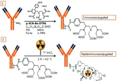
The production of radiolabeled monoclonal antibodies is a process that involves several steps (Cooper, Sabbah, Mather, 2006): (1) pre-purification of mAb to remove free metals; (2) conjugation of mAb with chelating groups; (3) immunoconjugate purification and concentration; (4) and radiolabeling.
Considering several steps and many different variables, it was imperative for our laboratory to establish appropriated working conditions to produce 111
In-DTPA-Cetuximab with high radiochemical yield and stability. In an unprecedented way, this research evaluated the influence of the specific activity (MBq/mg) on the radiochemical purity and stability of the preparation. The specific activity is an important property for specific target-labeled radiopharmaceuticals, since the untarget-labeled antibody will compete with the radiopharmaceutical for the same receptors. On the other hand, high specific activity could increase the effect of radiolysis on antibody structure.
I n m o s t c o u n t r i e s , t h e p r e p a r a t i o n o f radioimmunoconjugates is performed “in house”, therefore, radiopharmaceuticals for radioimmunodiagnostic (RID) and radioimmunotherapy (RIT) are prepared and applied at the same clinical imaging center. Currently, the production of these radiopharmaceuticals in Brazil
is carried out by the Institute of Energy and Nuclear
Research (IPEN-CNEN), in São Paulo, which guarantees
the radiopharmaceutical’s quality and stability during their transport and distribution to the clinical research centers. Therefore, the stability of the radiolabeled mAb was evaluated, in order to determine the radiopharmaceutical expiration date, and the optimal conditions for storage, that would allow the radioimmunoconjugate to be transported to different sites for future clinical studies.
Considering the first experience in Brazil, this study presents the procedures for 111In-DTPA-Cetuximab
preparation. We aimed to optimize the conjugation process to obtain a radioimmunoconjugate with high radiochemical yield and stability for future pre-clinical and clinical applications.
MATERIAL AND METHODS
Analysis of the structural integrity of the antibody
(Laemmli, 1970) in order to verify the integrity of the antibody. The electrophoresis was performed in a BioRad system, using a 4-20% Tris-Glycine gel. Then the antibody was diluted in non-reducing Laemmli buffer and the gel was run at 200V for 30 minutes. Afterwards, the gel was analyzed using a Packard instant image electronic autoradiography.
Conjugation of Cetuximab with p-SCN-Bn-DTPA
The production of radiolabeled monoclonal antibodies involves several steps which have been summarized in Figure 1. Cetuximab was conjugated to p-isothiocyanatobenzyl-diethylenetriaminepentaacetic acid (p-SCN-Bn-DTPA, Macrocyclis, Dallas, TX) in 0.2 M sodium bicarbonate pH 8.5 for 1 hour at room temperature, using 10 and 20 times molar excess of DTPA.
Purification of the immunoconjugate
The conjugation process was carried out with excess chelator which must be removed after conjugation, while avoiding interference with the radiolabeling process. After
the conjugation time, 111In-DTPA-Cetuximab was purified
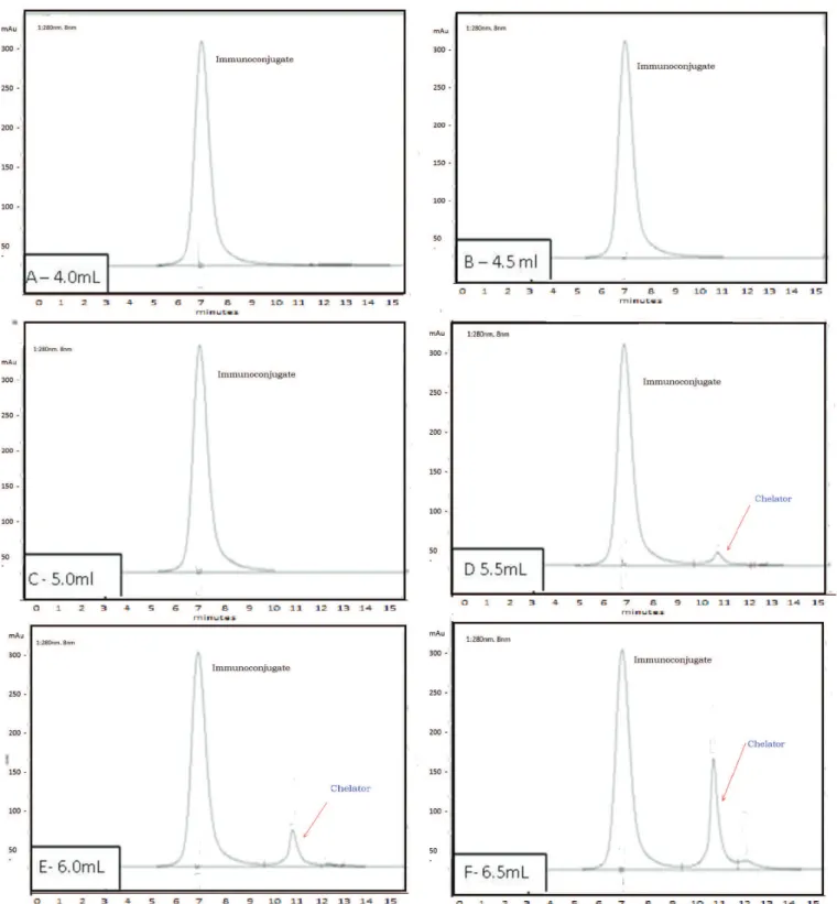
by molecular exclusion gel column (PD-10 column - GE Healthcare Bio-Sciences, Pittsburgh, USA). The column was equilibrated with 50 mL of 0.25 M ammonium acetate buffer pH 6.5. Ten aliquots of 0.5 mL were collected and the absorbance of each sample was analyzed in a spectrophotometer (160 Evolution - Thermo Scientific, USA, 280 nm). All fractions were subjected to analysis by high-performance liquid chromatography (HPLC) (Shimadzu BioSep SEC S-3000 column x 3007.8 mm5 µm, 1 mL/min flow, mobile phase 0.1M sodium phosphate buffer pH 7.0, 15 minute), with UV detection (280 nm), to
examine the structural integrity of the immunoconjugate.
Substances with higher molecular weight are first eluted in
a molecular exclusion column, thus, the immunoconjugate
has lower retention time (RT) when compared with the unconjugated antibody. The fractions that contained the pure immunoconjugate were selected and subsequently combined in an ultrafiltration concentrator tube (Amicon® Ultra 10,000 MWCO, Millipore, USA) (Mikro 220, Hettich Germany). Statistical analysis was performed using Prism software (GraphPad Software).
Determination of protein concentration of the immunoconjugate
The protein concentration of the purified immunoconjugate was determined by the bicinchoninic acid method (BCA), using Albumin standard 5mg/ml (Thermo Scientific, EUA), as previously described (Brady
et al., 2004).
Determination of the number of chelates per antibody by the spectrophotometric method
The number of chelates per antibody, was determined in purified immunoconjugate by the mass spectrometry method (MALDI / TOF) (Siegel et al., 1991), using
spectrophotometer Bruker Autoflex Speed (CEFAP-ICB/ USP). The molecular weight of the immunoconjugate, in different molar ratios, was obtained and the molecular weight of the antibody, equivalent to 150,000 g/mol, was subtracted. The difference corresponds to the chelating mass incorporated into Cetuximab. The chelating mass was divided by the molecular weight of the chelator (DTPA: 649.9 g/mol) and the result was expressed in a number of chelators coupled to the monoclonal antibody
molecule.
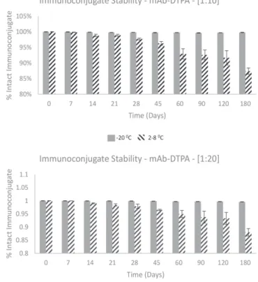
Stability of the Immunoconjugate
Aliquots of 20 µL of Cetuximab-DTPA(5 mg/mL),
conjugated at molar ratios 10:1 and 20:1, were fractionated in 1.5 mL microcentrifuge tubes (Eppendorf, Brazil) and
stored at 2 - 8 °C and - 20 °C. An aliquot was taken weekly
for analysis by HPLC, using the same system as described
above.
Radiolabeling development and analysis of specific activity
Cetuximab-DTPA (5 mg/mL) was radiolabeled with different 111-Indium activities (0.185 MBq/µL - 3.7 MBq/
µL) (Nordion/Canada) in 0.25 M ammonium acetate buffer
pH 6.5, the reaction time was 30 minutes at 42 °C (Lewis,
Raubitschek, Shively, 1994).
The radioimmunoconjugates were stored under refrigeration (2-8 °C) for 48 hours and radiochemical
purity was determined by CCD using 50 mM EDTA and 0.1 M ammonium acetate buffer pH 5.5 - 6.0 as a mobile phase and then a glass fiber plate was impregnated with silica gel (ITLC-SG, Merck, Germany) as stationary phase. The distribution of radioactivity in the chromatographic plate was performed by radiochromatography (radio-TLC Imaging Scanner AR-2000, Eckert & Ziegler / USA) (Chang, Smith, Lapi, 2013). The radiochemical purity was
also evaluated by HPLC (Agilent, detector UV 190-300 nm) (Cooper, Sabbah, Mather, 2006; Chang, Smith, Lapi,
2013), using the same methodology as described above.
Analysis of the stability of radiolabeled Cetuximab in human serum
The radioimmunoconjugate (18.5 MBq) was added to 1 mL of human serum. To obtain the human serum, 10 mL of blood was collected, without coagulant, from a healthy donor. The sample was then centrifuged at 1400 rpm for 10 minutes. After centrifugation, the fibrin clot was removed and a new centrifugation was performed to separate the serum. The samples were incubated at 37
°C, 350 rpm for 72 hours. After 1, 24, 48 and 72 hours
of incubation (Lewis, Raubitschek, Shively, 1994), the aliquots were analyzed by CCD as previously described.
Binding of the radiolabeled Cetuximab to serum protein in vitro
To determine the 111In-DTPA-Cetuximab binding
to serum proteins, 18.5 MBq of radiolabeled mAb was added to 1 mL of human serum in triplicate. After each incubation period, 1, 24, 48 and 72 hours, an aliquot (200 µL) was withdrawn and ethanol was added (1:1 v/v) to precipitate proteins. The sample was centrifuged at 10,000 rpm and the precipitate and supernatant radioactivity was determined using a gamma counter (D5002 Cobra II, Canberra Packard, USA) (Lewis, Raubitschek, Shively, 1994). The percent of serum protein binding was calculated (radioactivity of precipitate/radioactivity of precipitate + radioactivity of supernatant).
RESULTS AND DISCUSSION
S e v e r a l i m m u n o t h e r a p e u t i c a g e n t s a r e commercialized to receptor-specific cancer therapy,
including Cetuximab. However, there are no radiopharmaceuticals available for RID or RIT in Brazil. In this work, methodologies of the conjugation of MAb to bifunctional chelators were studied and the effectiveness
of radiolabeling methods of the immunoconjugates
with indium-111 was evaluated in order to obtain radiopharmaceuticals for diagnostic application.
Analysis of the antibody structural integrity
The cetuximab integrity was confirmed by electrophoretic analysis on a polyacrylamide gel (Figure 2). Cetuximab analyzed samples (A1 to A3) were compared to the protein molecular standard, showing a molecular weight compatible with the antibody
molecule.
Cetuximab conjugation with p-SCN-Bn-DTPA
Mabs labeled with radiometals are generally possible to prepare by the use of bifunctional chelating agents possessing a reactive portion for covalent attachment to proteins and a portion capable to bind tightly to the radiometal, thus forming a physiologically stable complex with the radionuclide. Such stability is essential for the agents used in RID and RIT, in order to reduce radiation damage and toxicity to normal organs and tissues (Rasaneh
et al., 2010).
According to the chromatographic profile (Figure 3), the fractions corresponding to 4.0 mL, 4.5 mL, 5.0 mL, 5.5 mL and 6.0 mL, obtained from immunoconjugate purification process in PD-10, were selected since they do not have or present a minimal amount of the free
FIGURE 2 - Electrophoresis in 4-20% Tris-Glicin gel: A1 / A3
chelator. Subsequently, the fractions were assembled in an
ultracentrifugation tube, in order to eliminate the residual
chelator and concentrate the immunoconjugate sample for radiolabeling procedure.
Cooper and colleagues (2006) used only concentrator tubes (ultrafiltration) for purification and concentration. This process has a disadvantage due to the high number of spin cycles, around eight cycles,
FIGURE 3 - HPLC Immunoconjugate chromatographic profile (UV, 280 nm) after the purification process through size exclusion
30 minutes each, to complete the purification of the
immunoconjugate.
The procedure used in this work, with preliminary purification in a PD-10 column and further purification
and concentration by ultrafiltration, reduced the time
expended on purification by 50%, since it took only two
centrifugation cycles to concentrate the immunoconjugate.
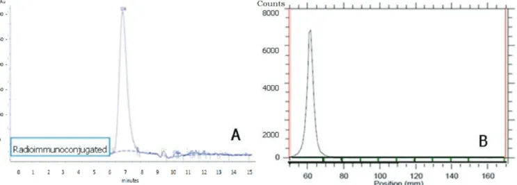
In addition, this method provided an immunoconjugate with purity exceeding 99% (N = 15), as showed in HPLC profile (Figure 3 and 7A).
The retention time (RT) for the unconjugated antibody was compared with the RT of the immunoconjugate showing significant differences for both molar ratios: DTPA 10:1 (P < 0.0001), DTPA 20:1 (P < 0.0001) (Table I). The same RT in HPLC profile was observed for the mAb conjugated with DTPA at both molar ratios 10:1 and 20:1, apparently indicating no greater incorporation of DTPA chelators to mAb when the molar ratio was
doubled.
The immunoconjugate was analyzed for stability and the samples remained stable when stored at -20 oC,
showing an single peak higher to 98% (m = 98.7% ± 0.9; N = 36) in HPLC profile, that corresponds to the Cetuximab-DTPA (Figure 4).
Analysis of the protein concentration of the immunoconjugate
The determination of the protein concentration after conjugation and purification is essential to calculate the radiopharmaceutical specific activity and analysis of
radiolabeling conditions.
The results of protein concentrations for the Cetuximab-DTPAimmunoconjugate, produced with molar ratios 10:1 and 20:1 were 4.43 ± 0.24 mg/mL and 4.54 ±
0.22 mg/mL, respectively. Considering an initial antibody concentration of 5 mg/mL, it obtained a protein recovery above 83±1.42% (N = 15). These results demonstrated that
the method applied for purification of immunoconjugate was more reproducible than the method described in the
literature (Cooper, Sabbah, Mather, 2006), which results are variable due to the loss of protein (10 - 50%).
Determination of number of chelators per antibody
The optimal number of chelators per antibody must be achieved in the process of conjugation, in order to guarantee
the stability of the immunoconjugate, under storage and after
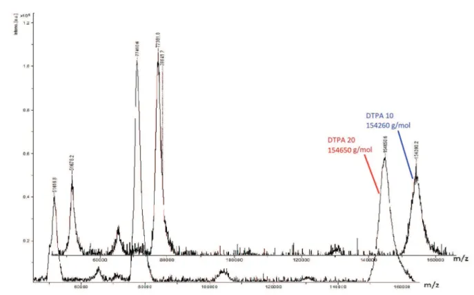
radiolabeling, to preserve the antibody immunoreactivity. The immunoconjugate molecular weight was determined by mass spectrometry (MALDI/TOF) (Figure 5), and the number of chelators coupled to the antibody were calculated through the difference of unconjugated antibody molecular weight (Table II). The mass spectrometry is the most accurate method for determining chelators coupled to mAbs. The results demonstrated that even with a higher amount of chelator (20:1), there was no change in the number of p-SCN-Bn-DTPA chelators coupled to Cetuximab (p-value>0.06). Approximately 6-7 chelators/ mAb were incorporated, confirming the results obtained in HPLC, in which there was no significant difference between the retention times of the peaks generated with the
immunoconjugate molar ratio 10:1 or 20:1.
TABLE I - HPLC analysis of the unconjugated antibody and
immunoconjugate. Retention time (RT) in minutes. Results
expressed as mean ± SD. Chromatograms obtained by Shimadzu
system
Molar ratio RT cetuximab
(min)
RT immunoconjugate
(min)
DTPA 10:1 7,33 ± 0,26 6,76 ± 0,05
DTPA 20:1 7,31 ± 0,29 6,72 ± 0,03
FIGURE 4 - HPLC stability study of immunoconjugate at
Radioimmunoconjugate development and analysis of specific activity
There are difficulties in radiolabeling conjugated mAbs, since the presence of trace metals (contaminants) can compete with the radiometal, interfering directly with the radiolabeling and consequently reducing radiochemical yield. This fact requires pre purification of all reagents employed in the labeling step for removing metals. Moreover, the purification and concentration of the conjugated protein are important steps to remove
the excess chelating agent added for the conjugation
process.
Figure 6 shows the percentage of radiochemical purity (% PR) for radioimmunoconjugate at different specific activities, obtained by TLC analysis immediately,
24 and 48 hours after labeling procedure. The samples were stored at 2 - 8 °C. The specific activity of 185
MBq/mg (5 mCi/mg) was adopted as reference since it showed superior activity, with higher radiochemical purity yields (Figure 7). The average of radiochemical purity, immediately after labeling, was greater than 95% for the specific activity 74 MBq/mg (95.9% ±
1.8), 111 MBq/mg (96.2% ± 1.6) and 185 MBq/mg
(97.3% ± 1.3). A significant difference in decreasing
radiochemical purity, was observed (P = 0.036), when comparing the results of radiochemical purity over per time. Nevertheless, radioimmunoconjugates with specific activity of 111 MBq/mg and 185MBq/mg were stable for 48 hours, which allows a possible distribution of the radiopharmaceutical, even in more distant regions of the production center.
FIGURE 5 - MALDI/TOF immunoconjugate spectrum (spectrometer Bruker Autoflex Speed, CEFAP ICB / USP). Molecular weight
(MW) of Cetuximab-DTPA produced at different molar ratios: DTPA 10 MW was 154.260 g/mol; DTPA 20 MW 154.650 g/mol.
TABLE II - Determination of number of chelators per antibody by spectrometry Autoflex MALDI-TOF/TOF
Molecular Weight Chelator DTPA (g/mol)
Chelation Molar Ratio
MALDI/TOF Molecular Weight
(g/mol)
Difference
[imunoconjugated (g/mol)-Cetuximab
(150.000g/mol)]
Number Chelator/mAb
649.9 DTPA 10 154260 4260 6.6
FIGURE 6 - Radioimmunoconjugate stability study (48 hours) - Radiochemical purity analysis of Cetuximab-DTPA (20:1)
radiolabeled with indium-111, at different specific activities (MBq/mg). Results obtained by CCD (n ≥ 3).
FIGURE 7 - HPLC chromatographic profile of the radioimmunoconjugate, 185MBq/mg (A). TLC scanner chromatographic profile
of the radioimmunoconjugate, 185MBq/mg (B).
Stability analysis of radiolabeled Cetuximab in human serum and binding to serum protein in vitro
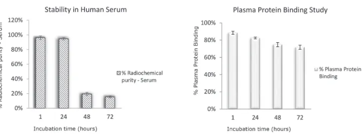
Considering the results obtained, the specific activity of 185 MBq/mg (5 mCi/mg) has been used to evaluate the
stability of the radiolabeled Cetuximab in human serum. According to stability study in human serum, no bound
plasma proteins 111In-DTPA-Cetuximab degraded in 48
hours, possibly due to plasma enzyme activity (Figure 8A).
In addition, a high fraction of radiolabeled mAbs bound
to serum protein (Figure 8B), which should contribute to the slow blood clearance in vivo.
Diagnostic information can assist in the selection of patients, and can potentially predict the risk-benefit balance of a planned therapy with radionuclides through
the dosimetric analysis. In addition, the imaging data can
This study demonstrated that 111In-DTPA-Cetuximab
has been efficiently developed. The conjugation with the chelator DTPA was successful and the immunoconjugate presented stability suitable for the routine production of radiopharmaceuticals. The labeled antibody was produced with high radiochemical yield and high specific activity, showing that 111In-DTPA-Cetuximab is feasible to produce
for its application in immuno-SPECT for the diagnosis of head-and- neck cancers and colorectal carcinoma. therefore, encouraged the progress of the clinical studies.
ACKNOWLEDGMENTS
The author is grateful to scientific cooperation of Marycel Rosa Felisa Figols Barboza and the National
Commission of Nuclear Energy - CNEN, for financial
support.
REFERENCES
Brady ED, Chong HS, Milenic DE, Brechbiel MW. Development of a spectroscopic assay is difunctional ligand-protein conjugates based on copper. Nucl Med Biol. 2004;31(6):795-802.
Chang AJ, Smith RA, Lapi S. Development and characterization
of 89Zr-Labeled panitumumab is immuno-positron emission tomographic imaging of the epidermal growth factor receptor. Mol Imaging. 2013;12(1):17-27.
Cooper MS, Sabbah E, Mather SJ. Conjugation of chelating agents to proteins and radiolabeling with trivalent metallic isotopes. Nature Protocols. 2006;1(1):314-317.
Denardo GL, Sysko VV, Denardo SJ. Cure of incurable lymphoma. Int J Radiat Oncol Biol. 2006;66(Suppl.2):46-56.
FIGURE 8 - Radioimmunoconjugate stability in human serum (A); % protein binding (B). Results expressed as mean ± SEM (n> 3).
Divgi CR, Welt S, Kris M, Real FX, Yeh SD, Gralla R, et
al. Phase I and imaging trial of indium-111 labeled
anti-epidermal growth factor receptor monoclonal antibody 225 in Patients with squamous cell lung carcinoma. J Natl Cancer Inst.1991;83(2):97-104.
Doerr RJ1, Abdel-Nabi H, Baker JM, Steinberg S. Detection of primary colorectal cancer with indium 111 monoclonal antibody B72.3. Arch Surg. 1990;125(12):1601-5.
Evans HL, Nguyen Q, Carroll LS, Kaliszczak M, Twyman FJ,
Spivey AC, et al. A bioorthogonal 68 Ga-labeling strategy is alive rapidin imaging. Chem Commun. 2014;50:9557-60.
Hoeben BAW, Molkenboer-Kuenen JDM, Oyen WJG, Peeters WJM, Bussink J, Boerman OC. Radiolabeled cetuximab: dose optimization for epidermal growth factor recept or imaging in the head-and-neck squamous cell carcinoma model. Int J Cancer. 2011;129(4):870-878.
Instituto Nacional do Cancer. INCA. Cancer incidence in Brazil
- estimate 2014. [cited 2016 May 11]. Available from: http://
www.inca.gov.br/estimativa/2014/estimativa-24042014.pdf.
Jalilian AR, Beiki D, Hassanzadeh-Rad A, Eftekhari A, Geramifar P, Eftekhari M. Production and clinical applications of radiopharmaceuticals and medical radioisotopes in Iran. Sem Nucl Med. 2016;46(4):340-358.
Laemmli, UK. Cleavage of structural proteins during
R. Benedetto, A. V. F. Massicano, J. J. Silva, C. A. W. Villas Boas, J. Mengatti, E. B. Araújo
Lewis MR, Raubitschek A, Shively JE. The facile, water-soluble method for modification of proteins with DOTA. Use of elevated temperature and optimized pH to Achieve high specific activity
and high chelate stability in radiolaleled immunoconjugates.
Bioconjug Chem. 1994;5(6):565-576.
Mccarron PA, Olwll SA, Marouf WM, Buick RJ, Walker B, Scott CJ. Antibody conjugates and therapeutic strategies. Mol Interv. 2005;5(6):368-380.
Morris MJ, Divgi CR, Pandit-Taskar N, Batraki M, Warren N, Nacca A, Smith-Jones P, et al. Pilot trial of unlabeled and indium-111-labeled anti-prostate-specific membrane antigen antibody J591 for castrate metastatic prostate cancer. Clin. Cancer Res. 2005;11(20):7454-61.
Niu G, Sun X, Cao Q, Courter D, Koong A, Le QT, et al. Cetuximab-based immunotherapy and radioimmunotherapy of head and neck squamous cell carcinoma. Clin Cancer Res. 2010;16(7):2095-2105.
Perik PJ, Lub-de HMN, Gietema JA, van der Graaf WT, Korte MA, Jonkman S, et al. Indium-111-labeled trastuzumab scintigraphy in Patients with human epidermal growth factor receptor-2 positive metastatic breast cancer. J Clin Oncol. 2006;24(15):2276-2282.
Ping LW, Meyer LA, Capretto DA, Sherman CD, Anderson CJ. Receptor-binding, Biodistribution, and metabolism studies of 64 Cu-DOTA-cetuximab, PET-imaging agent for epidermal receptor-positive tumors growth-factor. Cancer Biother Radiopharm. 2008;23(2):158-171.
Rasaneh S, RajabiI H, Babaei MH, Daha FJ. 177Lu labeling of Herceptin and preclinical validation as a new radiopharmaceutical for radioimmunotherapy of breast cancer. Nucl Med Biol. 2010;37(8):949-955.
Reilly RM, Lam K, Chan C, Levine M. Advancing Molecular novel imaging agents from preclinical studies to first-in-humans Phase I clinical trials in academia-a roadmap for overcoming perceived barriers. Bioconjug Chem. 2015;26(4):625-32.
Schechter NR, Wendt RE, Yang DJ, Azhdarinia A, Erwin WD, Stachowiak AM, et al. Radiation dosimetry of 99mTc-labeled C225 in Patients with squamous cell carcinoma of the head and neck. J Nucl Med. 2004;45(10):1683-1687.
Scott AM, Tebbutt N, Lee FT, Cavicchiolo T, Liu Z, Gill S, et al. The phase I and Biodistribution pharmacokinetic trial of humanized monoclonal antibody Hu3s193 in Patients with advanced epithelial cancers que express the Lewis Y antigen. Clin Cancer Res. 2007;13(11):3286-92.
Siegel MM, Hollander IJ, Hamann PR, James JP, Hinman L, Smith BJ. Matrix-assisted laser desorption-UV/ionization mass spectrometric analysis of monoclonal antibodies for
the determination of carbohydrate, conjugated chelator and
conjugated drug content. Anal Chem. 1991;63(21):2470-81.
Sihver W, Pietzsch J, Krause M, Baumann M, Steinbach J, Pietzsch H. Radiolabeled targeted EGFR cetuximab conjugates for cancer diagnostics and therapy. Pharmaceuticals. 2014;7(3):311-38.
Strome SE, Sausville EA, Mann D. Mechanistic perspective of monoclonal antibodies in cancer therapy beyond target-related effects. Oncologist. 2007;12(9):1084-95.
Van de Watering FJC, Rijpkema M, Perk L, Brinkmann U, Oyen WJG, Boerman OC. Zirconium-89 labeled antibodies: a new tool for molecular imaging in cancer patients. BioMed Res Int. 2014;14:203601.
Wiechec E, Hansson KT, Alexandersson L, Jönsson JI, Roberg K. Hypoxia Mediates Differential Response to Anti-EGFR Therapy in HNSCC Cells. Int J Mol Sci. 2017;943(18):1-12.
Received for publication on 07th February 2017