Avaliação do efeito de centrifugado de medula
óssea no processo de reparo ósseo ao redor de
implantes instalados em tíbia de coelho.
Análise histológica e histométrica.
Walter Betoni Junior
Avaliação do efeito de centrifugado de medula
óssea no processo de reparo ósseo ao redor de
implantes instalados em tíbia de coelho.
Análise histológica e histométrica.
Tese de Doutorado apresentada à Faculdade de Odontologia de Araçatuba da Universidade Estadual Paulista “Júlio de Mesquita Filho” – UNESP, como parte dos requisitos para obtenção do Título de Doutor em Odontologia – Área de Cirurgia e Traumatologia Bucomaxilofacial.
Orientador: Prof. Dr. Pedro Felício Estrada Bernabé
Agradecimentos
Ao meu eterno e amado, Jesus Cristo, por todas as bênçãos recebidas durante toda a minha vida, com presença marcante em todos os momentos, permitindo que mais uma etapa da minha Jornada Acadêmica fosse cumprida. Que Ele receba toda honra e glória.
Ao Orientador Professor Dr. Pedro Estrada Felício Bernabé pelo estímulo, amizade e companheirismo.
Ao Professor Dr. Idelmo Rangel Garcia Junior, pelo exemplo de profissionalismo e dedicação, que me possibilitaram adquirir novos conhecimentos. O meu sincero e eterno agradecimento.
Ao Professor Dr. Osvaldo Magro Filho pela competência, disponibilidade e amizade demonstrada durante todo este curso.
Ao Professor Dr. Wilson Roberto Poi, por servir de exemplo de professor a ser seguido.
Ao Professor Dr. Mário Jefferson Quirino Louzada, pela gentileza de ceder o Biotério do Curso de Medicina Veterinária da UNESP para que pudéssemos manter os animais utilizados em nosso experimento e pela atenção dada a todos seus alunos.
À Professora Dra. Roberta Okamoto, pela disponibilidade em todos os momentos necessários.
À secretária do departamento de cirurgia e amiga Cleide Lemes da Silva pela proteção e carinho demonstrados.
À Faculdade de Odontologia de Araçatuba – UNESP, sob direção do Prof. Dr. Pedro Felício Estrada Bernabé e Profª. Adj. Dra. Ana Maria Pires Soubhia, e à Comissão de Ética na Experimentação Animal (CEEA) da UNESP, pela oportunidade de realizar o curso de pós-graduação.
À Disciplina de Cirurgia e Traumatologia Buco Maxilo Facial, professores Tetuo Okamoto, Michel Saad Neto, Idelmo Rangel Garcia Júnior, Osvaldo Magro Filho, Alessandra Marcondes Aranega e Cristiane Mara Ruiz de Sousa Fattah.
companheirismo.
Às colegas de doutorado Jéssica Lemos Gulinelli e Thallita Pereira
Queiroz.
Aos colegas de pós-graduação: Abrahão, Albanir, Camila, Francisley,
Flávia, Marcos, Rodolpho, Heloísa, Cassiano, Pedro Ivo, Valter, Jônatas e Fernando pelos momentos compartilhados, pela ajuda, carinho e amizade.
Aos funcionários do Laboratório de Cirurgia Tetuo Okamoto, Bernadete, Dirce e Gilmar pela colaboração e amizade.
Aos funcionários da pós-graduação: Diogo, Marina e Valéria, pela ajuda e paciência nos problemas burocráticos.
Aos professores da disciplina de Cirurgia e Traumatologia Buco Maxilo Facial da Faculdade de Odontologia de Cuiabá: Fábio Lima, Gilberto Almeida Botelho, Luciana Amaral, João Milanez, Thiago Iafelice e Vinicius Canavarros Palma, pelo apoio e contribuição durante todo o período dessa pós-graduação.
Epígrafe
E não vos conformeis com este mundo, mas transformai-vos pela renovação do vosso entendimento para que experimenteis qual seja a boa, agradável
e perfeita vontade de Deus.
histológica e histométrica. 66f. Tese (Doutorado) – Faculdade de Odontologia,
Universidade Estadual Paulista, Araçatuba, 2008.
Resumo
O objetivo deste estudo experimental foi comparar histologicamente o reparo ósseo ao redor de implantes preenchidos por coágulo local com ou sem a associação de centrifugado de medula óssea, por meio de análises histológica e histométrica. Doze coelhos receberam dois implantes osseointegráveis em cada tíbia direita, onde a primeira cortical foi preparada com 5 mm e a segunda, ou cortical inferior, com 3 mm. Os implantes foram estabilizados na cortical inferior com 40 N, sendo um defeito preenchido por coagulo sanguíneo e o outro com coágulo e centrifugado de medula óssea. O sacrifício dos animais foi realizado nos períodos de 7, 21 e 60 dias. As peças foram submetidas a processamento laboratorial de rotina e coradas com vermelho de alizarina e azul de Stevenel. Tanto o grupo coágulo quanto o grupo coágulo e centrifugado de medula, após 60 dias, tiveram seus defeitos preenchidos por tecido ósseo, não apresentando diferença estatística entre os grupos quanto à extensão linear de contato entre tecido ósseo e implante.
BETONI JUNIOR, W. Evaluation of the effect of bone marrow centrifuged in the
bone repair around implant installed in tibia’s rabbit. Histological and histometric analysis. 66p. Thesis (PhD) - University of Dentistry, UNESP - São
Paulo State University, Araçatuba, 2008.
Abstract
The objective of this experimental study was to compare histologically the bone repair around implant filled out by local coagulum with or without association of bone marrow centrifuged through histological and histometric analysis. Twelve rabbits received two implants in the right tibia, where the first cortical it was prepared with 5 mm and the second or lower cortical, with 3 mm. The implants were stabilized in lower cortical with 40 N, being one defect filled out by blood coagulum and the other filled out by blood coagulum and bone marrow centrifuged. The sacrifice of the animals was accomplished in the periods of 7, 21 and 60 days. The pieces were submitted to routine laboratorial process and stained with alizarin red and Stevenel’s blue. Both groups had their defects filled by bone after 60 days, not presenting statistical difference between the groups about the linear extension of contact between bone tissue and implant.
coágulo sanguíneo preenchendo a área do defeito ósseo peri-implantar (vermelho de alizarina e azul de Stevenel, 45 X). ... 41 Figura 2 Grupo controle, 7 dias. Região apical do implante. Tecido ósseo
pré-existente próximo às roscas do implante, mas sem o estabelecimento de interface de contato (vermelho de alizarina e azul de Stevenel, 200X). 41 Figura 3 Grupo controle, 21 dias. Aspecto histológico total do implante. Os
defeitos ósseos peri-implantares não apresentaram completo preenchimento por tecido ósseo neoformado, que apresentou características de osso esponjoso (vermelho de alizarina e azul de Stevenel, 200X). ... 42 Figura 4 Grupo controle, 21 dias (região cervical do implante). Em algumas
áreas observou-se tecido conjuntivo em contato direto com a superfície do implante (vermelho de alizarina e azul de Stevenel, 200X). ... 42 Figura 5 Grupo controle, 21 dias (região apical). Nota-se neoformação óssea significativa na região das roscas do implante (vermelho de alizarina e azul de Stevenel, 200X). ... 43 Figura 6 Grupo controle, 21 dias (região medular). Poucas trabéculas ósseas
neoformadas são observadas no vale das roscas, sem o estabelecimento de uma interface com o implante osseointegrável (vermelho de alizarina e azul de Stevenel, 200X). ... 43 Figura 7 Grupo tratado, 21 dias (região cervical). Trabéculas de osso neoformado
preenchendo parcialmente a área do defeito ósseo peri-implantar, envoltas por tecido conjuntivo celularizado, com células semelhantes a osteoblastos (setas) (vermelho de alizarina e azul de Stevenel, 200X). 44 Figura 8 Grupo tratado, 21 dias (região medular). Trabéculas ósseas
neoformadas entremeadas por tecido conjuntivo celularizado e poucas áreas de contato entre o tecido ósseo e a superfície do implante (vermelho de alizarina e azul de Stevenel, 200X). ... 44 Figura 9 Grupo tratado, 21 dias (região apical). Tecido ósseo neoformado
áreas de contato entre o tecido ósseo e a superfície do implante
(vermelho de alizarina e azul de Stevenel, 200X). ... 45
Figura 10 Grupo tratado, 60 dias. Aspecto histológico total do implante. A integridade da cortical superior da tíbia dos coelhos foi restaurada na área dos defeitos ósseos confeccionados na região cervical peri-implantar (vermelho de alizarina e azul de Stevenel, 45X). ... 45
Figura 11 Grupo controle, 60 dias (região cervical). Neoformação óssea na região do defeito peri-implantar e presença de trabéculas ósseas circundadas por tecido conjuntivo celularizado com espaços de remodelação (setas) (vermelho de alizarina e azul de Stevenel, 200X). ... 46
Figura 12 Grupo tratado, 60 dias (região medular). Poucas trabéculas ósseas neoformadas, circundadas por tecido conjuntivo celularizado e pequenas extensões lineares de contato entre o tecido ósseo neoformado e a superfície do implante (vermelho de alizarina e azul de Stevenel, 200X).46 Figura 13 Grupo controle, 60 dias (região apical). Neoformação óssea preenchendo totalmente a área das roscas dos implantes. Não foi possível distinguir o tecido ósseo neoformado do osso pré-existente (vermelho de alizarina e azul de Stevenel, 200X). ... 47
Figura 14 Representação da mediana (coluna), mínimo e máximo (extremos da barra vertical) de porcentagem de ELCOI, nas regiões cervical e apical.47 Figura 15 Representação da mediana (coluna), mínimo e máximo (extremos da barra vertical) de porcentagem de AON. ... 48
Figura 16 Os animais foram mantidos, durante o experimento, em gaiolas individuais no Biotério do Curso de Medicina Veterinária da Faculdade de Odontologia da UNESP, Araçatuba, Brasil. ... 59
Figura 17 Agulha de mielograma, própria para punção de medula óssea de calibre 16 ... 59
Figura 18 Aspiração da medula óssea. ... 60
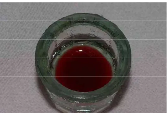
Figura 19 O centrifugado acondicionado em um frasco dappen. ... 60
Figura 20 Incisão na porção medial da tíbia direita... 61
Figura 21 Acesso ao tecido ósseo. ... 61
Figura 24 Broca piloto utilizada para alargar o orifício na segunda cortical de 2 para 3 milímetros. ... 63
Figura 25 Os dois defeitos confeccionados, com 5 milímetros na primeira cortical e 3 milímetros na segunda ... 63
Figura 26 Os implantes instalados, ancorados na cortical inferior. ... 64 Figura 27 Os implantes instalados, sendo que no lado esquerdo o defeito
peri-implantar foi preenchido com coágulo e no lado direito preenchido com centrifugado de medula. ... 64 Figura 28 As tíbias foram removidas em bloco, depois, através de osteotomias,
Lista de Tabelas
cm = centímetros mg = miligrama
mg/kg = miligramas por quilo N = Newton
ml = mililitros
RPM = rotações por minutos µm = micrômetro
SUMÁRIO
1 INTRODUÇÃO ... 16
2 PROPOSIÇÃO ... 18
3 MATERIAL E MÉTODO ... 20
4 RESULTADO ... 25
4.1 Análise Qualitativa ... 25
4.2 Análise Quantitativa ... 27
5 DISCUSSÃO ... 30
6 CONCLUSÃO ... 33
REFERÊNCIAS ... 35
FIGURAS ... 41
TABELA ... 49
ANEXOS ... 50
ANEXO A – CERTIFICADO DA COMISSÃO DE ÉTICA ... 51
1 Introdução
*A escolha de enxerto autógeno como recurso para reconstrução de
defeitos ósseos como fenestrações, defeitos residuais e alvéolos pós-exodônticos é
a mais adequada, no entanto, a limitada disponibilidade de tecido e o aumento do
tempo e número de intervenções a torna questionável, principalmente em casos de
grandes reconstruções (Gotfredsen et al., 1991; Piattelli et al., 1997; Becker et al.,
1994; Becker et al., 1998; Trejo et al., 2000).
Recentemente, técnicas de engenharia tecidual têm apresentado
resultados promissores buscando reparar, substituir ou regenerar órgãos e tecidos
específicos (Griffith et al., 2002). A cultura e transplante de células indiferenciadas
da medula óssea são procedimentos eficazes, porém complexos e caros. Como
alternativa, o uso de aspirados de medula óssea administrados no sítio a ser
regenerado tem sido pesquisado clínica e laboratorialmente, buscando preencher
defeitos ósseos, estimular a consolidação de fraturas e tratar pseudo-artroses.
(Conolly, 1995; Paley, 1998; Yamada, 2004; Ito, 2006).
Niemeyer et al (2004) relataram o uso de medula óssea aspirada e
inserida a arcabouços de colágeno e de fosfato tricálcico e observaram graus de
diferenciação e maturação de células-tronco mesenquimais de medula óssea em
células osteoprogenitoras e osteoblastos para reconstrução de defeitos ósseos.
Considerando o potencial osteogênico de algumas células da medula óssea, este
trabalho se propôs a avaliar histologicamente o efeito do centrifugado de medula
óssea no processo de reparo ósseo ao redor de implantes osseointegráveis
instalados em tíbia de coelho.
*
18
2 Proposição
Avaliar histologicamente o reparo ósseo em defeitos peri-implantares
preenchidos por coágulo local com ou sem associação de centrifugado de medula
3 Material e Método
Este estudo experimental foi submetido e aprovado pelo Comitê de Ética
na Experimentação Animal (CEEA) da Universidade Estadual Paulista - UNESP,
Campus de Araçatuba, Brasil.
Doze coelhos (Oryctolagus cuniculus) machos, com peso corporal médio
de 3.350 gramas, foram selecionados para o estudo. Os animais foram alimentados
com ração sólida triturada (Ração Sítios e Quintais fabricada pela Purina
Nutrimentos Ltda.) durante todo o experimento, tendo água à vontade, exceto no
período de oito horas antecedentes ao ato cirúrgico. A etapa cirúrgica foi realizada
sob anestesia geral, administrando-se por via intramuscular a combinação de
50mg/kg de Ketamina intramuscular (Francotar – Vibrac do Brasil Ltda., São Paulo,
Brasil) e 5mg/Kg de cloridrato de Xilazina (Rompum – Bayer AS – Saúde Animal,
São Paulo, Brasil). Após a anestesia local do animal, foi realizada tricotomia na
região de crista ilíaca e tíbia direitas, seguida de rigoroso protocolo asséptico,
incluindo a esterilização do instrumental a ser utilizado, delimitação da área a ser
operada com campos estéreis e uso de aventais e luvas cirúrgicas estéreis.
Com o coelho já anestesiado, uma agulha de mielograma de calibre 16,
própria para punção de medula óssea, foi introduzida na crista ilíaca posterior do
animal e, com movimentos de rotação delicados, penetrou-se a cavidade medular. O
trocater foi retirado e uma seringa plástica descartável de 20 ml, cuja parte interna
foi previamente embebida em solução de heparina a 1:1000, foi conectada à agulha.
Por meio de sucção por firme tração do êmbolo da seringa, foram aspirados 3 ml de
medula óssea. O material obtido foi colocado em um tubo de ensaio de 5 ml e
21
sobrenadante, de aproximadamente 1,2 ml, foi descartado com ajuda de seringa de
1 ml. Do material sedimentado no tubo, foram coletados 0,5ml, formados pela
camada intermediária entre o plasma descartado e as células sedimentadas no
fundo do tubo, onde se encontra maior quantidade de células nucleadas, cuja
densidade é maior. O material foi separado em um frasco dappen e adicionado a
0,2ml de cloreto de cálcio a 10% para que alcançasse a forma de gel.
Com uma lâmina número 15 (Feather Industries Ltda, Tókio, Japão)
montada em cabo de bisturi n° 3 (Hu-Friedy, Alemanha) , foi realizada uma incisão de
aproximadamente 3 cm de comprimento na porção medial da tíbia direita, até a base
óssea e, a seguir, o tecido mole foi divulsionado e afastado com o auxílio de
descoladores de periósteo, expondo o osso para receber os implantes.
O preparo dos leitos receptores foi iniciado com uma trefina de 5 mm para
delimitar a localização dos implantes e remover a primeira cortical óssea. Em
seguida, no centro do orifício de 5 mm, foram utilizadas as fresas lança, fresa de 2.0
mm, piloto de 2.0/3.0 mm e fresa de 3.0 mm passando pela segunda cortical,
seqüencialmente, na velocidade máxima de perfuração de 1600 RPM, com irrigação
por meio de solução de cloreto de sódio a 0,9% (Darrow, Rio de Janeiro, Brasil)
durante toda a preparação.
Posteriormente foram instalados dois implantes de 3,75 mm/8,5 mm, na
porção medial da tíbia direita, preservando-se uma distância de 5 mm entre os dois
orifícios, com o auxílio de motor e contra-ângulo. No defeito correspondente ao
grupo controle, o implante foi inserido no local da osteotomia, preenchido apenas
com coágulo sanguíneo, até que a plataforma ficasse ao nível da primeira cortical da
instalado implante de comprimento e diâmetro iguais ao grupo controle e o espaço
ao redor do implante foi preenchido com o centrifugado de medula óssea. Ambos os
implantes foram estabilizados na segunda cortical com 40 N. Os tecidos foram
suturados em planos, empregando-se fio reabsorvível (ácido polilático – Vycril 4.0,
Ethicon, Johnson Prod., São José dos Campos, Brasil) com pontos interrompidos e
com fio monofilamentar (Nylon 5.0, Mononylon, Ethicon, Johnson Prod., São José
dos Campos, Brasil). Após a sutura, foi realizada novamente a anti-sepsia da área
com Polivinil Pirrolidona Iodo Tópico (PVPI 10%, Riodeine, Rioquímica, São José do
Rio Preto).
Os animais foram sacrificados aos 7, 21 e 60 dias após as cirurgias. As
áreas operadas foram reabertas, os implantes expostos e osteotomias foram
realizadas na tíbia para remoção dos dois fragmentos contendo as áreas
experimentais, grupo tratado e grupo controle.
Imediatamente após a remoção, os fragmentos foram submetidos a
processo laboratorial de rotina para inclusão em resina acrílica hard-grade (LR
White®, London Resin Company Ltd, Berkshire, England). Os blocos plastificados
foram montados em lâminas e cortados numa espessura de 40 µm (Microslice 2®,
Ultratec Inc, Santa Ana, USA), lixados manualmente até uma espessura aproximada
de 15µm e finalmente corados com vermelho de alizarina e azul de Stevenel. Foi
realizada uma secção longitudinal em cada implante, para análise histológica.
Todas as lâminas foram examinadas e as imagens capturadas através de
um microscópio Leica DMLB® (Leica Microsystems Wetzlar ®, Germany), equipado
com câmera digital Leica DFC 300FX (Leica Microsystems Wetzlar ®, Germany),
acopladas a um computador. Com o auxílio do programa Imagelab 2000, versão 2.4,
23
entre tecido ósseo e o implante e a área de tecido ósseo neoformado ao redor do
implante (AON), de 3 regiões pré-selecionadas, correspondentes às regiões cervical,
média (medular) e o ápice dos implantes, para ambos os grupos.
Foram criadas duas variáveis para a análise da interface do defeito
ósseo/implante. A ELCOI, que é a extensão linear de contato entre o osso e
implante, e a AON, que é a área de osso neoformado em contato com o implante.
Duas regiões da interface osso/implante de cada grupo foram avaliadas – a região
cervical, que corresponde à porção da cortical superior do defeito; e a apical, que
corresponde à porção da cortical inferior. Os valores de extensão linear obtidos para
tecido ósseo (ELCOI) e a área de tecido ósseo neoformado (AON) nos grupos
controle e tratado foram convertidos em valores percentuais e comparados por meio
do teste não-paramétrico de Kruskal-Wallis. O coeficiente de correlação de
Spearman foi empregado para verificar a existência de associação entre as variáveis
4 Resultado
Os animais tiveram boa recuperação pós-operatória e nenhuma alteração
nas condições de saúde durante todo o período da pesquisa.
4.1 Análise Qualitativa
Os resultados obtidos foram descritos após analise qualitativa da interface
osso/implante, conforme descrição a seguir:
Aos 7 dias pós-operatórios
Em ambos os grupos, aos 7 dias, observou-se a presença do coágulo
sanguíneo preenchendo a área do defeito ósseo peri-implantar confeccionado na
área cervical do implante e no espaço medular adjacente (figura 1). Na região apical,
notou-se tecido ósseo pré-existente próximo às roscas do implante, mas sem o
estabelecimento de interface de contato, principalmente na região dos vales das
roscas (figura 2).
Aos 21 dias pós-operatórios
Todos os implantes apresentaram-se osseointegrados nesse período.
No grupo controle, observou-se que os defeitos ósseos peri-implantares
não se apresentavam totalmente preenchidos por tecido ósseo neoformado (corado
pelo vermelho de alizarina). O osso neoformado apresentou características de osso
esponjoso (figura 3). As trabéculas ósseas foram circundadas por tecido conjuntivo
celularizado (corado pelo azul de Stevenel). Em algumas áreas, observou-se tecido
conjuntivo em contato direto com a superfície do implante (figura 4). Os implantes
encontraram-se parcial ou totalmente recobertos por tecido ósseo neoformado,
26
na região das roscas (figura 5), que progrediu cervicalmente, em direção à área de
osso medular, onde poucas trabéculas ósseas neoformadas foram observadas no
vale das roscas, sem o estabelecimento de uma interface com o implante (figura 6).
No grupo tratado, também foi possível observar a presença de trabéculas
de osso neoformado preenchendo parcialmente a área do defeito ósseo
peri-implantar, na região cervical. As trabéculas estavam envoltas por tecido conjuntivo
celularizado, com células semelhantes a osteoblastos (figura 7). Em algumas áreas
da plataforma do implante e das roscas presentes nessa região houve o
estabelecimento de uma superfície de contato entre o osso neoformado e a
superfície do implante. Alguns implantes deste grupo também se apresentavam
parcialmente recobertos por tecido ósseo neoformado. Na área medular, notou-se
presença de trabéculas ósseas neoformadas entremeadas por tecido conjuntivo
celularizado e poucas áreas de contato entre o tecido ósseo e a superfície do
implante (figura 8). Nas roscas do implante localizadas mais apicalmente na área
medular, notou-se significativa neoformação óssea e estabelecimento de interface
de contato entre o tecido ósseo e a superfície do implante. Na área apical
observou-se tecido ósobservou-seo neoformado preenchendo a área das roscas, com inúmeras
trabéculas ósseas e áreas de contato entre o tecido ósseo e a superfície do implante
(figura 9).
Aos 60 dias pós-operatórios
Todos os implantes mostraram-se osseointegrados neste período. A
integridade da cortical superior da tíbia dos coelhos foi restaurada na área dos
defeitos ósseos confeccionados (figura 10). Em ambos os grupos, observou-se
neoformação óssea na região do defeito peri-implantar e presença de trabéculas
(figura 11). Em alguns implantes houve preenchimento ósseo do defeito em apenas
um dos lados do implante, e em outros, houve a regeneração do osso cortical
superior na área do defeito ósseo criado. Houve o estabelecimento de interface
óssea com a superfície dos implantes em algumas áreas, principalmente na região
cervical e apical do implante. Alguns implantes apresentavam-se recobertos por
tecido ósseo neoformado na parte superior (acima da plataforma do implante). Na
área medular, nos dois grupos analisados, observou-se presença de poucas
trabéculas ósseas neoformadas, circundadas por tecido conjuntivo celularizado e
pequenas extensões lineares de contato entre o tecido ósseo neoformado e a
superfície do implante (figura 12). Na região apical, notou-se neoformação óssea
preenchendo a área das roscas dos implantes, estendendo-se cervicalmente em
direção à região medular. Em algumas roscas não foi possível distinguir o tecido
ósseo neoformado do osso pré-existente (figura 13).
4.2 Análise Quantitativa
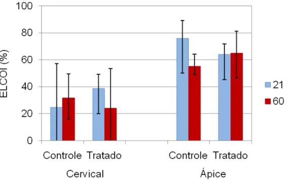
Observando-se a Tabela 1, nota-se variação grande das porcentagens de
ELCOI, tanto na cervical como no ápice, que, aliado ao pequeno número de animais
disponíveis, levou à utilização de procedimentos estatísticos não-paramétricos de
análise dos dados. O teste de Kruskal-Wallis foi aplicado e não identificou diferença
significativa entre as quatro combinações de grupos e períodos quanto à ELCOI, na
cervical (p= 0,897) e no ápice (p=0,461).
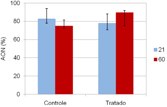
Em relação à variável AON, a variação das medidas é menor, mas por
prudência e padronização das análises, também se empregou o teste de
Kruskal-Wallis. Não houve evidência de diferença significativa entre os grupos nos dois
28
Nas figuras 15 e 16 estão representadas graficamente a mediana, mínimo
e máximo de ELCOI e AON, respectivamente. Essas figuras permitem uma visão
global dos resultados e corroboram com os resultados da análise estatística.
O coeficiente de correlação de Spearman foi calculado e apontou
associação entre a ELCOI no ápice e a AON, com coeficiente igual a 0,64. A variável
ELCOI cervical ficou comprometida pela sua maior variação, dando coeficientes de
5 Discussão
A utilização da tíbia de coelhos para avaliar o reparo ao redor de
implantes de diferentes tipos tem sido recorrente na literatura, com variações quanto
à área receptora. O implante pode ser instalado na articulação do joelho e metáfise
tibial (Sennerby et al.1992), na porção medial-proximal da tíbia (Kong et al. 2002), ou
na metáfise tibial (Cordiolli et al, 2000; Margonar et al. 2003), dentre outros sítios
descritos em vários trabalhos que avaliaram a osseointegração de diferentes tipos
de implantes. Neste estudo, foi definida a utilização da porção medial da tíbia direita
do coelho como área receptora, considerando o fato desta área estar distante do
centro de crescimento, localizado na metáfise tibial.
Devido à sua conicidade intrínseca, após a extração dentária, o alvéolo
freqüentemente apresenta dimensões maiores que o diâmetro de um implante,
formando um espaço entre a região cervical do implante e o tecido ósseo. Neste
estudo, confeccionou-se defeito cujo diâmetro cervical (na cortical superior da tíbia)
era 1,25 mm maior que o diâmetro do implante, simulando a situação encontrada na
clínica. Akimoto et al, em 1999, demonstraram que a largura do defeito ósseo
influencia diretamente a porcentagem do contato osso/implante, destacando a
importância da utilização dos biomateriais para o preenchimento desses defeitos.
A não-observação de osso neoformado nas áreas cervicais e médias
(medulares), aos 7 dias pós-operatórios, em ambos os grupos, pode ser justificada
pela precocidade do período. Por esta ausência, este período foi retirado da análise
31
A similaridade do reparo ósseo dos defeitos ao reparo ósseo alveolar
justifica o fato de a neoformação óssea seguir direção centrípeta e de apical para
cervical, conforme visto nos dois grupos, aos 21 dias pós-operatórios.
O restabelecimento da cortical superior não garantiu a formação de osso
maduro na região medular de ambos os grupos, mesmo aos 60 dias
pós-operatórios.
Em nosso estudo, pudemos observar que nos defeitos confeccionados,
apenas o coágulo foi suficiente para um bom reparo ósseo ao redor dos implantes.
Estes resultados também foram observados por Botticelli et al em estudos de 2003,
2004, 2005 e 2006, onde obtiveram preenchimento por tecido ósseo neoformado em
amplos defeitos peri-implantares confeccionados em cães, fornecendo alto grau de
osseointegração. Niemeyer et al, em 2004 e Yamada et al, em 2003, demonstraram
melhora na formação óssea quando utilizada medula óssea aspirada e inserida a
algum tipo de arcabouço para tratamento de defeitos ósseos em cães.
A não-observação de diferença estatisticamente significante entre os
grupos deste trabalho pode estar relacionada ao leito receptor. A tíbia é um osso
longo, com amplo canal medular. Após a confecção dos defeitos, que requerem
remoção das trabéculas ósseas medulares para a instalação dos implantes, pôde-se
observar a comunicação entre os defeitos do grupo tratado e grupo controle, via
canal medular, mesmo respeitando-se a distância de cinco milímetros entre os
preparos. Apesar da adição de cloreto de cálcio a 10% para geleificação da medula,
não houve a certeza de que esta não chegou ao leito do grupo controle, ou mesmo
6 Conclusão
Dentro das condições da presente pesquisa, concluímos que:
- ao final de 60 dias o defeito de 0,625 mm ao redor de implantes em tíbia
de coelhos é totalmente preenchido;
- histologicamente, não houve diferença no reparo ósseo com o uso ou
35
REFERÊNCIAS**
1. Akimoto, K., Becker, W., Persson, R., Baker, D.A., Rohrer, M.D. & Oಿneal, R.B.
(1999) Evaluation of titanium implants placed into simulated extraction sockets: a
study in dogs. The International Journal of Oral and Maxillofacial Implants 14:
351-360.
2. Becker, W., Becker, B.E. & Cafesse, R. (1994) A comparision of demineralized
freeze-dried bone and autologous bone to induce bone formation in human extraction
sockets. J. Periodontol. 65: 1128-1133.
3. Becker, W., Clokie, C., Sennerby, L., Urist, M.R. & Becker, B.E. (1998) Histologic
findings after implantation and evaluation of different grafting materials and titanium
micro screws into extraction sockets:case reports. J. Periodontol. 69: 414-421.
4. Botticelli, D., Berglundh, T., Buser, D. & Lindhe, J. (2003) The jumping distance
revisted: an experimental study in the dog. Clinical Oral Implants Research 14:35-42.
5. Botticelli, D., Berglundh, T. & Lindhe, J. (2004) Resolution of bone defects of
varying dimension and configuration in the marginal portion of the peri-implant bone.
An experimental study in the dog. Journal of Clinical Periodontology 31: 309-317.
6. Botticelli, D., Berglundh, T., Persson, L.G. & Lindhe, J. (2005) Bone regeneration
at implants with turned or rough surfaces in self-contained defects. An experimental
study in dog. Journal of Clinical Periodontology 32:448-455.
7. Botticelli, D., Persson, L.G., Lindhe, J. & Berglundh, T. (2006) Bone tissue
formation adjacent to implants placed in fresh extraction sockets: an experimental
study in dogs. Clinical Oral Implants Research 17: 351-358.
**
8. Buser, D., Dahlin, C., Schenk, R.K. (1996) Regeneração óssea guiada na
implantodontia. Quintessence.
9. Campagnoli, C., Chen, N.J., Hsieh, S.L., Bennett, P.R., Bellantuono, I. & Fisk,
N.M. (2001) Identification of mesenchymal stem/progenitor cells in human
first-trimester fetal blood, liver, and bone marrow. Blood 98: 2396-402.
10. Clarke, D.L., Jphansson, C., Wilbertz, J., Veress, B., Nilssln, E., Karlstrom, N.,
Lendahl, U. & Frisén, J. (2000) Generalized potencial of adult neural stem cells.
Science 288: 1660-1663.
11. Connoly, J.F. (1995) Injectable bone marrow preparations to stimulate
osteogenic repair. Clin Orthop Relat Res. 313: 8-18.
12. Cordioli, G., Majzoub, Z., Piatelli, A. & Scarano, A. (2000) Removal torque and
histomorphometric investigation of 4 different titanium surfaces: an experimental
study in the rabbit tibia. The International Journal of Oral and Maxillofacial Implants
15: 668-674.
13. Ferrari, G., Cusella, G., Coletta, M., Paolucci, E., Stornaiuolo, A., Cossu, G. &
Mavilio, F. (1998) Muscle Regeneration by bone-marrow derived myogenic
progenitors. Science 279: 1528-2530.
14. Gasper, P.W. & Tthrall, M.A. (2000) Hemopoietic stem cell. In: Feldman, B.F. et
al. Schalms veterinary hematology, cap.11, p. 63-68. Philadelphia: Williams &
Willians.
15. Gotfredsen, K., Warrer, K., Hjorting-Hansen, E. & Karring, T. (1991) Effect of
membranes and hydroxyapatite on healing in bone defects around titanium implants.
37
16. Griffith, L.G. & Naughton, G. (2002) Tissue engineering – current challenges and
expanding opportunites. Science 295: 1009-14.
17. Ito, K., Yamada, Y., Naiki, T. & Ueda, M. (2006) Simultaneous implant placement
and bone regeneration around dental implants using tissue-engineered bone with
fibrin glue, mesenchymal stem cells and platelet-rich plasma. Clin. Oral Impl. Res.
17: 579–586.
18. Kong, Y.M., Kim, D.H., Kim, H.E., Heo, S.J. & Koak, J.Y. (2002)
Hydroxyapatite-based composite for dental implants: an in vivo removal torque experiment. Journal
of Biomedical Materials Research 63: 714-721.
19. Korbling, M. & Estrov, Z. (2003) Adult stem cells for tissue repair – a new
therapeutic concept? N Engl J Med 349: 570–582.
20. Margonar, R., Sakakura, C.E., Holzhaussen, M., Pepato, M.T., Alba-Júnior, R.C.
& Marcantonio-Júnior, E. (2003) The influence of diabetes mellitus and insulin
therapy on biomechanical retention around dental implants: a study in rabbits.
Implant Dentistry 12: 333-339.
21. Niemeyer, P., Krause, U., Fellenberg, J., Kasten, P., Seckinger, A., Ho, A.D. &
Simank, H.G. (2004) Evaluation of mineralized collagen and tricalcium Phosphate as
scaffolds for tissue engineering of bone using human mesenchymal stem cells. Cells
Tissues Organs 177: 68-78.
22. Otsuki, D.A. (2004) Hemodiluição normovolêmica aguda: estudo experimental
comparativo utilizando amido hidroxietilico a 6% ou solução de Ringer lactato. Tese
apresentada a Faculdade de Medicina Veterinária e Zootecnia da Universidade de
23. Paley, D. (1998) Percutaneos bone marrow grafting on fractures and bone
defects and experimental study in rabbit. Clin Orthop Relat Res. 208: 300-12.
24. Piattelli, A., Podda, G. & Scarano, A. (1997) Clinical and histological results in
alveolar ridge enlargement using coralline calcium carbonate. Biomaterials 18:
623-627.
25. Reddy, R.L. (2005) Mobilization and collection of peripheral blood progenitor cells
for transplantation: transfusion and apheresis. Science 32: 63-72.
26. Repetti, E., Soerensen, B., Barros, A.R., Christovão, F.G., Vieira, V., Moreira,
M.B., Polegato, E.P.S., Pacchini, C.E., Rubia, W.J. & Fuentes, D.B. Valores normais
do quadro hematológico dos animais domésticos.
http://www.unimar.br/ciencias/6-2-1.htlm
27. Santos, R.B., Soares, M.B.P., Carvalho, A.C.C. (2004) Transplante de células da
medula óssea no tratamento da cardiopatia chagássica crônica. Revista da
Sociedade Brasileira de Medicina Tropical, 37(6): 490-495.
28. Sennerby, L., Thomsen, P. & Ericson, L.E. (1992) A morphometric and
biomechanic comparision of titanium implants inserted in rabbit cortical and 75
cancellous bone.The International Journal of Oral and Maxillofacial Implants 7:62-71.
29. Storb ,R., Thomas, E.D., Buckner, C.D., Clift, R.A., Hohnson, F.L., Fefer, A.,
Glucksberg, H., Gilblett, E.R., Lemer, K.G. & Neimam, P. (1974) Allogeneic marrow
grafting for treatament of aplastic anemia. Blood 43: 157-180.
30. Trejo, P.M., Weltman, R. & Cafesse, R. (2000) Treatment of intraosseous defects
with bioabsorbable barriers alone or in combination with decalcified freeze-dried
39
31. Urist, M. & Strates, B.S. (1971) Bone morphogenetic protein. Journal Dent. Res.
50:1392.
32. Yamada, Y., Boo, J.S., Ozawa, R., Nagasaka, T., Okazaki, Y., Hata, K. & Ueda,
M. (2003) Bone regeneration following injection of mesenchmal stem cells and fibrin
glue with a biodegradable scaffold. J. Cranio-maxillofacial Surg 31: 27–33.
33. Yamada, Y., Ueda, M., Naiki, T. & Nagasaka, T. (2004) Tissue-engineered
injectable bone regeneration for osseointegrated dental implants. Clin. Oral Impl.
Figuras
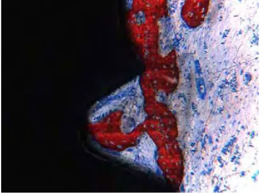
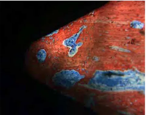
Figura 1 – Grupo tratado, 7dias. Visão histológica total do implante. Presença do coágulo sanguíneo preenchendo a área do defeito ósseo peri-implantar (vermelho de alizarina e azul de Estevenel, 45X).
42
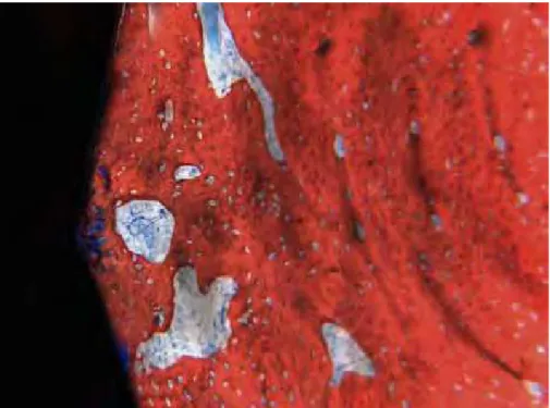
Figura 3 – Grupo controle, 21 dias. Visão histológica total do implante. Os defeitos ósseos peri-implantares não apresentaram completo preenchimento por tecido ósseo neoformado, que apresentou características de osso esponjoso (vermelho de alizarina e azul de Stevenel, 45X).
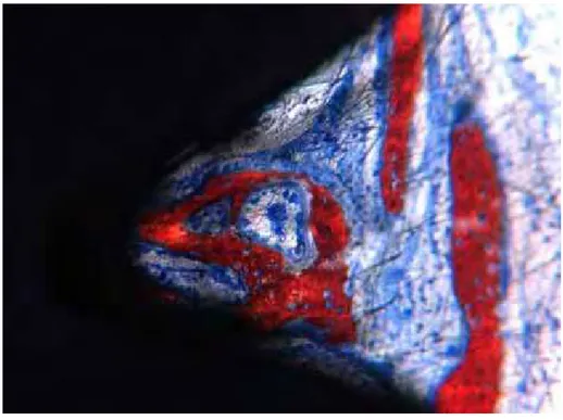
Figura 5 – Grupo controle, 21 dias (região apical). Nota-se neoformação óssea significativa na região das roscas do implante (vermelho de alizarina e azul de Stevenel, 200X).
44
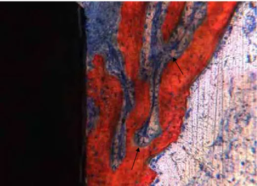
Figura 7 – Grupo tratado, 21 dias (região cervical). Trabéculas de osso neoformado preenchendo parcialmente a área do defeito ósseo peri-implantar, envoltas por tecido conjuntivo celularizado, com células semelhantes a osteoblastos (setas) (vermelho de alizarina e azul de Stevenel, 200X).
Figura 9 – Grupo tratado, 21 dias (região apical). Tecido ósseo neoformado preenchendo a área das roscas, com inúmeras trabéculas ósseas e áreas de contato entre o tecido ósseo e a superfície do implante (vermelho de alizarina e azul de Stevenel, 200X).
46
Figura 11 – Grupo controle, 60 dias (região cervical). Neoformação óssea na região do defeito peri-implantar e presença de trabéculas ósseas circundadas por tecido conjuntivo celularizado com espaços de remodelação (setas) (vermelho de alizarina e azul de Stevenel, 200X).
Figura 13 – Grupo controle, 60 dias (região apical). Neoformação óssea preenchendo totalmente a área das roscas dos implantes. Não foi possível distinguir o tecido ósseo neoformado do osso pré-existente (vermelho de alizarina e azul de Stevenel, 200X).
48
Tabela
Tabela 1 - Estatísticas descritivas de porcentagem de Extensão linear de contato entre tecido ósseo e implante (ELCOI), nas regiões cervical e apical e de Área de osso neoformado (AON).
Grupo Período Estatística ELCOI AON (dias) Cervical Ápice
Controle 21 Mínimo 0,0 50,2 78,1
Máximo 57,3 89,3 94,1
Mediana 24,8 75,8 82,5
Média 22,7 72,8 87,0
60 Mínimo 16,2 49,1 72,7
Máximo 49,6 64,2 81,5
Mediana 31,6 55,3 74,8
Média 32,3 56,0 76,0
Tratado 21 Mínimo 20,1 45,2 70,7
Máximo 49,4 71,8 88,2
Mediana 38,9 63,9 77,9
Média 36,8 61,2 78,7
60 Mínimo 0,0 46,7 75,0
Máximo 53,5 81,3 91,9
Mediana 24,0 64,9 89,7
Anexo A
52
Anexo B
Normas da Revista Clinical Oral Implants Research
1. GENERAL
Clinical Oral Implants Research conveys scientific progress in the field of implant dentistry and its related areas to clinicians,
teachers and researchers concerned with the application of this information for the benefit of patients in need of oral implants. The journal addresses itself to clinicians, general practitioners, periodontists, oral and maxillofacial surgeons and prosthodontists, as well as to teachers, academicians and scholars involved in the education of professionals and in the scientific promotion of the field of implant dentistry.
Clinical Oral Implants Research publishes:
Original research articles of high scientific merit in the field of material sciences, physiology of wound healing, biology of tissue integration of implants, diagnosis and treatment planning, prevention of pathologic processes jeopardizing the longevity of implants, clinical trials on implant systems, stoma-tognathic physiology related to oral implants, new developments in therapeutic concepts and prosthetic rehabilitation.
Review articles by experts on new developments in basic sciences related to implant dentistry and clinically applied concepts.
Case reports and case series only if they provide or document new fundamental knowledge.
Novel developments if they provide a technical novelty for any implant system.
Short communications of important research findings in a concise format and for rapid publication.
Treatment rational by experts with evidence-based treatment approach.
Please read the instructions below carefully for details on the submission of manuscripts, the journal's requirements and standards as well as information concerning the procedure after a manuscript has been accepted for publication in Clinical Oral
Implants Research. Authors are encouraged to visit Blackwell Publishing Author Services for further information on the preparation and submission of articles and figures.
2. ETHICAL GUIDELINES
Clinical Oral Implants Research adheres to the below ethical guidelines for publication and research.
2.1. Authorship and Acknowledgements
Authors submitting a paper do so on the understanding that the manuscript have been read and approved by all authors and that all authors agree to the submission of the manuscript to the Journal. ALL named authors must have made an active contribution to the conception and design and/or analysis and interpretation of the data and/or the drafting of the paper and ALL must have critically reviewed its content and have approved the final version submitted for publication. Participation solely in the acquisition of funding or the collection of data does not justify authorship.
Clinical Oral Implants Research adheres to the definition of authorship set up by The International Committee of Medical Journal
Editors (ICMJE). According to the ICMJE authorship criteria should be based on 1) substantial contributions to conception and design of, or acquisition of data or analysis and interpretation of data, 2) drafting the article or revising it critically for important intellectual content and 3) final approval of the version to be published. Authors should meet conditions 1, 2 and 3.
Up to 6 authors are accepted without need for justification. In the case of a specific and detailed justification of the role of every author, up to 8 authors may be mentioned. It is a requirement that all authors have been accredited as appropriate upon submission of the manuscript. Contributors who do not qualify as authors should be mentioned under Acknowledgements.
Acknowledgements: Under acknowledgements please specify contributors to the article other than the authors accredited. Acknowledge only persons who have made substantive contributions to the study. Authors are responsible for obtaining written permission from everyone acknowledged by name because readers may infer their endorsement of the data and conclusions.
2.2. Ethical Approvals
reviewed and approved by an ethical board should also be included. Editor reserve the right to reject papers if there are doubts as to whether appropriate procedures have been used.
When experimental animals are used the methods section must clearly indicate that adequate measures were taken to minimize pain or discomfort. Experiments should be carried out in accordance with the Guidelines laid down by the National Institute of Health (NIH) in the USA regarding the care and use of animals for experimental procedures or with the European Communities Council Directive of 24 November 1986 (86/609/EEC) and in accordance with local laws and regulations.
2.3 Clinical Trials
Clinical trials should be reported using the CONSORT guidelines available at www.consort-statement.org. A CONSORT checklistshould also be included in the submission material.
Clinical Oral Implants Research encourages authors submitting manuscripts reporting from a clinical trial to register the trials in
any of the following free, public clinical trials registries: www.clinicaltrials.gov, http://clinicaltrials-dev.ifpma.org/, http://isrctn.org/. The clinical trial registration number and name of the trial register will then be published with the paper.
2.4 Conflict of Interest and Source of Funding
Clinical Oral Implants Research requires that sources of institutional, private and corporate financial support for the work within
the manuscript be fully acknowledged, and any potential conflicts of interest noted. Suppliers of materials should be named and their location (town, state/county, country) included. Information concerning conflict of interest and sources of funding should be included under Acknowledgements.
2.5 Appeal of Decision
The decision on a paper is final and cannot be appealed.
2.6 Permissions
If all or parts of previously published illustrations are used, permission must be obtained from the copyright holder concerned. It is the author's responsibility to obtain these in writing and provide copies to the Publishers.
2.7 Copyright Assignment
Authors submitting a paper do so on the understanding that the work and its essential substance have not been published before and is not being considered for publication elsewhere. The submission of the manuscript by the authors means that the authors automatically agree to assign exclusive copyright to Blackwell Publishing if and when the manuscript is accepted for publication. The work shall not be published elsewhere in any language without the written consent of the publisher. The articles published in this journal are protected by copyright, which covers translation rights and the exclusive right to reproduce and distribute all of the articles printed in the journal. No material published in the journal may be stored on microfilm or videocassettes or in electronic database and the like or reproduced photographically without the prior written permission of the publisher.
Correspondence to the journal is accepted on the understanding that the contributing author licences the publisher to publish the letter as part of the journal or separately from it, in the exercise of any subsidiary rights relating to the journal and its contents.
Upon acceptance of a paper, authors are required to assign the exclusive licence to publish their paper to Blackwell Publishing. Assignment of the exclusive licence is a condition of publication and papers will not be passed to the publisher for production unless licence has been assigned. (Papers subject to government or Crown copyright are exempt from this requirement; however, the form still has to be signed). A completed Exclusive Licence Formmust be sent to the address or email address specified on the Exclusive Licence Form, before any manuscript can be published. Authors must send the completed Exclusive Licence Form upon receiving notice of manuscript acceptance, i.e., do not send the Exclusive Licence form at submission. Faxing or e-mailing the form does not meet requirements.
For questions concerning copyright, please visit Blackwell Publishing's Copyright FAQ
3. SUBMISSION OF MANUSCRIPTS
Manuscripts should be submitted electronically via the online submission site http://mc.manuscriptcentral.com/coir. The use of an online submission and peer review site enables immediate distribution of manuscripts and consequentially speeds up the review process. It also allows authors to track the status of their own manuscripts. Complete instructions for submitting a paper is available online and below. Further assistance can be obtained from the Editorial Assistant Ms. Brigitte Baur. E-mail:
coir@zmk.unibe.ch
54
Launch your web browser (supported browsers include Internet Explorer 6 or higher, Netscape 7.0, 7.1, or 7.2, Safari 1.2.4, or Firefox 1.0.4) and go to the journal's online Submission Site: http://mc.manuscriptcentral.com/coir
• Log-in or click the "Create Account" option if you are a first-time user.
• If you are creating a new account.
- After clicking on "Create Account", enter your name and e-mail information and click "Next". Your e-mail information is very important.
- Enter your institution and address information as appropriate, and then click "Next."
- Enter a user ID and password of your choice (we recommend using your e-mail address as your user ID), and then select your area of expertise. Click "Finish".
• If you have an account, but have forgotten your log in details, go to Password Help on the journals online submission system
http://mc.manuscriptcentral.com/coir and enter your e-mail address. The system will send you an automatic user ID and a new temporary password.
• Log-in and select Corresponding Author Center.
3.2. Submitting Your Manuscript
• After you have logged in, click the "Submit a Manuscript" link in the menu bar.
• Enter data and answer questions as appropriate. You may copy and paste directly from your manuscript and you may upload your pre-prepared covering letter.
• Click the "Next" button on each screen to save your work and advance to the next screen.
• You are required to upload your files.
- Click on the "Browse" button and locate the file on your computer.
- Select the designation of each file in the drop-down menu next to the Browse button.
- When you have selected all files you wish to upload, click the "Upload Files" button.
• Review your submission (in HTML and PDF format) before sending to the Journal. Click the "Submit" button when you are
finished reviewing.
3.3. Manuscript Files Accepted
Manuscripts should be uploaded as Word (.doc) or Rich Text Format (.rft) files (not write-protected) plus separate figure files. GIF, JPEG, PICT or Bitmap files are acceptable for submission, but only high-resolution TIF or EPS files are suitable for printing. The files will be automatically converted to HTML and PDF on upload and will be used for the review process. The text file must contain the entire manuscript including title page, abstract, text, references, tables, and figure legends, but no embedded figures. In the text, please reference figures as for instance "Figure 1", "Figure 2" etc to match the tag name you choose for the individual figure files uploaded. Manuscripts should be formatted as described in the Author Guidelines below. Please note that any manuscripts uploaded as Word 2007 (.docx) will be automatically rejected. Please save any .docx file as .doc before uploading.
3.4. Blinded Review
All manuscripts submitted to Clinical Oral Implants Research will be reviewed by two experts in the field. Clinical Oral Implants
Research uses single blinded review. The names of the reviewers will thus not be disclosed to the author submitting a paper.
3.5. Suggest a Reviewer
Clinical Oral Implants Research attempts to keep the review process as short as possible to enable rapid publication of new
scientific data. In order to facilitate this process, please suggest the names and current email addresses of one potential international reviewer whom you consider capable of reviewing your manuscript. In addition to your choice the journal editor will choose one or two reviewers as well.
You may suspend a submission at any phase before clicking the "Submit" button and save it to submit later. The manuscript can then be located under "Unsubmitted Manuscripts" and you can click on "Continue Submission" to continue your submission when you choose to.
3.7. E-mail Confirmation of Submission
After submission you will receive an e-mail to confirm receipt of your manuscript. If you do not receive the confirmation email after 24 hours, please check your e-mail address carefully in the system. If the e-mail address is correct please contact your IT department. The error may be caused by some sort of spam filtering on your e-mail server. Also, the e-mails should be received if the IT department adds our email server (uranus.scholarone.com) to their whitelist.
3.8. Manuscript Status
You can access Manuscript Central any time to check your "Corresponding Author Center" for the status of your manuscript. The Journal will inform you by e-mail once a decision has been made.
3.9. Submission of Revised Manuscripts
To submit your revised manuscript, locate your manuscript under "Manuscripts with Decisions" and click on "Submit a Revision". Please remember to delete any old files uploaded when you upload your revised manuscript.
4. MANUSCRIPT TYPES ACCEPTED
Original research articles of high scientific merit in the field of material sciences, physiology of wound healing, biology of tissue integration of implants, diagnosis and treatment planning, prevention of pathologic processes jeopardizing the longevity of implants, clinical trials on implant systems, stomatognathic physiology related to oral implants, new developments in therapeutic concepts and prosthetic rehabilitation.
Review articles by experts on new developments in basic sciences related to implant dentistry and clinically applied concepts. Reviews are generally by invitation only and have to be approved by the Editor-in-Chief before submission.
Case reports and case series, but only if they provide or document new fundamental knowledge and if they use language understandable to the clinician.
Novel developments if they provide a technical novelty for any implant system.
Short communications of important research findings in a concise format and for rapid publication.
Treatment rational by experts with evidence-based treatment approach.
Proceedings of international meetings may also be considered for publication at the discretion of the Editor.
5. MANUSCRIPT FORMAT AND STRUCTURE
5.1. Page Charge
Articles exceeding 10 pages (including figures, tables and references) are subject to a charge of USD$160 per additional page. One published page equates to approximately 3 manuscript pages or 650 words (excluding figures and tables).
5.2. Format
Language: The language of publication is English. Authors for whom English is a second language might choose to have their manuscript professionally edited by an English speaking person before submission to make sure the English is of high quality. A list of independent suppliers of editing services can be found at www.blackwellpublishing.com/bauthor/english_language.asp. All services are paid for and arranged by the author, and use of one of these services does not guarantee acceptance or preference for publication.
Abbreviations, Symbols and Nomenclature: The symbol % is to be used for percent, h for hour, min for minute, and s for second. In vitro, in vivo, in situ and other Latin expressions are to be italicised. Use only standard abbreviations. All units will be metric. Use no roman numerals in the text. In decimals, a decimal point and not a comma will be used. Avoid abbreviations in the title. The full term for which an abbreviation stands should precede its first use in the text unless it is a standard unit of measurement. In cases of doubt, the spelling orthodoxy of Webster's third new international dictionary will be adhered to.
56
with the first letter of the genus (e.g., S. sanguis). If abbreviation of the generic name could cause confusion, the full name should be used. If the vernacular form of a genus name (e.g., streptococci) is used, the first letter of the vernacular name is not capitalised and the name is not underlined. Use of two letters of the genus (e.g., Ps. for Peptostreptococcus) is incorrect, even though it might avoid ambiguity. With regard to drugs, generic names should be used instead of proprietary names. If a proprietary name is used, it must be attached when the term is first used.
5.2. Structure
All manuscripts submitted to Clinical Oral Implants Research should include Title Page, Abstract, Main Text and Acknowledgements, Tables, Figures and Figure Legends as appropriate.
Title Page: should contain the title of the article, full name(s) of the authors (no more than 6) and institutional affiliation(s), a running title not exceeding 60 letters and spaces, and the name, telephone and fax numbers, email and complete mailing address of the author responsible for correspondence. The author must list appropriate key words for indexing purposes.
Abstract: should not to exceed 250 words. This should be structured into: objectives, material and methods, results, conclusions, and no other information.
Main Text of Original Research Article should include Introduction, Material and Methods, Results and Discussion.
Introduction: Summarise the rationale and purpose of the study, giving only strictly pertinent references. Do not review existing literature extensively. State clearly the working hypothesis.
Material and Methods: Material and methods should be presented in sufficient detail to allow confirmation of the observations. Published methods should be referenced and discussed only briefly, unless modifications have been made. Indicate the statistical methods used, if applicable.
Results: Present your results in a logical sequence in the text, tables, and illustrations. Do not repeat in the text all data in the tables and illustrations. The important observations should be emphasised.
Discussion: Summarise the findings without repeating in detail the data given in the Results section. Relate your observations to other relevant studies and point out the implications of the findings and their limitations. Cite other relevant studies.
Main Text of Short Communications: Short communications are limited to two printed pages including illustrations and references and need not follow the usual division into material and methods, etc., but should have an abstract.
Acknowledgements: Acknowledge only persons who have made substantive contributions to the study. Authors are responsible for obtaining written permission from everyone acknowledged by name because readers may infer their endorsement of the data and conclusions. Sources of financial support should be acknowledged.
5.3. References
References should quote the last name(s) of the author(s) and the year of publication (Black & Miller 1988). Three or more authors should always be referred to as, for example, (Fox et al. 1977).
A list of references should be given at the end of the paper and should follow the recommendations in Units, symbols and abbreviations: a guide for biological and medical editors and authors (1988), p. 52, London: The Royal Society of Medicine.
a) The arrangement of the references should be alphabetical by author's surname.
b) The order of the items in each reference should be:
(i) for journal references:
name(s) of author(s), year, title of paper, title of journal, volume number, first and last page numbers.
(ii) for book references:
name(s) of author(s), year, title of book, edition, volume, chapter and/ or page number, town of publication, publisher.
c) Author's names should be arranged thus: Daniels, J.A., Kelly, R.A. & Til, T.C.
Note the use of the ampersand and omission of comma before it. Author's names when repeated in the next reference are always spelled out in full.
c) The title of the paper should be included, without quotation marks.
f) The journal title should be written in full, italicised (single underlining on typescript), and followed by volume number in bold type (double underlining on typescript), and page numbers.
Examples:
Tonetti, M. S., Schmid, J., Hämmerle,C. H. & Lang, N. P. (1993) Intraepithelial antigen-presenting cells in the keratinized mucosa around teeth and osseointegrated implants. Clinical Oral Implants Research 4: 177-186.
Poole, B., Ohkuma, S. & Warburton, M. (1978)Some aspects of the intracellular breakdown of erogenous and endogenous proteins. In: Segal, H.S. & Doyle, D.J., eds. Protein turnover and lysosome function, 1st edition, p. 43. New York: Academic Press.
We recommend the use of a tool such as EndNote or Reference Managerfor reference management and formatting. EndNote reference styles can be searched for here: www.endnote.com/support/enstyles.asp . Reference Manager reference styles can be searched for here: www.refman.com/support/rmstyles.asp
5.4. Tables, Figures and Figure Legends
Tables: Tables should be numbered consecutively with Arabic numerals. Type each table on a separate sheet, with titles making them self-explanatory. Due regard should be given to the proportions of the printed page.
Figures: All figures should clarify the text and their number should be kept to a minimum. Details must be large enough to retain their clarity after reduction in size. Illustrations should preferably fill a single-column width (81 mm) after reduction, although in exceptional cases 120mm (double-column) and 168 mm (full page) widths will be accepted. Micrographs should be designed to be reproduced without reduction, and they should be dressed directly on the micrograph with a linear size scale, arrows, and other designators as needed. Each figure should have a legend
Preparation of Electronic Figures for Publication: Although low quality images are adequate for review purposes, print publication requires high quality images to prevent the final product being blurred or fuzzy. Submit EPS (lineart) or TIFF (halftone/photographs) files only. MS PowerPoint and Word Graphics are unsuitable for printed pictures. Do not use pixel-oriented programmes. Scans (TIFF only) should have a resolution of 300 dpi (halftone) or 600 to 1200 dpi (line drawings) in relation to the reproduction size (see below). EPS files should be saved with fonts embedded (and with a TIFF preview if possible). For scanned images, the scanning resolution (at final image size) should be as follows to ensure good reproduction: lineart: >600 dpi; half-tones (including gel photographs): >300 dpi; figures containing both halftone and line images: >600 dpi.
Further information can be obtained at Blackwell Publishing's guidelines for figures:
www.blackwellpublishing.com/bauthor/illustration.asp.
Check your electronic artwork before submitting it: www.blackwellpublishing.com/bauthor/eachecklist.asp
Permissions: If all or parts of previously published illustrations are used, permission must be obtained from the copyright holder concerned. It is the author's responsibility to obtain these in writing and provide copies to the Publishers.
6. AFTER ACCEPTANCE
Upon acceptance of a paper for publication, the manuscript will be forwarded to the Production Editor who is responsible for the production of the journal.
6.1 Proof Corrections
The corresponding author will receive an email alert containing a link to a web site. A working email address must therefore be provided for the corresponding author. The proof can be downloaded as a PDF (portable document format) file from this site. Acrobat Reader will be required in order to read this file. This software can be downloaded (free of charge) from the following Web site: www.adobe.com/products/acrobat/readstep2.html . This will enable the file to be opened, read on screen, and printed out in order for any corrections to be added. Further instructions will be sent with the proof. Hard copy proofs will be posted if no e-mail address is available; in your absence, please arrange for a colleague to access your e-mail to retrieve the proofs. Proofs must be returned to the Production Editor within three days of receipt.
Excessive changes made by the author in the proofs, excluding typesetting errors, will be charged separately. Other than in exceptional circumstances, all illustrations are retained by the publisher. Please note that the author is responsible for all statements made in his work, including changes made by the copy editor.
Articles should not normally exceed 10 printed pages, including illustrations and references. Additional pages will be charged to the author(s) at the rate of USD 160 per page.
58
Clinical Oral Implants Research is covered by Blackwell Publishing's Early View service. Early View articles are complete
full-text articles published online in advance of their publication in a printed issue. Early View articles are complete and final. They have been fully reviewed, revised and edited for publication, and the authors' final corrections have been incorporated. Because they are in final form, no changes can be made after online publication. The nature of Early View articles means that they do not yet have volume, issue or page numbers, so Early View articles cannot be cited in the traditional way. They are therefore given a Digital Object Identifier (DOI), which allows the article to be cited and tracked before it is allocated to an issue. After print publication, the DOI remains valid and can continue to be used to cite and access the article.
6.3 Author Services
Online production tracking is available for your article through Blackwell's Author Services. Author Services enables authors to track their article - once it has been accepted - through the production process to publication online and in print. Authors can check the status of their articles online and choose to receive automated e-mails at key stages of production. The author will receive an e-mail with a unique link that enables them to register and have their article automatically added to the system. Please ensure that a complete e-mail address is provided when submitting the manuscript. Visit
www.blackwellpublishing.com/Bauthor/ for more details on online production tracking and for a wealth of resources including FAQs and tips on article preparation, submission and more.
For more substantial information on the services provided for authors, please see Blackwell Publishing Author Services
6.4 Author Material Archive Policy
Please note that unless specifically requested, Blackwell Publishing will dispose of all hardcopy or electronic material submitted two months after publication. If you require the return of any material submitted, please inform the editorial office or production editor as soon as possible.
6.5 Offprints
A PDF offprint of the online published article will be provided free of charge to the corresponding author, and may be distributed subject to the Publisher's terms and conditions. Additional paper offprints may be ordered online. Please click on the following link, fill in the necessary details and ensure that you type information in all of the required fields: Offprint.Cosprinters. If you have queries about offprints please e-mail offprint@cosprinters.com
6.6 Author Services
For more substantial information on the services provided for authors, please see Blackwell Publishing Author Services
6.7 Note to NIH Grantees
Anexo C
Figuras Complementares
Figura 16 – Os animais foram mantidos, durante o experimento, em gaiolas
individuais no Biotério do Curso de Medicina Veterinária da Faculdade de Odontologia da UNESP, Araçatuba, Brasil.
60
Figura 18 – Aspiração da medula óssea.
Figura 19 – O centrifugado acondicionado em um frasco dappen.
Figura 20 – Incisão na porção medial da tíbia direita.
62
Figura 22 – Trefina de 5 mm para delimitar a localização dos implantes e remover a primeira cortical óssea.
Figura 24 – Broca piloto utilizada para alargar o orifício na segunda cortical de 2 para 3 milímetros.
64
Figura 26 – Os implantes instalados, ancorados na cortical inferior.
Figura 28 – Após o sacrifício, as tíbias foram removidas em bloco, depois, através de osteotomias, com broca 701, foram separadas em dois fragmentos, cada um contendo um implante.