UNIVERSIDADE ESTADUAL PAULISTA “JÚLIO
DE MESQUITA FILHO”
FACULDADE DE MEDICINA
Carolina Sanitá Tafner Ferreira
Avaliação proteômica do conteúdo vaginal em resposta ao
tratamento da vaginose bacteriana
Dissertação apresentada à Faculdade de Medicina, Universidade Estadual Paulista “Júlio de Mesquita Filho”, Câmpus de Botucatu, para obtenção do título de Mestra em Patologia.
Orientadora: Profa. Dra. Camila Marconi Coorientadora: Profa. Dra. Márcia Guimarães da Silva
Carolina Sanitá Tafner Ferreira
Avaliação proteômica do conteúdo vaginal em resposta
ao tratamento da vaginose bacteriana
Dissertação apresentada à Faculdade de Medicina, Universidade Estadual Paulista “Júlio de Mesquita Filho”, Câmpus de Botucatu, para obtenção do título de Mestra em Patologia.
Orientadora: Profa.Dra. Camila Marconi
Coorientadora:Profa.Dra. Márcia Guimarães da Silva
Dedicatória
Á minha irmã Maiara, meu orgulho e exemplo de pessoa, na qual eu me espelhei e me motivei a
batalhar e aprender a viver, que sempre me apoiou me dando motivos para continuar e ter paciência
mesmo nas horas mais difíceis.
Á minha avó, Maria Helena que também sempre esteve ao meu lado me ajudando sempre que
possível e que possibilitou meu aprendizado e todas as minhas conquistas.
Á todos aqueles que sempre me apoiaram e acreditaram em mim e nos meus esforços, em especial
Agradecimentos especiais
À minha orientadora Dra. Camia Marconi, pelo exemplo a ser seguido, por todo conhecimento transmitido e por me dar a oportunidade de realizar essa conquista e pela grande ajuda todos esses anos.
À minha co-orientadora, Profa. Dra. Márcia Guimarães da Silva, com quem iniciei à pesquisa e permitiu que eu fizesse parte desse grupo de estudo e pela confiança.
Às minhas queridas, Dra. Lucilene Delazari dos Santos e Letícia Gomes de Pontes pela orientação e apoio para a realização da metodologia desse trabalho e compreensão.
Agradecimentos
Á Coordenação de Aperfeiçoamento de Pessoal de Nível Superior (CAPES), pela bolsa e à fundação de Amparo à Pesquisa do Estado de São Paulo (FAPESP) pelo auxílio (2012/16800-3, 2012/10403-2) e à Pró-Reitoria de Pós-Graduação (ProPG) da UNESP pelo auxílio financeiro para a participação em eventos nacionais.
A todos os colaboradores e funcionários dos serviços de ginecologia que participaram da inclusão de mulheres nesse estudo.
Aos funcionários da Seção de Pós-Graduação da Faculdade de Medicina de Botucatu e à secretária da Pós-Graduação em Patologia, Vânia Soler, pela gentileza, atenção e trabalho prestado.
Aos funcionários do Departamento de Patologia da Faculdade de Medicina de Botucatu. Ao Gabriel Vitor e as meninas do laboratório pela aprendizagem e companheirismo durante todos esses anos, Ana Carolina Pereira Martins, Bruna Ramos, Jossimara Polettini, Laura Martin, Natália Prearo Moço e em especial às queridas Larissa Doddi Marcolino e Nathália Noda.
Sumário
1. Revisão da literatura... 05
1.1 Fluido cérvico-vaginal... 05
1.2 Vaginose bacteriana... 07
1.3 Tratamento da vaginose bacteriana... 09
1.4 Proteoma do fluido cérvico-vaginal... 11
2. Referências bibliográficas... 15
3. Resumo... 26
4. Artigo científico ... 27
1. Revisão da literatura
1.1 Fluido cérvico-vaginal
O ambiente vaginal é um ecossistema complexo e dinâmico, sendo seu conteúdo
constituído por água, colesterol, lipídeos, mucina, carboidratos, aminoácidos, proteínas e
sais inorgânicos, que hidratam a mucosa criando uma barreira física contra a invasão de
patógenos. Além disso, são encontrados nesse ambiente mediadores do sistema imune
provenientes do transudato local, muco cervical, secreção das glândulas de Bartholin e
Skene, células endometriais, epiteliais, inflamatórias, de descamação, e os metabólitos da
microbiota local.1,2 Esses componentes associados à comunidade bacteriana presente no
trato genital inferior feminino contribuem para a manutenção da homeostase local. Dessa
forma, o fluido cérvico-vaginal constitui a primeira linha de defesa contra a invasão de
microrganismos patogênicos e, portanto, o equilíbrio desse ecossistema é fundamental para
a saúde reprodutiva da mulher.3
A microbiota vaginal é considerada normal quando as espécies de Lactobacillus
predominam em relação às outras espécies bacterianas que podem ser encontradas
colonizando esse ambiente.4 A importância da manutenção do predomínio de lactobacilos
vaginais se dá pelo fato de serem encontradas inúmeras linhagens produtoras de peróxido
de hidrogênio, metabólito com ação antimicrobiana contra outros microrganismos.5 Além
disso, o epitélio vaginal, sob ação estrogênica, acumula glicogênio que posteriormente será
hidrolisado em glicose que, por sua vez, é metabolizada pelos lactobacilos levando à
produção e liberação de ácido lático.6 O ácido lático derivado dessa via metabólica leva à
redução do pH vaginal entre 3,8 e 4,5, sendo que a manutenção desse pH ácido também
confere importância aos lactobacilos vaginais é a produção de bacteriocinas que tem a
função de impedir a proliferação de patógenos.6,8-10 Um exemplo de bacteriocina produzida
por lactobacilos é a gassericina A da linhagem LA39 de Lactobacillus gasseri11,12 e de
lactocina 160 produzida por Lactobacillus rhamnosus13. Da mesma forma, algumas linhagens
de Lactobacillus acidophilus também são capazes de produzir bacteriocinas, cujo efeito
inibitório já foi comprovado in vitro em linhagens de Gardnerella vaginalis.14
Já foi demonstrado que a diminuição ou mesmo depleção dos lactobacilos vaginais
promovem significativas alterações no sistema imune do hospedeiro e aumentam o risco
para importantes complicações ginecológicas e obstétricas como doença inflamatória
pélvica,15 infecções pós-cirúrgicas16, rotura prematura de membranas pré-termo,17 parto
prematuro e baixo peso ao nascimento.18 Outra séria consequência do desequilíbrio da
microbiota vaginal é o aumento do risco de aquisição de infecções sexualmente
transmissíveis como tricomoníase, infecção clamidiana e gonorreia, além do vírus da
imunodeficiência humana (HIV).19-21 De fato, os mecanismos relacionados ao efeito protetor
dos lactobacilos contra a aquisição de infecções sexualmente transmissíveis tem sido
demonstrados in vitro. Autores demonstraram que a produção de peróxido de hidrogênio
por lactobacilosé capaz de inibir o crescimento de Neisseria gonorroeae e HIV.22-24 Já estudo
realizado por Graver & Wade (2011) demonstrou que a acidificação do ambiente provocada
pelos lactobacilosinibe o crescimento de Neisseria gonorroeae.25
1.2 Vaginose bacteriana
Dentre as alterações de microbiota vaginal, a vaginose bacteriana é a mais comum
dentre as mulheres em idade reprodutiva, com prevalência de 30%.26,27 Tal condição é
sua maioria anaeróbias.26 Os sintomas mais reportados por mulheres com vaginose
bacteriana incluem o aumento do corrimento vaginal e mal odor vaginal.26 No entanto, já foi
demonstrado que até 50% das mulheres com vaginose bacteriana não reportam tais
sintomas, o que constitui um desafio para o diagnóstico e subsequente tratamento dessa
alteração de microbiota.28,29
A alta taxa de mulheres assintomáticas representa um problema na prática clínica,
visto as sérias consequências ginecológicas e obstétricas que já foram associadas à vaginose
bacteriana.15-30 Já foi demonstrado que mulheres com vaginose bacteriana apresentam
maior risco para desenvolver doença inflamatória pélvica,15 infecções pós-cirúrgicas,16 além
do aumento do risco de aquisição e transmissão de infecções sexualmente transmissíveis.
19-21
Somado a tais complicações, em gestantes a vaginose bacteriana assume ainda maior
importância, visto que é associada à ocorrência de aborto espontâneo,30 corioamnionite,17
rotura prematura de membranas,17 parto pré-termo e baixo peso ao nascimento.18
Portanto, o diagnóstico da vaginose bacteriana é fundamental na prática ginecológica e
obstétrica para prevenção de tais complicações associadas.
O Ministério da Saúde31 preconiza que o diagnóstico de vaginose bacteriana seja
realizado através da presença de três ou mais dos critérios estabelecidos por Amsel et al.,32
que incluem: corrimento vaginal branco, fino e homogêneo, pH vaginal maior que 4,5, whiff
test positivo e presença de clue-cells na microscopia a fresco do conteúdo vaginal. Ou ainda,
utilizando esfregaços vaginais corados pelo método de Gram, pela classificação
microscópica da microbiota vaginal descrita por Nugent et al.33 que é considerada o
padrão-ouro para o diagnóstico da vaginose bacteriana. Tal método baseia-se na atribuição de
escores de acordo com quantidade de morfotipos bacterianos, lactobacilos ou não,
(escores de 4 a 6) e vaginose bacteriana (escores de 7 a 10).
A vaginose bacteriana é considerada uma entidade polimicrobiana, visto que
inúmeras espécies bacterianas são associadas a ela. Além disso, a composição bacteriana
pode variar entre os diferentes casos.34,35 Muitas espécies já foram identificadas como
associadas à vaginose bacteriana utilizando métodos de cultura do conteúdo vaginal, dentre
as quais destaca-se a G. vaginalis.36 No entanto, a recente utilização de técnicas moleculares
não só confirmou confirmação da presença de espécies já identificadas por métodos de
cultura37, tais como G. vaginalis38, Prevotella bivia39, Mobiluncus curtisii39, Mycoplasma
hominis40, como também possibilitou a detecção de inúmeras espécies até então não
identificadas como Atopobium vaginae, BVAB 1-3, Leptotrichia sp, Megasphaera sp., entre
outros.41,42 Além do aspecto polimicrobiano da vaginose bacteriana, trabalhos também
demonstraram importantes relações sinérgica entre as espécies associadas a essa condição.
Um exemplo de sinergismo bacteriano presente na vaginose bacteriana é a relação entre P.
bivia e G. vaginalis, na qual a primeira produz amônia necessária para a estimulação do
crescimento de G. vaginalis que, por sua vez, produz aminoácidos importantes para o
metabolismo da P. bivia.43 Ainda, outro exemplo importante de associação entre diferentes
espécies na vaginose bacteriana é aquela demonstrada pela forte correlação entre as cargas
de G. vaginalis e A. vaginae no conteúdo vaginal.41Além disso, já foi demonstrado que tais
espécies são os principais componentes dos biofilmes vaginais, já que 60 a 95% da massa do
biofilme é constituído por G. vaginalis e até 40% por A. vaginae.44 Esse forte sinergismo
entre espécies bacterianas na vaginose bacteriana, bem como a formação dos biofilmes
vaginais, podem estar associados à maior dificuldade de tratamento dessa condição.45
1.3 Tratamento da vaginose bacteriana
(CDC), o tratamento da vaginose bacteriana pode ser realizado com 500 mg de metronidazol
via oral duas vez ao dia durante sete dias consecutivos.46 Embora já tenham sido
demonstrados benefícios do tratamento com metronidazol,47,48 o tratamento da vaginose
bacterina ainda é um grande desafio na prática clínica.49 A resposta ao tratamento a curto
prazo é aceitável, embora seja observada persistência ou recorrência da vaginose bacteriana
em 11 a 29% das mulheres em um mês.50-52 Apesar de poucos estudos a longo prazo terem
sido realizados, alguns demonstraram taxa de recorrência acima de 70% quando as
mulheres foram avaliadas em até um ano da realização do tratamento.53-55
Alguns fatores como a grande diversidade das bactérias presentes, a presença de
linhagens com capacidade de formação de biofilmes ou então resistentes ao antibiótico tem
sido propostos como a possível causa da baixa eficiência ao tratamento da vaginose
bacteriana com metronidazol. Além disso, algumas espécies recentemente identificadas
como bactérias associadas à vaginose bacteriana, denominadas BVAB 1, BVAB 2 e BVAB 3 já
foram associadas à falha ao tratamento dessa condição.56 Tendo em vista que tais espécies
são da ordem Clostridiales, sugere-se que elas tenham a capacidade de produzir esporos o
que resultaria numa rápida recolonização após o tratamento com o antibiótico.56
Conjuntamente, essas espécies foram positivamente associadas com a detecção dos
morfotipos sugestivos de Mobiluncus sp. pelo método de coloração de Gram, cujas
linhagens resistentes ao metronidazol já foram demonstradas.39,56,57 Outra espécie
associada à vaginose bacteriana, o A. vaginae, também possui várias linhagens resistentes
ao metronidazol.58 Além da resistência já descrita ao metronidazol, o A. vaginae possui a
capacidade de formação de biofilmes que também deve contribuir para a baixa eficiência da
antibioticoterapia.44
imune do hospedeiro também podem influenciar o padrão de resposta ao tratamento da
vaginose bacteriana. De fato, estudos utilizando imunoensaios demonstraram que a
vaginose bacteriana causa significativas alterações nos níveis cérvico-vaginais de vários
mediadores inflamatórios, tais como Interleucina (IL)-1beta, IL-4, IL-6, IL-10, IL-8, IL-3, IL-7 e
IL-12, fator estimulador de colônias de granulócitos e macrófagos, além dos níveis de
peptídeos antimicrobianos, como as beta defensinas humanas produzidas pelas células
epiteliais e lactoferrina.59 Desse modo, vale ressaltar que a maioria dos estudos realizados
até agora têm utilizado imunoensaios comparando mulheres com microbiota vaginal normal
com aquelas com vaginose bacteriana para a pesquisa de biomarcadores.60-65 No entanto,
tais estudos permitiram a identificação de um número limitado de proteínas, ou seja,
apenas daquelas para quais existem anticorpos disponíveis comercialmente para a
realização dos ensaios. Além disso, muitas vezes tal metodologia não é capaz de discriminar
entre variantes de algumas proteínas. Sendo assim, técnicas dependentes de anticorpos
específicos não possibilitam a determinação do perfil proteômico completo e avaliação da
expressão diferencial de múltiplas proteínas, portanto, estudos proteômicos com o objetivo
de caracterizar o proteoma da vaginose bacteriana podem contribuir para a identificação de
biomarcadores para o diagnóstico e prognóstico de infecções ginecológicas e complicações
obstétricas e também predição dos casos com sucesso e falha terapêutica da vaginose
bacteriana.
1.4 Proteoma do fluido cérvico-vaginal
Recentemente estudos com o objetivo de determinar a composição proteômica do
conteúdo cérvico-vaginal têm sido realizados e já identificaram grande parcela das proteínas
presentes nesse material.1,3,66-73
classificação funcional de acordo com os processos biológicos nos quais estão envolvidas, a
maioria está relacionada ao metabolismo e à imunidade e defesa contra patógenos.66
Quando avaliadas em relação à localização, a maioria das proteínas é proveniente do
citoplasma ou da região extracelular.66 Das proteínas identificadas em maior abundância
nesse material, podemos citar as proteínas S100A9 e S100A8, que formam o heterodímero
calprotectina, um peptídeo que sequestra o íon zinco, inibindo o crescimento bacteriano,
sendo observada expressão dessas proteínas tanto em processos fisiológicos como gravidez
e parto quanto em condições patológicas, como no câncer cervical.74 Além dessas proteínas
provenientes do transudato local, também é encontrada grande quantidade de proteínas
provenientes da descamação de células epiteliais e, portanto, intracelulares e derivadas dos
processos metabólicos celulares e do citoesqueleto.75
No entanto, a composição do proteoma cérvico-vaginal difere entre os estudos já
realizados e, conforme revisado por Zegels et al.,75 varia de acordo com a população
estudada e metodologia empregada. A quantidade das proteínas extracelulares e
relacionadas à imunidade tende a ser relativamente menor em estudos que avaliaram um
maior tamanho amostral, já que o número total de proteínas identificadas é maior. Dessa
forma, tendo em vista a diminuição proporcional das proteínas relacionadas à imunidade
nesses trabalhos dificulta a identificação de biomarcadores específicos.75
Outro aspecto que contribui negativamente para a determinação de biomarcadores
fluido cérvico-vaginal em condições patológicas do trato genital inferior é a grande
diferença nas abundâncias relativas das proteínas identificadas nesse material. Tendo em
vista que algumas proteínas no fluido cérvico-vaginal são muito abundantes, o
sequenciamento desse grande número de peptídeos em espectrômetro de massas diminui a
exigindo um constante aperfeiçoamento das técnicas proteômicas visando o aumento da
sensibilidade e o poder de resolução desta metodologia. De fato, o desenvolvimento de
espectrômetros de massas de alta sensibilidade e resolução permitiu a identificação de mais
proteínas, possibilitando caracterizar grande parte do proteoma associado a condições
patológicas e consequentemente a descoberta de biomarcadores para tais condições, como
por exemplo, para o HIV.70,76,77
Alguns estudos de proteômica foram realizados com o objetivo de caracterizar o
perfil proteômico associado a condições patológicas que comumente afetam o trato genital
e possuem sérias consequências já bem conhecidas e estabelecidas, como as infecções
sexualmente transmissíveis pelo Papilomavirus humano (HPV) e HIV. Tais estudos
demonstraram significativas mudanças no perfil proteômico de mulheres com tais
condições em relação àquelas com ausência de infecção.No caso do HIV, Buergener et. al76
avaliaram indivíduos soronegativos que são expostos frequentemente ao HIV, como por
exemplo, profissionais do sexo, hemofílicos, filhos de mães infectadas por HIV, usuários de
drogas intravenosas e soronegativos que possuem parceiros soropositivos e demonstram
diferenças significativas na expressão de 15 proteínas, dentre as quais algumas envolvidas
na resposta imune como antiproteinases da família B de serpina e também cistatina A. A
identificação dessas proteínas contribuiu para outros trabalhos que tem como objetivo a
identificação de biomarcadores para prevenção do HIV.78 Já com relação ao HPV, a maioria
dos estudos de proteômica foram realizados a partir de biópsias dos tecidos de mulheres
com câncer cervical para avaliação do prognóstico.79-82 Bae et. al79 detectaram um total de
35 proteínas no carcinoma de células escamosas, dessa 12 já eram conhecidamente
associadas à presença do tumor, enquanto que as demais 21 foram descritas pela primeira
condição.
Considerando as recentes evidências da associação entre o perfil proteômico
cérvico-vaginal e as condições patológicas do trato genital inferior, bem como as sérias
implicações da vaginose bacteriana para a saúde reprodutiva da mulher, mais estudos são
necessários para a caracterização completa do proteoma associado a essa alteração de
microbiota e o padrão de resposta ao seu tratamento. Tais estudos deverão contribuir com
a determinação de biomarcadores para identificação de falha terapêutica da vaginose
2. Referências bibliográficas
1. Klein LL, Jonscher KR, Heerwagen MJ, Gibbs RS, McManaman JL. Shotgun proteomic
analysis of vaginal fluid from women in late pregnancy. Reprod Sci 2008;15:263-73.
2. Huggins GR, Preti G. Vaginal odors and secretions. Clin Obstet Gynecol 1981;24:355-77.
3. Dasari S, Pereira L, Reddy AP, Michaels JE, Lu X, Jacob T, et al. Comprehensive proteomic
analysis of human cervical-vaginal fluid. J Proteome Res 2007;6:1258-68.
4. Spiegel CA. Bacterial vaginosis. Clin Microbiol Rev 1991;4:485-502.
5. Ronnqvist PD, Forsgren-Brusk UB, Grahn-Hakansson EE. Lactobacilli in the female genital
tract in relation to other genital microbes and vaginal pH. Acta Obstet Gynecol Scand
2006;85:726-35.
6. Hillier SL. The vaginal microbial ecosystem and resistance to HIV. AIDS Res Hum
Retroviruses 1998;14:S17–21.
7. Boskey ER, Cone RA, Whaley KJ, Moench TR. Origins of vaginal acidity: high D/L lactate
ratio is consistent with bacteria being the primary source. Hum Reprod 2001;16:1809–13.
8. Quayle AJ. The innate and early immune response to pathogen challenge in the female
genital tract and the pivotal role of epithelial cells. J Reprod Immunol 2002;57:61-79.
9. Cole AM. Innate host defense of human vaginal and cervical mucosae. Curr Top Microbiol
Immunol 2006;306:199-230.
10. Valore EV, Park CH, Igreti SL, Ganz T. Antimicrobial components of vaginal fluid. Am J
11. Kawai Y, Saito T, Toba T, Samant SK, Itoh T. Isolation and characterization of a highly
hydrophobic new bacteriocin (gassericin A) from Lactobacillus gasseri LA39. Biosci
Biotechnol Biochem 1994;58:1218-21.
12. Pandey N, Malik RK, Kaushik JK, Singroha G, Gassericin A. A circular bacteriocin
produced by Lactic acid bacteria Lactobacillus gasseri. World J Microbiol Biotechnol
2013;29:1977-87.
13. Li J, Aroutcheva AA, Faro S, Chikindas ML. Mode of action of lactocin 160, a bacteriocin
from vaginal Lactobacillus rhamnosus. Infect Dis Obstet Gynecol 2005;13:135–40.
14. Aroutcheva AA, Simoes JA, Faro S. Antimicrobial protein produced by vaginal
Lactobacilus acidophilus that inhibits Gardnerela vaginalis. Infect Dis Obstet Gynecol
2001;9:33–9.
15. Sweet RL. Gynecologic conditions and bacterial vaginosis implications for the
non-pregnant patient. Infect Dis Obstet Gynecol 2000;8:184-90.
16. Guaschino S, De Santo D, De Seta F. New perspectives in antibiotic prophylaxis ofr
obstetric and gynecology surgery. J Hosp Infect 2002;50:S13-6.
17. Platz-Christensen JJ, Pernevi P, Hagmar B, Andersson E, Brandberg A, Wiqvist N. A
longitudinal follow-up of bacterial vaginosis during pregnancy. Acta Obstet Gynecol Scand
1993;72:99-102.
18. Leitich H, Bodner-Adler B, Brunbauer M, Kaider A, Egarter C, Husslein P. Bacterial
vaginosis as a risk factor for preterm delivery: a meta-analysis. Am J Obstet Gynecol
2003;189:139-47.
predictor of Neisseria gonorrhoeae and Chlamydia trachomatis infection. Clin Infect Dis
2003;36:663-8.
20. Myer L, Denny L, Telerant R, Souza M, Wright TC Jr, Kuhn L. Bacterial vaginosis and
susceptibility to HIV infection in South African women: a nested case-control study. J Infect
Dis 2005;192:1372-80.
21. Sewankambo N, Gray RH, Wawer MJ, Paxton L, McNaim D, Wabwire-Mangen F, et al.
HIV-1 infection associated with abnormal vaginal flora morphology and bacterial vaginosis.
Lancet 1997;350:546-50.
22. Zheng H, Alcorn TM, Cohen MS. Effects of H2O2-producing lactobacilli on Neisseria
gonorrhoeae growth and catalase activity. J Infect Dis 1994;170:1209-15.
23. Saigh JH, Sanders CC, Sanders WE. Inhibition of Neisseria gonorrhoeae by aerobic and
facultatively anaerobic components of the endocervical flora: evidence for a protective
effect against infection. Infect Immun 1978;19:704-10.
24. Klebanoff SJ, Coombs RW. Viricidal effect of Lactobacillus acidophilus on human
immunodeficiency virus type I: possible role in heterosexual transmission. J Exp Med
1991;174:289-92.
25. Graver MA, Wade JJ. The role of acidification in the inhibition of Neisseria gonorrhoeae
by vaginal lactobacilli during anaerobic growth. Ann Clin Microbiol Antimicrob 2011;10:8.
26. Sobel JD. Bacterial vaginosis. Annu Rev Med 2000;51:349-56.
27. Mengistie Z, Woldeamanuel Y, Asrat D, Adera A. Prevalence of bacterial vaginosis among
pregnant women attending antenatal care in Tikur Anbessa University Hospital, Addis
28. Koumans EH, Sternberg M, Bruce C, McQuillan G, Kendrick J, Sutton M, et al. The
prevalence of bacterial vaginosis in the United States, 2001-2004; associations with
symptoms, sexual behaviors, and reproductive health. Sex Transm Dis 2007;34:864-9.
29. Klebanoff MA, Schwebcke JR, Zhang J, Nansel TR, Yu KF, Andrews WW. Vulvovaginal
symptoms in women with bacterial vaginosis. Obstet Gynecol 2004;104:267-72.
30. Goldenberg RL, Hauth JC, Andrews WW. Intrauterine infection and preterm delivery. N
Engl J Med 2000;342:1500-7.
31. Gestação de Alto Risco / Secretaria de Políticas, Área Técnica da Saúde da Mulher.
Brasília: Ministério da Saúde, 2000.
32. Amsel R, Totten PA, Spiegel CA, Chen KCS, Eschenbach DA, Holmes KK. Nonspecific
vaginitis Diagnostic criteria and microbial and epidemiologic associations. Am J Med
1983;74:14-22.
33. Nugent RP, Krohn MA, Hillier SL. Reliability of diagnosing bacterial vaginosis is improved
by a standardized method of gram stain interpretation. J Clin Microbiol 1991;29:297-301.
34. Pereira L, Culhane J, McCollum K, Agnew K, Nyirjesy P. Variation in microbiologic profiles
among pregnant women with bacterial vaginosis. Am J Obstet Gynecol 2005;193:746-51.
35. Ravel J, Gajer P, Abdo Z, Schneider GM, Koenig SSK, McCulle SL, et al. Vaginal
microbiome of reproductive-age women. Proc Natl Acad Sci 2011;108:4680–87.
36. Forsum U, Holst E, Larsson PG, Vasquez A, Jakobsson T, Mattsby-Baltzer I. Bacterial
vaginosis—a microbiological and immunological enigma. Apmis 2005;113:81–90.
37. Larsen B, Monif GR. Understanding the bacterial flora of the female genital tract. Clin
38. Gardner HL, Dukes CD. Haemophilus vaginalis vaginitis: a newly defined specific
infection previously classified non-specific vaginitis. Am J Obstet Gynecol 1955;69:962-76.
39. Spiegel CA, Eschenbach DA, Amsel R, Holmes KK. Curved anaerobic bacteria in bacterial
(nonspecific) vaginosis and their response to antimicrobial therapy. J Infect Dis
1983;148:817-22.
40. Thorsen P, Jensen IP, Jeune B, Ebbesen N, Arpi M, Bremmelgaard A, et al. Few
microorganisms associated with bacterial vaginosis may constitute the pathologic core: a
population-based microbiologic study among 3596 pregnant women. Am J Obstet Gynecol
1998;178:580-7.
41. Verhelst R, Verstraelen H, Claeys G, Verschraegen G, Delanghe J, Simaey LV, et al.
Cloning of 16S rRNA genes amplified from normal and disturbed vaginal microflora suggests
a strong association between Atopobium vaginae, Gardnerella vaginalis and bacterial
vaginosis. BMC Microbiol 2004;21:16-26.
42. Fredricks DN Fiedler TL, Marrazzo JM. Molecular identification of bacteria associated
with bacterial vaginosis. N Engl J Med 2005;353:1899-911.
43. Pybus V, Onderdonk AB. Evidence for a commensal, symbiotic relationship between
Gardnerella vaginalis and Prevotella bivia involving ammonia: potential significance for
bacterial vaginosis. J Infect Dis 1997;175:406-13.
44. Swidsinski A, Mendling W, Loening-Baucke V, Ladhoff S, Swidsinski S, Hale LP, et al.
45. Swidsinski A, Mendling W, Loening-Baucke V, Swidsinski S, Dörffel Y, Scholze J, et al. An
adherent Gardnerella vaginalis biofilm persists on the vaginal epithelium after standard
therapy with oral metronidazole. Am J Obstet Gynecol 2008;198:97.e1-6.
46. Workowski KA, Berman S. Centers for Disease Control and Prevention (CDC). Sexually
transmitted diseases treatment guidelines, 2010. MMWR Recomm Rep
2010;59(RR-12):1-110.
47. Hauth JC, Goldenberg RL, Andrews WW, DuBard MB, Copper RL. Reduced incidence of
preterm delivery with metronidazole and erythromycin in women with bacterial vaginosis. N
Engl J Med 1995;333:1732-6.
48. Hendler I, Andrews WW, Carey CJ, Klebanoff MA, Noble WD, Sibai BM, et al. The
relationship between resolution of asymptomatic bacterial vaginosis and spontaneous
preterm birth in fetal fibronectin-positive women. Am J Obstet Gynecol 2007;197:488.e1-5.
49. Larsson PG, Forsum U. Bacterial vaginosis–a disturbed bacterial flora and treatment
enigma. APMIS 2005;113:305-16.
50. Hillier SL, Lipinski C, Briselden AM, Eschenbach DA. Efficacy of intravaginal 0.75%
metronidazole gel for the treatment of bacterial vaginosis. Obstet Gynecol 1993;81:963–7.
51. Hanson JM, McGregor JA, Hillier SL, et al. Metronidazole for bacterial vaginosis. A
comparison of vaginal gel vs. oral therapy. J Reprod Med 2000;45:889–96.
52. Livengood CH 3rd, McGregor JA, Soper DE, Newton E, Thomason JL. Bacterial vaginosis:
efficacy and safety of intravaginal metronidazole treatment. Am J Obstet Gynecol
53. Bradshaw CS, Morton AN, Hocking J, Garland SM, Morris MB, Moss LM, et al. High
recurrence rates of bacterial vaginosis over the course of 12 months after oral
metronidazole therapy and factors associated with recurrence. J Infect Dis
2006;193:1478-86.
54. Sanchez S, Garcia P, Thomas KK, Catlin M, Holmes KK. Intravaginal metronidazole gel
versus metronidazole plus nystatin ovules for bacterial vaginosis: a randomized controlled
trial. Am J Obstet Gynecol 2004;191:1898–906.
55. Sobel JD, Ferris D, Schwebke J, Nyirjesy P, Wiesenfeld HC, Peipert J. Suppressive
antibacterial therapy with 0.75% metronidazole vaginal gel to prevent recurrent bacterial
vaginosis. Am J Obstet Gynecol 2006;194:1283–89.
56. Marrazzo JM, Thomas KK, Fiedler TL, Ringwood K, Fredricks DN. Relationship of specific
vaginal bacteria and bacterial vaginosis treatment failure in women who have sex with
women: A Cohort Study. Ann Intern Med 2008;149:20–8.
57. Skarin A, Holst E, Mardh PA. Antimicrobial susceptibility of comma-shaped bacteria
isolated from the vagina. Scand J Infect Dis Suppl 1983;40:81–4.
58. De Backer E, Verhelst R, Verstraelen H, Claeys G, Verschraegen G, Temmerman M, et al.
Antibiotic susceptibility of Atopobiumvaginae. BMC Infect Dis 2006;6:51.
59. Spear GT, Kendrick SR, Chen HY, Thomas TT, Bahk M, Balderas R, et al. Multiplex
immunoassay of lower genital tract mucosal fluid from women attending an urban STD clinic
60. Balu RB, Savitz DA, Ananth CV, Hartmann KE, Miller WC, Thorp JM, et al. Bacterial
vaginosis, vaginal fluid neutrophil defensins, and preterm birth. Obstet Gynecol
2003;101:862-8.
61. Novak RM, Donoval BA, Graham PJ, Boksa LA, Spear G, Hershow RC, et al. Cervicovaginal
levels of lactoferrin, secretory leukocyte protease inhibitor, and RANTES and the effects of
coexisting vaginoses in human immunodeficiency virus (HIV)-seronegative women with a
high risk of heterosexual acquisition of HIV infection. Clin Vaccine Immunol 2007;14:1102-7.
62. Rein MF, Shih LM, Miller JR, Guerrant RL. Use of a lactoferrin assay in the differential
diagnosis of female genital tract infections and implications for the pathophysiology of
bacterial vaginosis. Sex Transm Dis 1996,23:517-21.
63. Valore EV, Wiley DJ, Ganz T. Reversible deficiency of antimicrobial polypeptides in
bacterial vaginosis. Infect Immun 2006,74:5693-702.
64. Wiesenfeld HC, Heine RP, Krohn MA, Hillier SL, Amortegui AA, et al. Association between
elevated neutrophil defensin levels and endometritis. J Infect Dis 2002;186:792-7.
65. Xu J, Holzman CB, Arvidson CG, Chung H, Goepfert AR. Midpregnancy vaginal fluid
defensins, bacterial vaginosis, and risk of preterm delivery. Obstet Gynecol
2008;112:524-31.
66. Zegels G, Van Raemdonck GA, Coen EP, Tjalma WA, Van Ostade XW. Comprehensive
proteomic analysis of human cervical-vaginal fluid using colposcopy samples. Proteome Sci
67. Panicker G, Ye Y, Wang D, Unger ER. Characterization of the Human Cervical Mucous
Proteome. Clin Proteomics 2010;6:18-28.
68. Tang LJ, De SF, Odreman F, Venge P, Piva C, Guaschino S, et al. Proteomic analysis of
human cervical-vaginal fluids. J Proteome Res 2007;6:2874-83.
69. Andersch-Bjorkman Y, Thomsson KA, Holmen Larsson JM, Ekerhovd E, Hansson GC.
Large scale identification of proteins, mucins, and their oglycosylation in the endocervical
mucus during the menstrual cycle. Mol Cell Proteomics 2007;6:708-16.
70. Venkataraman N, Cole AL, Svoboda P, Pohl J, Cole AM. Cationic polypeptides are
required for anti-HIV-1 activity of human vaginal fluid. J Immunol 2005;175:7560-7.
71. Di Quinzio MK, Oliva K, Holdsworth SJ, Ayhan M, Walker SP, Rice GE, et al. Proteomic
analysis and characterisation of human cervico-vaginal fluid proteins. Aust N Z J Obstet
Gynaecol 2007;47:9-15.
72. Pereira L, Reddy AP, Jacob T, Thomas A, Schneider KA, Dasari S, et al. Identification of
novel protein biomarkers of preterm birth in human cervical-vaginal fluid. J Proteome Res
2007;6:1269-76.
73. Shaw JL, Smith CR, Diamandis EP. Proteomic analysis of human cervicovaginal fluid. J
Proteome Res 2007;6:2859-65.
74. Kostakis ID, Cholidou KG, Kallianidis K, Perrea D, Antsaklis A. The role of calprotectin in
75. Zegels G, Van Raemdonk GAA, Tjalma WAA, Ostade XWM. Use of cervicovaginal fluid for
the identification of biomarkers for pathologies of the female genital tract. Proteome
Science 2010;8:63.
76. Burgener A, Boutilier J, Wachihi C, Kimani J, Carpenter M, Westmacott G, et al.
Identification of differentially expressed proteins in the cervical mucosa of HIV-1-resistant
sex workers. J Proteome Res 2008;7:4446-54.
77. Iqbal SM, Ball TB, Levinson P, Maranan L, Jaoko W, Wachihi C, et al. Elevated
elafin/trappin-2 in the female genital tract is associated with protection against HIV
acquisition. AIDS 2009,23:1669-77.
78. Hogarth PM, A a ia JC, Wi es BD. The F γR of hu a s a d o -human primates and
their interaction with IgG: implications for induction of inflammation, resistance to infection
and the use of therapeutic monoclonal antibodies. Curr Top Microbiol Immunol
2014;382:321-52.
79. Bae SM, Lee CH, Cho YL, Nam KH, Kim YW, Kim CK, et al. Two-dimensional gel analysis of
protein expression profile in squamous cervical cancer patients. Gynecol Oncol 2005;99:26–
35.
80. Wong YF, Cheung TH, Lo KW, Wang VW, Chan CS, Ng TB, et al. Protein profiling of
cervical cancer by protein-biochips: proteomic scoring to discriminate cervical cancer from
normal cervix. Cancer Lett 2004;211:227–34.
81. Zhu X, Lv J, Yu L, Zhu X, Wu J, Zou S, et al. Proteomic identification of
82. Bae SM, Min HJ, Ding GH, Kwak SY, Cho YL, Nam KH, et al. Protein expression profile
using two-dimensional gel analysis in squamous cervical cancer patients. Cancer Res Treat
3. RESUMO
A vaginose bacteriana é o tipo mais comum de flora vaginal anormal e pode ser definida
pela diminuição, ou mesmo depleção, dos lactobacilos vaginais. Tal condição está associada
ao aumento do risco de parto prematuro e aquisição de diversas infecções sexualmente
transmissíveis. A eficácia a curto prazo do tratamento da vaginose bacteriana com
metronidazol é baixa. Portanto, o objetivo desse trabalho foi caracterizar o proteoma do
fluido cérvico-vaginal de mulheres com vaginose bacteriana e comparar o perfil proteômico
entre as mulheres que foram tratadas com sucesso em relação àquelas que falharam em
restabelecer a microbiota lactobacilar após 7 dias de metronidazol. A presença da vaginose
bacteriana foi definida de acordo com os critérios de Nugent após coloração de Gram dos
esfregaços vaginais. Os perfis proteômicos do fluido cérvico-vaginal foram determinados
utilizando a metodologia de shotgun LC MS/MS. As análises comparativas do proteoma das
38 mulheres com vaginose bacteriana e 39 com microbiota vaginal normal identificaram e
determinaram a abundância relativa de 116 proteínas. Entre elas, catepsina G e a região
BRO da cadeia pesada V-III de imunoglobulina foram exclusivas de vaginose bacteriana e o
inibidor de elastase de leucócitos, involucrina e a proteína associada a diferenciação de
neuroblastos AHNAK exclusivas de microbiota vaginal normal. Além disso, 20 (17.2%)
proteínas foram diferencialmente expressas na vaginose bacteriana, das quais 9 são
envolvidas na resposta imune. Entretanto, a comparação do proteoma do fluido
cérvico-vaginal das 24 mulheres com vaginose bacteriana que foram tratadas com sucesso e 11 que
persistiram com esta condição após o tratamento com metronidazol não apresentou
diferença. Portanto, pudemos demonstrar que a vaginose bacteriana altera
significantemente o proteoma local, mas o perfil proteômico cérvico-vaginal do hospedeiro
Artigo Científico
Proteomic aspects of bacterial vaginosis and response to
metronidazole treatment
*
Proteomic aspects of bacterial vaginosis and response to
metronidazole treatment
Carolina Sanitá Tafner Ferreira,BSc1, Márcia Guimarães Da Silva, PhD1, Letícia Gomes de Pontes
MSc2, Camila Marconi, PhD1*
1
Department of Pathology, Botucatu Medical School, UNESP – Univ. Estadual Paulista, Botucatu, São Paulo,
Brazil.
2
Center of Study of Poisons and Poisonous Animals, Faculty of Agricultural Sciences, UNESP – Univ. Estadual
Paulista, Botucatu, São Paulo, Brazil.
*Reprint requests and corresponding author:
Camila Marconi, e-mail: marconi.cml@gmail.com
Departamento de Patologia, Faculdade de Medicina de Botucatu, Distrito de Rubião Junior s/n. Botucatu-SP,
ABSTRACT
Bacterial vaginosis is the most common type of abnormal vaginal flora and defined by the
depletion of vaginal lactobacilli. This condition increases the risk of premature labor and
acquisition of several sexually transmitted infections. Short-term efficacy of bacterial
vaginosis metronidazole treatment is low. Thus, we aimed to characterize the cervicovaginal
fluid proteome of women with bacterial vaginosis and to compare the proteomic profile
between women who were successfully treated with those who failed to reestablish
lactobacillar flora after the 7-days course of metronidazole. Presence of bacterial vaginosis
was defined according to Nugent criteria on Gram-stained vaginal smears. Proteomic profile
of cervicovaginal fluids were determined using shotgun LC MS/MS. Comparative analysis of
the proteome of 38 women with bacterial vaginosis and 39 with normal vaginal flora
identified and determined the relative abundance of 116 proteins. Among them, cathepsin
G and Ig heavy chain V-III region BRO were exclusive of bacterial vaginosis while leukocyte
elastase inhibitor, involucrin and neuroblast differentiation-associated protein AHNAK of
normal flora. Moreover, 20 (17.2%) proteins were differentially expressed in bacterial
vaginosis, of which 9 are involved in immune response. However, the cervicovaginal
proteome of 24 women with bacterial vaginosis who were successfully treated and 11 who
persisted with this condition did not differ between each other. Therefore,we showed that
bacterial vaginosis significantly changes the local proteome, but cervicovaginal host’s
proteins do not influence the response to metronidazole treatment.
INTRODUCTION
Bacterial vaginosis is the most common type of abnormal vaginal flora in
reproductive aged women.1,2 This condition is characterized by replacement of the normal
lactobacilli-dominated flora by an overgrowth of anaerobic bacteria.3 Prevalence ofbacterial
vaginosis is approximately 30%, but only half of the women affected is symptomatic
reporting an abnormal vaginal discharge with odor.1 Several gynecological and obstetric
complications are associated with bacterial vaginosis, such as the increased risk of pelvic
inflammatory disease, post-surgical infection, acquisition of sexually transmitted infections
(STI), including human immunodeficiency virus (HIV), preterm labor, preterm premature
rupture of membranes and low birth weight.4-9
The etiology of bacterial vaginosis is defined as polymicrobial due to the high
heterogeneity of bacterial species associated with this condition, among which important
synergistic relations are observed.10-12 Despite its polymicrobial feature, the conventional
treatment for bacterial vaginosis recommended by Centers for Disease Control and
Prevention (CDC) consists in 500 mg oral metronidazole twice a day for 7 consecutive days.13
Although benefits of the treatment for bacterial vaginosis have been demonstrated, it is still
challenging in the clinical practice.14,15 Short-term effectiveness of metronidazole treatment
is approximately 70%.16 Only few studies assessed long term response to treatment, but
they found cure rates of 30% after 6 months of end of therapy and only 42% after one
year.16,17
Although no conclusive evidence has been demonstrated, the low efficacy of the
treatment of bacterial vaginosis may be associated with the high diversity of bacterial
composition among the cases.18 Another factor that remains undetermined is whether the
presence of bacterial vaginosis leads to changes in cervicovaginal levels of several immune
mediators, such as interleukin (IL)-1beta, IL-4, IL-6, IL-10, IL-8, IL-3, IL-7, IL-12, human beta
defensins, granulocyte-macrophage colony-stimulating factor and lactoferrin, when
compared to women with normal vaginal flora.19 However, none of these mediators are
associated with the response to the treatment of bacterial vaginosis. Therefore, studies
focusing on a broader combination of immune mediators in bacterial vaginosis may
contribute to find biomarkers that could be used to identify those cases of bacterial
vaginosis treatment failure.
In fact, some studies have been recently performed aiming to determine the
proteome of cervicovaginal fluid.20-22 They were able to identify a large number of proteins
present in this type of sample and also revealed that a significant portion of the proteins
identified is related to host’s immune response and defense against pathogens.20-22
Currently knowledge on cervicovaginal fluid proteome of bacterial vaginosis by shotgun
proteomic analysis is limited and the relation between its proteome and treatment outcome
has not been evaluated so far.
Thus, in this study we aimed to assess the proteome cervicovaginal fluid of women
with bacterial vaginosis and to compare with those with normal vaginal flora. Secondarily,
we compared the cervicovaginal proteome of those cases in which treatment of bacterial
vaginosis was effective with those with treatment failure.
MATERIAL AND METHODS
Study design and subjects
one unit of Primary Health Care (Botucatu, São Paulo, Brazil) for routine cervical cancer
screening were invited to participate of this prospective study that was reviewed and
approved by the Ethics Committee Board of Botucatu Medical School, São Paulo State
University (Protocol 478.483). Women were not included if they reported urinary loss,
menstrual period, antibiotics in the previous 30 days and sexual intercourse in the last 48
hours. Considering the exclusion criteria for this study, from the total of 309 women initially
recruited, those with Nugent intermediate vaginal flora (n=33, 10.7%), vaginal candidosis
(n=23, 7.4%), trichomoniasis (n=6, 1.9%), chlamydia endocervicitis (n=22, 7.1%), gonorrhea
(n=2, 0.6%), concurrent chlamydia and gonorrhea (n=1, 0.3%) and whose low-quality vaginal
smears (n=9, 2.9%) were not considered for proteomic analyses. All subjects were informed
about the study aims and signed a consent form. Sociodemographic, behavioral and clinical
history data were acquired by applying individually a standardized questionnaire.
Sampling procedures and screening of co-infections
During the physical exam for collecting pap-smear, additional vaginal samples were
taken to assess the pH and to perform 10% KOH test (v/v). Microscopic analyses of vaginal
smears were performed to detect, by wet-mount, the presence of Candida sp. pseudo
hyphae and Trichomonas vaginalis and after, Gram-staining to classify the vaginal flora using
the Nugent scoring system in normal, intermediate and bacterial vaginosis.23 Assessment of
Chlamydia trachomatis and Neisseria gonorrhoeae infection was performed by PCR,
according to methods previously described in cervical brush samples.24,25 Additionally, for
proteomic analyses, cervicovaginal fluid samples were obtained with 3mL of sterile 0.9%
NaCl solution (w/v). The solutions, after being allowed contact with the vaginal wall, were
recovered using a sterile plastic pipette, centrifuged at 800 x g for 10 minutes and
Constitution of study groups and treatment
Microscopic classification of vaginal flora of the 213 women enrolled in the study
showed that 81 (38.0%) had bacterial vaginosis and 132 (62.0%) had normal
lactobacilli-dominated vaginal flora. Among the 81 women with bacterial vaginosis at enrollment, 38
(46.9%) completed the 7-days regimen of metronidazole and returned at the clinics for
follow-up. Thus, we included in the proteomic analyses only the cervicovaginal samples
from those 38 women who had bacterial vaginosis at enrollment, performed correctly the
treatment with metronidazole and returned at follow-up. As the control group, 39 out of
132 women with normal flora were randomly selected and evaluated concomitantly.
Treatment for bacterial vaginosis was performed with two daily doses of 500 mg
metronidazole during 7 days, according to the preconized by CDC.13 Medication was
provided free of charge to all bacterial vaginosis-positive women who were properly
instructed by the practitioner on how to perform the treatment. Follow-up visits were
scheduled for 45 to 60 days of the end of metronidazole treatment according to the
menstrual cycle of each subject. At follow up, before physical examination, another
standardized questionnaire was applied and women were asked in different ways if they
performed the treatment during the 7 days, had sexual intercourse in the progress of
treatment, skipped any daily doses due to gastric discomfort or had forgotten the
medication at some point, in order to assure that treatment was correctly performed in all
subjects.
Additionally, at follow-up, mid-third vaginal swabs were obtained for Nugent
classification of the vaginal flora in order to identify the cases in which bacterial vaginosis
metronidazole treatment was successful and those women who persisted with bacterial
identify 24 (63.2%) women who restored the lactobacilli-dominated flora after treatment
and 11 (28.9%) persisted with bacterial vaginosis. The remaining 3 (7.9%) women who
completed the treatment had vaginal candidosis and therefore were also excluded from
proteomic analyses. Cervicovaginal fluid proteome between the groups with treatment
success and failure were compared using the fluid samples obtained at the time of
enrollment.
Total protein quantitation
Total protein concentration of cervicovaginal fluid was determined using Bradford
method with the commercial kit BioRad Protein Assay (Bio-Rad Laboratories, Inc.), using a
two-fold dilution standard-curve of bovine serum albumin starting at a 1mg/mL
concentration.26
In-solution digestion
At digestion step, a solution of 8M urea was added to the sample at 1:1 and,
subsequently, dithiothreitol (DTT) was added at 5mM final concentration. After incubating
for 25 minutes at 56oC, an alkylation step was performed with 14 mM iodoacetamide (IAA), followed by another incubation step of 30 minutes at room temperature avoiding light exposure. To quench the free IAA, samples were subjected to a new incubation step of 15 minutes with 5mM DTT. Finally, calcium chloride 1mM was added to the samples, as well as trypsin (Promega,
Madison, WI) at 1:5 enzyme:substrate proportion. Digestion was completed after incubation
for 16h at 37oC, followed by enzyme inactivation with 0.4% (v/v) trifluoracetic acid (TFA). Digested cervicovaginal samples were centrifuged at 2500g for 10 minutes and supernatants
were recovered.
Mass spectrometry analyses
columns (Waters, Mildford, MA). A liquid chromatrography-tandem mass spectrometry
(LC-MS/MS) was performed using Q-Tof PREMIER API mass spectrometer (MicroMass/Waters).
An aliquot of . μl of digested protei s as i je t a al ti olu C . μ BEH
μ RP-UPLC (nanoAcquity UPLC, Waters) coupled with the a o
-electrospray tandem mass spectrometer at a flow rate of 600 nl/min. The instrument was
operated in MS positive mode, data continuum acquisition from m/z 100–2,000Da at a scan
rate of 1 s and an interscan delay of 0.1 s. Search for peptide identity from LC-MS/MS
experiments was performed against Human Uniprot Database (88,820 sequences;
35,204,890 residues) using Mascot Distiller v.2.3.2.0, 2009 (Matrix Science, Boston, MA) set
with carbamidomethyl–cys as fixed modification (monoisotopic mass 57.0215Da),
methionine oxidation as variable modification (monoisotopic mass 15.9949) and 0.1Da MS
and MSMS fragment tolerances.
Statistical analysis
Comparison of discrete and continuous sociodemographics and clinical variables
between the women with normal vaginal flora and bacterial vaginosis was performed,
respectively, by Chi-squared and the non-parametric Mann-Whitney test using GraphPad
Prism 5.0 software (GraphPad, San Diego, CA) with P<0.05 considered as significant. For the
protein quantitation, the .dat files from Mascot were analyzed using Scaffold Q+ software
(version 4.3.4, Proteome Software) and the quantitative value (normalized spectral counts)
obtained was used to compare by T-test the relative abundance of peptides between the
groups (P<0.05 as significant). Scaffold Q+ software was also used for gene ontology
analysis, considering, minimum similarity of 95%.
RESULTS
study are displayed in Table 1. When variables were compared between the two groups
most of them did not differ according to the type of flora. However, we found that
self-reporting as white colored-skin (P=0.03) and living in a marital union (P<0.001) were
protective against bacterial vaginosis. Some characteristics were close to reach statistical
significance, such as previous episode of bacterial vaginosis that tend to be more frequent in
women with bacterial vaginosis (P=0.05) and use of hormonal contraception in the last 12
months that was closely protective against bacterial vaginosis (P=0.05). Regarding the
clinical findings at time of enrollment, vaginal pH differed between the groups (P<0.001),
with highest values in bacterial vaginosis. The number of samples with positive or doubtful
result at 10% KOH test and the number of women who complained of abnormal vaginal
discharge and odor also differed between the groups, being more frequent in bacterial
vaginosis (P<0.001).
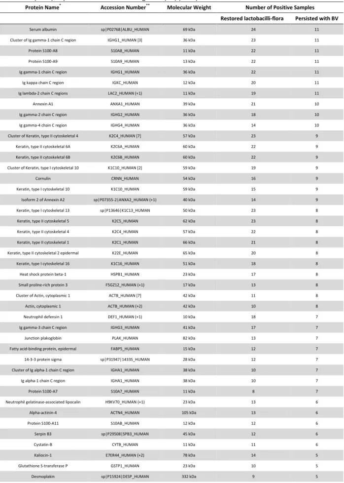
A total of 116 proteins were identified by proteomic analysis of cervicovaginal fluid
from 38 women with bacterial vaginosis at enrollment and 39 with normal vaginal flora. ,
(Table S1). For determining the proteomic profile of bacterial vaginosis, we compared the
group of women with this condition with those with normal flora. We found that among the
116 proteins identified, 2 of them, Cathepsin G and Ig heavy chain V-III region BRO were
exclusively detected in bacterial vaginosis, while 3 (leukocyte elastase inhibitor, involucrin
and neuroblast differentiation-associated protein AHNAK) were only found in normal
vaginal flora (Table 2). Cathepsin G and Ig heavy chain V-III region BRO were found in,
respectively, 7 (18.4%) and 4 (10.5%) women with bacterial vaginosis, while leukocyte
elastase inhibitor was detected in 7 (17.9%), involucrin in 3 (7.7%) and neuroblast
differentiation-associated protein AHNAK in 2 (5.1%) of the 39 women with normal vaginal
Quantitative analysis of normalized spectra between bacterial vaginosis and normal
flora showed a total of 20 (17.2%) proteins that significantly differed between the groups
(Table 2). We could observe that among the total of proteins differently expressed, 9
(45.0%) play a role in the immune response and defense against pathogens. Among these
immunity-related proteins that were differently expressed we found haptoglobin 25 times
more expressed in bacterial vaginosis, kaliocin-1 and neutrophil elastase 4 times more
expressed, cathepsin G, Ig lambda-2 chain, neutrophil elastase, protein S100-A8 and Ig
heavy chain V-III that were also overexpressed in bacterial vaginosis, while cluster of serpin
B3 and leukocyte elastase inhibitor were more abundant in normal flora. These
differently-expressed proteins were also classified according to biological process, cellular component
and molecular function (Figure 1). We observed that most of them are involved in cellular
processes (16%.0), are located in the extracellular region (21.0%) or cytoplasm (20.0%) and
present molecular function (35.0%) or binding activity (31.0%).
The 38 women with bacterial vaginosis that completed the treatment with
metronidazole and returned for follow-up were separated into two groups according to the
pattern of vaginal flora exhibited after 45 days of the end of treatment, except 3 women
that were excluded as they returned with vaginal candidosis. Thus, considering the
proteomic analysis of cervicovaginal fluids of 11 women who persisted with bacterial
vaginosis and 24 who were successfully treated, a total of 87 proteins could be identified
(Table S2). No statistically significant difference was detected between the relative
abundance of proteins in these 2 groups. However, we found that apoliprotein A-I and
cathepsin G were exclusively detected in, respectively, 3 (12.5%) and 5 (20.8%) women
Table 1. Sociodemographic, behavioral and clinical characteristics of 213 women whose
samples were included in the study analysis, distributed according to their pattern
of vaginal flora.
Variables Normal flora
(n=132)
Bacterial vaginosis (n=81)
P
Age, median (range), years£ 34 (17-51) 31 (17-48) 0.47
Race (self-defined), n (%)∫
Nonwhite (n=90) 48 (53.3) 42 (46.7) 0.03
White (n=123) 84 (68.3) 39 (31.7)
Marital status, n (%)∫
Single (n=70) 35 (50.0) 35 (50.0) <0.001
Married (n=143) 97 (67.8) 46 (32.2)
Years at school, median (range)£ 9 (0 - 16) 8 (0 - 15) 0.10
Remunerated activity, n (%)∫
Yes (n=121) 75 (62.0) 46 (38.0) 0.99
No (n=92) 57 (62.0) 35 (38.0)
Smoking habit, n (%)∫
Yes (n=43) 22 (51.2) 21 (48.8) 0.10
No (n=170) 110 (64.7) 60 (35.2)
Sex partners, prior 12 months, n (%)∫
0 or 1 (n=188) 120 (63.8) 68 (36.2) 0.13
2 or more (n=25) 12 (48.0) 13 (52.0)
Number of vaginal intercourse/week, median (range) £ 2 (0-7) 2 (0-7) 0.90
Previous BV, n (%)∫
Yes (n=100) 55 (55.0) 45 (45.0) 0.05
No (n=113) 77 (68.1) 36 (31.9)
Previous STD, n (%)∫
Yes (n=17) 10 (58.8) 7 (41.2) 0.78
No (n=196) 122 (62.2) 74 (37.8)
Consistent condom use, n (%)∫
Yes (n=71) 43 (60.7) 28 (39.4) 0.76
No (n=142) 89 (62.7) 53 (37.3)
Hormonal contraception use, prior 12 months, n (%)∫
Yes (n=94) 65 (69.1) 29 (30.9) 0.05
No (n=119) 67 (56.3) 52 (43.7)
Parity, n (%)∫
0 (n=45) 30 (66.7) 15 (33.3) 0.53
≥ (n=168) 102 (60.7) 66 (39.3)
Vaginal pH, median (range)£ 4.7 (4.0-5.0) 4.7 (4.0-7.0) <0.001
KOH test, n (%)∫
Positive or doubtful (n=136) 66 (48.5) 70 (51.4) <0.001
Negative (n=77) 66 (85.7) 11 (14.3)
Complaints, n (%)∫ Discharge (n=90) Odor (n=54) Itching (n=31) 42 (46.7) 22 (40.7) 19 (61.3) 48 (53.3) 32 (59.2) 12 (38.7) <0.001 <0.001 0.93 £
Mann Whitney test, p<0.05 considered as significant; ∫
Table 2. Proteins differentially expressed in the cervicovaginal fluids of women with
bacterial vaginosis and normal flora.
Proteins Normal
Mean (SD) Bacterial vaginosis Mean (SD) P** Fold Change
Leukocyte elastase inhibitor Involucrin
Neuroblast differentiation-associated protein AHNAK Cathepsin G
Ig heavy chain V-III region BRO
0.22 (0.51) 0.42 (1.52) 0.15 (0.85) - - - - - 0.30 (0.72) 0.56 (1.75) 0.01 0.09 0.28 0.01 0.04 - - - - -
Small proline-rich protein 3 17.27 (11.51) 3.34 (6.42) <0.0001 0.20
Periplakin 2.98 (2.67) 0.62 (1.31) <0.0001 0.20
Cluster of Cornifin-B 7.14 (8.25) 1.54 (3.51) <0.01 0.20
Cellular retinoic acid-binding protein 2 (Fragment) 0.31 (0.47) 0.02 (0.13) <0.01 0.07
Cluster of Fatty acid-binding protein, epidermal 3.07 (1.93) 1.53 (2.18) <0.01 0.50
Cluster of Serpin B3 3.67 (3.98) 1.31 (2.20) <0.01 0.40
Serum albumin 26.59 (25.10) 48.24 (33.77) <0.01 1.80
Glyceraldehyde-3-phosphate dehydrogenase 0.84 (1.22) 0.16 (0.56) <0.01 0.20
Kaliocin-1 1.07 (1.71) 4.48 (6.90) <0.01 4.20
Cystatin-A 1.92 (2.72) 0.39 (1.87) <0.01 0.20
Protein S100-A11 1.80 (1.53) 1.00 (1.23) 0.01 0.60
Junction plakoglobin 2.57 (2.32) 1.41 (1.86) 0.02 0.50
Ig lambda-2 chain C regions 1.49 (1.77) 2.45 (1.88) 0.02 1.60
Neutrophil elastase 0.14 (0.41) 0.59 (1.13) 0.02 4.20
Isoform 2 of Annexin A2 3.83 (4.01) 2.17 (2.76) 0.04 0.60
Haptoglobin 0.02 (0.11) 0.46 (1.34) 0.04 25.00
Protein S100-A8 2.93 (1.83) 4.74 (5.14) 0.04 1.60
*
Mean (SD: standard deviation) of normalized total spectra of peptides.
**
Figure 1. Pie charts representing the gene ontology categorization of the proteins
differentially expressed identified by LC-MS/MS between women with normal
vaginal flora and with bacterial vaginosis. Classification according to: A) biological
process, B) cellular component and C) molecular function.
DISCUSSION
This study is the first to describe the full proteomic profile of cervicovaginal fluid of
women with bacterial vaginosis comparing the proteome of women who were successfully
regimen, as recommended by CDC.13 The inclusion criteria of women in this study were very
strict to assure that none of the samples had any concurrent genital tract infection that
could interfere in the profile of proteins. Although we were able to successfully identify a
large number of differently expressed proteins in the cervicovaginal fluid of women with
bacterial vaginosis, no peptide was associated with bacterial vaginosis treatment failure
with metronidazole.
The majority of proteins identified in cervicovaginal fluids were from plasma
transudate and epithelial cells secretion, which is in agreement with previous study that also
evaluated cervicovaginal fluid samples by shotgun proteomic analysis.27 In fact, our data
show that a high proportion of proteins identified in the cervicovaginal fluid is located in the
extracellular space and cytoplasm. Intracellular proteins, as well as those proteins involved
in epidermis development and keratinization, were also frequently identified, which is
explained by the constant desquamation the vaginal epithelium undergoes.20
When comparing bacterial vaginosis cervicovaginal fluids with normal vaginal flora,
we found that a significant portion of the differently expressed proteins are involved with
immune response and defense against pathogens, which is in agreement with the
literature.28 Among proteins with immune function that were differently expressed, most of
them were overexpressed in bacterial vaginosis. Additionally, all proteins that were
exclusively found in bacterial vaginosis also play a role in immune response. Taken together,
these findings demonstrate the cervicovaginal fluid proteome is influenced by the type of
flora and that dysregulated proteins are mostly immunity-related.
When evaluating the function of those proteins that were overexpressed or
exclusively detected in women with normal vaginal flora, we found two proteins that act as
elastase inhibitor.29,30 In addition to these immune-related proteins, we also found that
normal vaginal flora has increased abundance of isoform 2 of annexin A2 that is involved in
heat-stress response.31 The remaining proteins that were overexpressed in normal vaginal
flora are responsible by epidermis development and differentiation of keratinocytes
(involucrin, protein S100 A11, small proline-rich protein 3, periplakin, cornifin A, cornifin B,
cellular retinoic acid-binding protein 2 and cluster of fatty acid-binding protein,
epidermal).32-36 So, these data indicate that normal vaginal flora is associated with
overexpression of proteins involved in epithelium structure. This finding can be explained by
the fact that women with normal lactobacilli-dominated flora have lower vaginal pH, which
leads to lysis of epithelial cells and consequently increased desquamation of epithelium,
which does not happen in bacterial vaginosis. We also noted that some overexpressed
protein in normal flora are localized in the cell junctions, such as junction of plakoglobin,
periplakin and cystatin A.32,34,36 These proteins not only are responsible for signaling in cell
to cell communication, but also are essential for epithelial cell adhesion playing an
important role as epithelial barrier against the entry of microorganisms.
Of those proteins exclusively found in bacterial vaginosis or overexpressed in this
condition, they were all derived from plasma transudate. Among these, serum albumin is
the main protein of plasma with binding activity to several molecules.37 Other
overexpressed proteins in bacterial vaginosis include: haptoglobin, which results from
hemolysis and have antimicrobial and antioxidant activity modulating many aspects of the
acute phase response;38,39 cathepsin G, a serine protease that have antibacterial activity
mainly against lipopolysaccharide (LPS) of Gram-negative bacterium;40-42 neutrophil
elastase, involved in the regulation of innate immunity, inflammation and infection, can