Marcos André dos Santos da Silva1, Edmundo Médici Filho2, Julio Cezar de Melo Castilho3, Cássia T. Lopes de Alcântara Gil4
Assessment of divine proportion in the cranial structure
of individuals with Angle Class II malocclusion on
lateral cephalograms
Introduction: The study of the Divine Proportion (φ = 1.618) began with the Greeks, having as main researchers the
mathematician Pythagoras and the sculptor Phidias. In Dentistry, Ricketts (1981-82) was an early to study this issue.
Objective: This study proposed to evaluate how some cephalometric measures are presented in relation to the
Divine Proportion, with the total of 52 proportions, formed by 28 cephalometric landmarks.
Methods: Lateral cephalograms of 40 Class II adults patients aging from 17 to 45 years (13 male and 27 female)
were evaluated. The linear distances between the landmarks were measured using Radiocef Studio software.
Results: After statistical analysis, the data shown an average of 65,48% in the Divine Proportion, 17,5% in the
rela-tion Ans-Op/V1S-DM16 and 97,5% in the relarela-tions Na-Me/Na-PoNa e Na-PoNa/Na-Gn.
Conclusion: Among all cephalometric measurements investigated, the lower facial third and the dental arches
showed the smallest percentages of Divine Proportion.
Keywords: Divine Proportion. Class II malocclusion. Cephalometry.
How to cite this article: Silva MAS, Médici Filho E, Castilho JCM, Gil CTLA. As-sessment of divine proportion in the cranial structure of individuals with Angle Class II malocclusion on lateral cephalograms. Dental Press J Orthod. 2012 May-June;17(3):88-97.
Submitted: March 9, 2009 - Revised and accepted: August 16, 2009
» The authors report no commercial, proprietary, or financial interest in the products or companies described in this article.
Contact address: Marcos André dos Santos da Silva Centro Universitário do Maranhão – UniCEUMA
R. Josué Montello, 1 – Renascença II – Zip code: 65.075-120 – São Luís/MA – Brazil E-mail: [email protected]
1 Post-Graduation Student, UNICEUMA.
2 Full Professor, School of Dentistry of São José dos Campos, UNESP.
3 Associate Professor, School of Dentistry of São José dos Campos, UNESP.
INTRODUCTION
At this moment human beings are increasingly concerned about esthetics, beauty and harmonious shapes, specially facial ones.4,23,24 Such concern ex-ists since pre-historic times, from the Paleolithic period until now.2,4 Beauty is a vital force that acts on the development of our lives and the human mind has been relentlessly searching for beauty in the different populations and periods.4,23 The search for prettier shapes that may satisfy the in-dividual represents the endless desire for perfec-tion and balance, leading to the concept of design and esthetics. However, the evaluation of beauty may be relative and abstract, i.e. something that is inside the mind of each person.
The dental treatment should follow artistic and scientific regulations. The teeth must be estheti-cally pleasant and fully functional with other facial structures. Orthodontists should not solely move teeth and gingiva by the fast techniques or strictly apply conventional methods. There is no univer-sal treatment for all patients, since this might not be in accordance with nature and arts. The final goal after achieve a normal occlusion should be an improvement in facial esthetics. If the propor-tions are distorted instead of being reestablished, the employed method may have been unsuccessful and shall affect the final outcome. The association of scientific knowledge, meticulous and system-atic observation, application of beauty rules, daily training and effort to improve health of the patient and beauty allows the clinicians to promote the health and happiness of patients.18,24
The study of Divine Proportion was initiated by the Greeks, being the main researchers the math-ematician Pythagoras and the sculptor Phidias. These investigators noticed that some findings were related to certain standards and numbers, which might explain the beauty and harmony ob-served in nature.9,10,11 The Divine Proportion is one of the most effective resources of esthetic propor-tionality available. It has been widely employed throughout the art history. The ancient Egyp-tians already knew the golden ratio and applied it in the construction of the pyramids. The Greeks employed it in their temples, the great artists in their paintings and sculptures, and even the great
composers applied it in their works. The Divine Proportion may be used for morphological analy-sis and esthetic evaluation of the teeth and facial skeleton and soft tissues, since many proportions found and defined as beautiful from human point of view, or comfortable and pleasant from a physi-cal standpoint, display this proportion. Therefore, it was indicated for analysis of the structural har-mony and may be applied in the orthodontic treat-ment planning, as well as in the planning of maxil-lofacial and plastic surgeries.14,19 Thus, the search for an ideal esthetics might be scientifically con-ducted instead using subjective perceptions.18
The investigation of this issue calls the inter-est of different areas such as Orthodontics, Maxil-lofacial Surgery, Plastic Surgery and Esthetics. It has also been applied in cephalometric analyses by authors such as Ricketts,18 Zietsman et al,25 Gil,8 Gil and Medici Filho7 and Medici Filho at al14 who dem-onstrated the existence of Divine Proportion be-tween different measurements of the human skull. According to Baker and Woods2, few studies have been published on the Divine Proportion observed in the measurements of human skull. This demon-strates the importance of the present study, which aimed at evaluating the Divine Proportion in lateral cephalograms of Class II adult subjects, who were not submitted to previous orthodontic treatment.
MATERIAL AND METHODS
The sample comprised lateral cephalograms of 40 untreated Class II adult individuals (13 males and 27 females), aging from 17 to 45 years, with an ANB angle larger than 6° and no craniofacial defor-mities, syndromes or cleft lip and palate.
The work was carried out as follows:
besides some other landmarks suggested by Gil and Medici Filho12 and demonstrated in Figure 1 and Table 1. The linear measure-ments were measured on the Radiocef Studio software. The analyses LDA1 and LDA2 com-prised 52 factors each, and each factor of the LDA1 was divided by the corresponding fac-tor on the LDA2. For example, the facfac-tor #1 of the LDA1 was divided by factor #1 of the LDA2 and so on up to factor #52 for verification of the presence or absence of Divine proportion in each radiograph. It should be highlighted that the larger value is always divided by the smaller value in order to facilitate the statis-tical calculations, i.e. the factors presented in LDA1 would be in Divine Proportion with their corresponding factors in LDA2 if this division yielded values ranging from 1.431 to 1.853, as advocated by Gil8 in 2001.
» As an attempt to eliminate possible mark-ing errors, each radiograph was traced twice, with a 15-day interval between them. Error calculation was conducted by the Intraclass Correlation Coefficient (ICC), which repre-sents the total estimate of variability induced by individual variations. This coefficient esti-mates the degree of agreement between two values achieved in distinct moments.12 The examinations were individually analyzed by the author by means of the LDA1 and LDA2, applied for each patient (Tables 2 and 3). » Statistical analysis of the linear
measure-ments achieved by means of the LDA1 and LDA2 calculated on the Radiocef Studio soft-ware were conducted in order to observe the presence or absence of Divine Proportion in the human skull.
STATISTICAL ANALYSIS
Statistical analysis of the data was based on the fol-lowing concept of divine proportion: One pair of mea-surements (A, B) is in Divine Proportion if A/B = 1.618, where A>B. The range from 1.431 to 1.853 was em-ployed to assess the pairs of measurements in Divine Proportion, as suggested by Gil.7
The Minitab 13 software (Minitab Inc, State College, USA) was employed for calculation of the
divisions of the factors of LDA1 by those of LDA2 for each radiograph. It should be highlighted that this division was also performed by division of the largest value by the smallest value. After calculation of these proportions, the Statistix for Windows 7.0 software (Analytical Software, Tallahassee, USA) was used to submit the data to Descriptive Statisti-cal Analysis (mean, standard deviation and median) at a confidence interval of 95%. This software also allowed calculation of the frequency distribution in order to establish how many factors in each radio-graph were within the range established and, there-fore, in Divine Proportion (Tables 1 and 2) (Fig 2).
RESULTS
Results are shown in Figure 2 and Tables 1 and 2.
DISCUSSION
The study of Divine Proportion in Dentistry was initiated in the 70s and 80s and was mainly con-ducted by Torres22 and Ricketts.18,19 Investigation of this subject has provided important contribu-tions to the improvement and enhancement of the diagnosis and treatment planning of the patients, providing dentists a further instrument to evalu-ate whether shape, harmony, esthetics and cranio-facial proportion are present.7,8,14,18,19,22,23,24
The sample of the present study comprised 40 lateral cephalograms of 40 untreated Class II adult subjects (13 males and 27 females) with more than 17 years of age. Ricketts5 employed a sample of 30 lateral cephalograms of adult Peruvian male pa-tients with normal occlusion and no admixture of races for assessment of the presence of Divine Pro-portion. Gil8 and Gil and Medici Filho7 observed the Golden Proportion in the cranial structures on a population of 23 untreated adult subjects with normal occlusion, of both genders, by means of lat-eral, frontal and axial cephalograms.
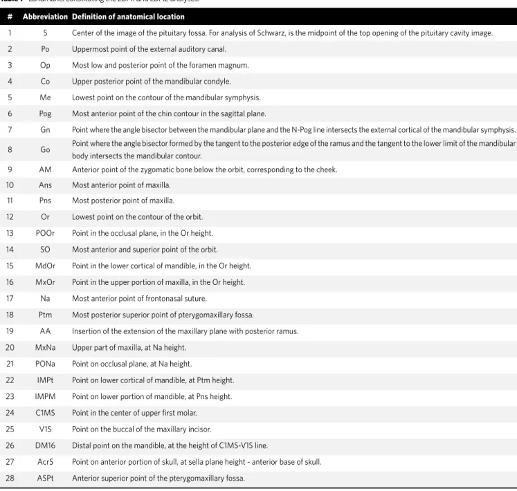
Figure 1 - Landmarks constituting the LDA1 and LDA2 analyses.
# Abbreviation Definition of anatomical location
1 S Center of the image of the pituitary fossa. For analysis of Schwarz, is the midpoint of the top opening of the pituitary cavity image.
2 Po Uppermost point of the external auditory canal.
3 Op Most low and posterior point of the foramen magnum.
4 Co Upper posterior point of the mandibular condyle.
5 Me Lowest point on the contour of the mandibular symphysis.
6 Pog Most anterior point of the chin contour in the sagittal plane.
7 Gn Point where the angle bisector between the mandibular plane and the N-Pog line intersects the external cortical of the mandibular symphysis.
8 Go Point where the angle bisector formed by the tangent to the posterior edge of the ramus and the tangent to the lower limit of the mandibular body intersects the mandibular contour.
9 AM Anterior point of the zygomatic bone below the orbit, corresponding to the cheek.
10 Ans Most anterior point of maxilla.
11 Pns Most posterior point of maxilla.
12 Or Lowest point on the contour of the orbit.
13 POOr Point in the occlusal plane, in the Or height.
14 SO Most anterior and superior point of the orbit.
15 MdOr Point in the lower cortical of mandible, in the Or height.
16 MxOr Point in the upper portion of maxilla, in the Or height.
17 Na Most anterior point of frontonasal suture.
18 Ptm Most posterior superior point of pterygomaxillary fossa.
19 AA Insertion of the extension of the maxillary plane with posterior ramus.
20 MxNa Upper part of maxilla, at Na height.
21 PONa Point on occlusal plane, at Na height.
22 IMPt Point on lower cortical of mandible, at Ptm height.
23 IMPM Point on lower portion of mandible, at Pns height.
24 C1MS Point in the center of upper first molar.
25 V1S Point on the buccal of the maxillary incisor.
26 DM16 Distal point on the mandible, at the height of C1MS-V1S line.
27 AcrS Point on anterior portion of skull, at sella plane height - anterior base of skull.
28 ASPt Anterior superior point of the pterygomaxillary fossa.
Table 2 - Lateral Divine Analysis 1.
Computerized Cephalometrics – Lateral Divine Analysis 1
Patient: Age: Gender:
Orthodontist: Date:
Factors Landmarks 1 Value found Landmarks 2
1 Na-Me Na 0.00 Me
2 Na-Me Na 0.00 Me
3 Na-Me Na 0.00 Me
4 Ans-Me Ans 0.00 Me
5 Ans-Me Ans 0.00 Me
6 Na-Ans Na 0.00 Ans
7 Na-Ans Na 0.00 Ans
8 Na-Ans Na 0.00 Ans
9 Na-Ans Na 0.00 Ans
10 Na-Ans Na 0.00 Ans
11 Na-Ans Na 0.00 Ans
12 Na-Ans Na 0.00 Ans
13 Na-Poor Na 0.00 Poor
14 Na-Poor Na 0.00 Poor
15 Na-Poor Na 0.00 Poor
16 Na-Poor Na 0.00 Poor
17 Na-Poor Na 0.00 Poor
18 Pt-IMPt Pt 0.00 IMPt
19 Pt-IMPt Pt 0.00 IMPt
20 Pt-IMPt Pt 0.00 IMPt
21 Pns-ImPm Pns 0.00 ImPm
22 Pns-ImPm Pns 0.00 ImPm
23 SO-Or SO 0.00 Or
24 SO-Or SO 0.00 Or
25 SO-Or SO 0.00 Or
26 A-Pog A 0.00 Pog
27 A-Pog. A 0.00 Pog.
28 A-Pog. A 0.00 Pog.
29 A-Pog. A 0.00 Pog.
30 A-Pog. A 0.00 Pog.
31 Ans-Pns Ans 0.00 Pns
32 Ans-Pns Ans 0.00 Pns
33 Ans-Pns Ans 0.00 Pns
34 Ans-Pns Ans 0.00 Pns
35 Ans-Pns Ans 0.00 Pns
36 Ans-Pns Ans 0.00 Pns
37 Pog-Op Pog 0.00 Op
38 Pog-Op Pog 0.00 Op
39 Pog-Op Pog 0.00 Op
40 Pog-Op Pog 0.00 Op
41 Na-Op Na 0.00 Op
42 Na-Op Na 0.00 Op
43 Na-Op Na 0.00 Op
44 Na-Op Na 0.00 Op
45 Na-Op Na 0.00 Op
46 Ans-Op Ans 0.00 Op
47 Ans-Op Ans 0.00 Op
48 Ans-Op Ans 0.00 Op
49 V1S-C1MS V1S 0.00 C1MS
50 V1S-C1MS V1S 0.00 C1MS
51 Mdor-Poor Mdor 0.00 Poor
52 Mdor-Poor Mdor 0.00 Poor
Computerized Cephalometrics – Lateral Divine Analysis 2
Patient: Age: Sex:
Orthodontist: Date:
Factors Landmarks 1 Value found Landmarks 2
1 Ans-Me Ans 0.00 Me
2 Na-PoNa Na 0.00 PoNa
3 Pt-IMPt Pt 0.00 IMPt
4 Na-Gn Na 0.00 Gn
5 Co-Gn Co 0.00 Gn
6 Ans-AA Ans 0.00 AA
7 Go-Pog Go 0.00 Pog
8 Na-PoNa Na 0.00 PoNa
9 Or-Me Or 0.00 Me
10 V1S-AA V1S 0.00 AA
11 Pns-Op Pns 0.00 Op
12 S-Acrs S 0.00 Acrs
13 Co-Gn Co 0.00 Gn
14 Na-Gn Na 0.00 Gn
15 Pns-IMPM Pns 0.00 IMPM
16 Na-MxN Na 0.00 MxN
17 Or-Poor Or 0.00 Poor
18 Co-Gn Co 0.00 Gn
19 Na-Gn Na 0.00 Gn
20 Pns-IMPM Pns 0.00 IMPM
21 Go-Pog Go 0.00 Pog
22 Co-Am Co 0.00 Am
23 Mxor-So Mxor 0.00 So
24 Mxor-Mdor Mxor 0.00 Mdor
25 Ans-Pog Ans 0.00 Pog
26 Or-Me Or 0.00 Me
27 Po-Na Po 0.00 Na
28 V1S-C1MS V1S 0.00 C1MS
29 V1S-AA V1S 0.00 AA
30 V1S-AA V1S 0.00 AA
31 V1S-C1MS V1S 0.00 C1MS
32 Op-Pns Op 0.00 Pns
33 Or-Me Or 0.00 Me
34 SO-Poor SO 0.00 Poor
35 Ans-AA Ans 0.00 AA
36 Op-Pns Op 0.00 Pns
37 Op-ASPt Op 0.00 ASPt
38 Or-Me Or 0.00 Me
39 Go-Pog Go 0.00 Pog
40 V1S-AA V1S 0.00 AA
41 Op-Pns Op 0.00 Pns
42 SO-Poor SO 0.00 Poor
43 Or-Me Or 0.00 Me
44 Go-Pog Go 0.00 Pog
45 V1S-AA V1S 0.00 AA
46 Op-Pns Op 0.00 Pns
47 Go-Pog Go 0.00 Pog
48 V1S-AA V1S 0.00 AA
49 Ans-Pns Ans 0.00 Pns
50 Ans-Pog Ans 0.00 Pog
51 Mxor-Mdor Mxor 0.00 Mdor
52 Mxor-Poor Mxor 0.00 Poor
A
D
G
J
B
E
H
K
C
F
I
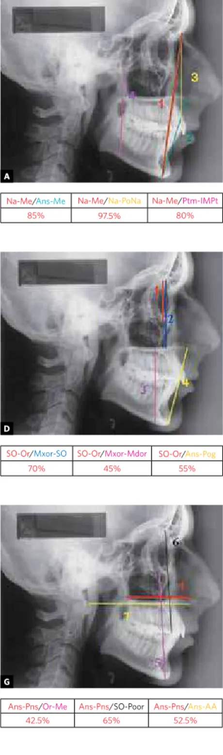
Figure 2 - Proportions found relating the cephalo-metric factors.
Na-Me/Ans-Me Na-Me/Na-PoNa Na-Me/Ptm-IMPt
85% 97.5% 80%
Na-Ans/Ans-AA Na-Ans/Go-Pog Na-Ans/Na-PONa
60% 47.5% 90%
Na-PoNa/Na-MxN Na-PoNa/Or Poor
95% 85%
SO-Or/Mxor-SO SO-Or/Mxor-Mdor SO-Or/Ans-Pog
70% 45% 55%
A-Pog/V1S-C1MS A-Pog/V1S-DM16
65% 77.5%
Ans-Pns/V1S-DM16Ans-Pns/V1s-CaMSAns-Pns/Op-Pns
52.5% 30% 42.5%
Ans-Pns/Or-Me Ans-Pns/SO-Poor Ans-Pns/Ans-AA
42.5% 65% 52.5%
Pog-Op/Op-Pns Pog-Op/Go-Pog Pog-Op/V1S-DM16
67.5% 82.5% 45%
Na-Op/Op-Pns Na-Op/Go-Pog Na-Op/V1S-DM16
75% 60% 55%
V1S-C1MS/Ans-Pns V1S-C1MS/Ans-Pog
30% 62.5%
Ans-Op/Op-Pns Ans-Op/Go-Pog Ans-Op/V1S-DM16
1 2 3 4 5 6 7 8 9 10 11 12 13 14 15 16 17 18 19 20 21 22 23 24 25 26 27 28 29 30 31 32 33 34 35 36 37 38 39 40 100%
90% 80%
70% 60% 50% 40% 30% 20% 10% 0% 1 3 5 7 9 11 13 15 17 19 2123 25 27 29 31 33 35 37 39 41 43 45 47 49 51
100% 90% 80% 70% 60% 50% 40% 30% 20% 10% 0%
including some of Divine Proportion, whereas the non-pretty subjects displayed a correspondence of just 38.33%. The present study did not evaluate the patient’s attractiveness, since our sample suggests the presence of a facial esthetic imbalance second-ary to the Angle Class II malocclusion present.
Ricketts,18 Zietsman et al,25 Garbin,5,6 Piccin,16 Snow,21 Araújo et al1 and Oliveira Junior15 conduct-ed specific investigations on the oromaxillofacial structures and also found Divine Proportion. For example, Ricketts18 observed this proportion in horizontal and vertical measurements. Gil,8 Gil and Medici Filho7 and Medici Filho14 found the presence of several measurements in Golden Pro-portion, which were related to each other in sev-eral manners and provided the human skull with an effective balance. These findings strongly sug-gested that the skull, as well as other structures in nature, follows the laws of conservation of en-ergy and thus is a very effective structure in both shape and composition. In the present study, many structures were found to be in Divine Proportion, as demonstrated on the tables and figures.
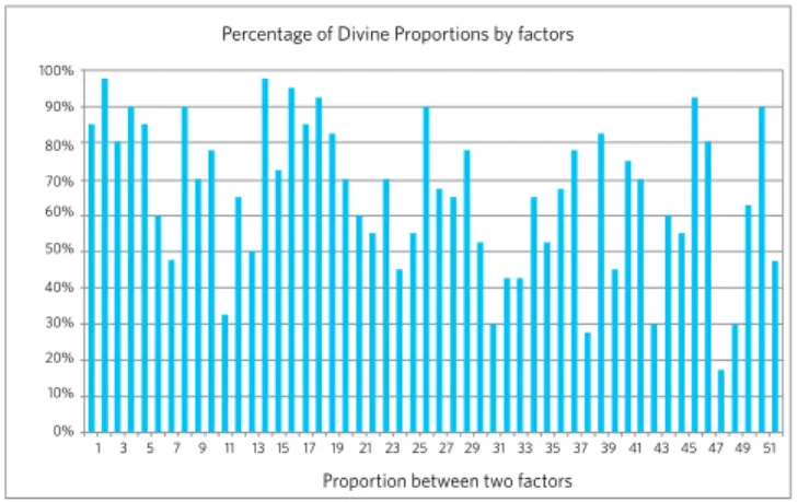
Radiographic cephalometrics consists on the measurement of physical, linear and angular di-mensions in skull radiographs. It is a very good auxiliary and supplementary instrument for diag-nosis and may even be regarded as essential for ob-servation of growth and evaluation of orthodontic treatments. This technique has been and still is the most widely employed for assessment of the facial growth, facial profile and also of the relationship Figure 3 - Graphic of percentages of divine proportion between factors.
Percentage of Divine Proportions by factors
Proportion between two factors
Figure 4 - Graphic of percentages of divine proportion between patients. Percentage of divine proportions by patients
Radiographs
between maxilla and mandible in human beings. Some authors have employed it to investigate the presence of Divine Proportion in the oromaxillofa-cial structures and achieved satisfactory outcomes (Ricketts,18 Zietsman et al,25 Garbin,5,6 Araújo et al,1 Baker and Woods,4 Gil and Medici Filho,7 Medici Filho et al14). The present study comprised evalua-tion of measurements of the human skull structure by means of landmarks and factors measured on lateral cephalograms, by means of a computerized cephalometric software called Radiocef Studio. Ac-cording to Martins13 and Brangeli,3 the advent of informatics and its application in clinical cephalo-metrics has provided high-technology resources for the achievement of elements of diagnosis and also for manipulation of such elements, for the accom-plishment of projections, analyses and treatment simulations, enhancing and facilitating selection of the best therapeutic approach. On the other hand, there may be errors in the cephalometric analy-ses with employment of the computer, leading to doubtful measurements with employment of this method. Error control is fundamental for the out-comes of cephalometric investigations to be valid.10
Now we are going to discuss the results of Di-vine Proportions observed in the present study, which shall be divided by groups of factors of ceph-alometric measurements in order to make inter-pretation of such outcomes easier.
than 80% of the sample, suggesting that even in the presence of Class II malocclusions the muscle forces that define the vertical dimension were pres-ent and could provide balance, harmony and even a proper facial proportion. It should be noticed that Na-Me represents the anterior facial height of the patient in frontal view and was in Divine Proportion with the intermaxillary distance (ANS-Me) when in occlusion. In 1982, Ricketts18 found Divine Propor-tion when related similar measurementes to Na-Me and ANS-Me in soft tissue, using photographs of beautiful women (models) of different races.
The present results are also in agreement with Gil8 and Gil and Medici Filho,7 who also observed a percentage of Golden Proportion above 80% in an evaluation of lateral cephalograms of patients with normal occlusion.
Relationship between measurements compris-ing just one point at the maxilla and another at the skull, Na-ANS / Na-PONa, (Fig 2B), revealed the presence of Divine Proportion in 90% of the sam-ple. However, the observation of the correlation Na-ANS / ANS-AA, on which one cephalometric point is located at the mandible (AA), the percent-age of Divine Proportion was decreased to 60% of the sample. Moreover, the correlation Na-ANS / Go-Pog, which related one factor with one point at the maxilla and another at the skull to another factor measured just in the mandible, revealed the presence of Divine Proportion in just 47.5% of the cases. These values are different from the findings of Gil,8 and Gil and Medici Filho,7 which observed Divine Proportion in such relationship in more than 80% of the sample. This difference might be assigned to a retruded mandible in relation to the maxilla as observed in Class II patients.
Figure 2C demonstrates the presence of Divine Proportion in 95% of the patients for Na-PoNa / Na-MxN and 85% of the patients for Na-PoNa / Or-Poor; these factors are located just at the maxilla and facial bones and therefore are not influenced by the disproportion existing between maxilla and mandible of Class II patients. These observations were in agreement with Gil,8 Gil and Medici Filho.7
The measurements SO-Or / Mxor-SO (Fig 2D), which are measurements of the maxilla and upper facial third, displayed a higher percentage of Divine
Proportion (70%) than the measurements SO-Or / Mxor-Mdor and SO-Or / ANS-Pog, 45% and 55% re-spectively, which comprise maxillary and mandibular measurements and therefore are more susceptible to the alterations observed in subjects with malocclu-sion. For that reason, these outcomes disagree with the findings of Gil,8 and Gil and Medici Filho.7
Divine Proportion was observed in 65% of cases for the A-Pog / V1S-C1MS and in 77.5% for A-Pog /V1S-DM16 (Fig 2E). These factors are prone to variations that are directly related to occlusal dis-turbances, since they are horizontal factors on the maxilla and thus may vary with the mandibular retraction in relation to the maxilla. Another pos-sible explanation for this reduced ratio of Divine Proportion might be the involvement of factors based on points on the teeth, which are similarly influenced by malocclusions. Thus, these per-centages of Divine Proportions were smaller than those observed by Gil8 and Gil and Medici Filho,7 who found the presence of Divine Proportion in more than 80% of the subjects in skeletal and den-tal measurements and also on denden-tal and skeleden-tal measurements on the maxillary incisors.
The comments on Figure 2E are confirmed in Figure 2F, which demonstrates presence of Di-vine Proportion for the horizontal measurements in 42.5% for the ANS-PNS / Op-Pns and 52.5% for the ANS-PNS / V1S-DM16, i.e., factors influenced by the anterior posterior relationship between maxilla and mandible, and in 30% for ANS-PNS / V1S-C1MS, which also involved the teeth.
Araújo et al1 observed that the patients pre-sented different responses to treatment and found statistical differences in the outcomes between the pre- and post-operative data in the proportions A-1 / 1-Pm and Co-Xi / Xi-Pm. Yet this did not occur for the proportion Pfr-A / A-Pm, which presented a significant difference, revealing no alterations with surgery from an esthetic point of view. The authors explained that the vertical measurements, compared to the Co-Xi / Xi-PM measurement, dis-played a smaller alteration with the mandibular advancement, which provides a larger change in anterior posterior than in vertical direction.
base ANS-PNS in relation to the arch size V1S-C1MS, which leads to such disharmony. Similarly, Figure 2G reveals presence of Divine Proportion in 42.5% for ANS-PNS / Or-Me, 65% for ANS-PNS / SO-Poor and 52.5% for ANS-Pns / ANS-AA. There-fore, the ratios between cephalometric factors dis-played a smaller percentage of Divine Proportion than reported by Gil8 and Gil and Medici Filho.7
According to Gil,8 when one factor in the groups of measurements Pog-Op, Na-Op and ANS-Op is in proportion with one of these measurements, it shall also be in proportion with the other two mea-surements. The three measurements were regard-ed as equal in his study. However, in the present study the relationship between the factors Pog-Op, Na-Op and ANS-Op with each of the factors Op-PNS, Go-Pog and V1S-DM16 (Fig 2H, I and J) presented different results, as shown in Table 4.
Figure 2L represents positioning of the maxil-lary incisor and maxilmaxil-lary first molar, which refer to the anterior posterior positioning of the tooth, an important aspect for Class II patients. Corre-lation between factors of horizontal dimensions, (V1S-C1MS/ANS-PNS) revealed Divine Propor-tion in 30% of the patients, yet the correlaPropor-tion between one horizontal and one vertical factor (V1S-C1MS / ANS-Pog) displayed a percentage of Divine Proportion of 62.5%. These relationships displayed a larger percentage of Divine Proportion in the study of Gil8 and Gil and Medici Filho.7
In general, calculation of the mean of percentag-es of the 52 correlations between the cephalomet-ric factors investigated revealed a rate of 65.48% of Divine Proportion, different from the outcomes of Gil8 and Gil and Medici Filho,7 who found a per-centage above 80%. Moreover, Divine Proportion was observed in 17.5% for the ANS-Op/V1S-DM16 relationship and 97.5% for the Na-Me/Na-PoNa and Na-PoNa/Na-Gn correlations, which were the
lowest and highest percentages of Divine Propor-tion observed in the present sample, respectively.
During the development of this study and in agreement with the literature review, it could be no-ticed that even though the discovery of the Divine Proportion is very old, its study and application in health specialties and mainly in Dentistry are based on few studies. Investigations on this subject have been conducted since the ancient Greece, yet just in 1982 Ricketts18 demonstrated the presence of Divine Proportions in lateral cephalograms. As de-scribed, the Divine Proportion may play a very im-portant role in the evaluation of diagnosis and also as an auxiliary therapeutical tool in Dentistry.
CONCLUSIONS
Based on these methods and on the analysis of the results achieved, the following could be con-cluded on the cranial structure of untreated Class II adult subjects:
» There was a mean percentage of 65.48% of the cephalometric measurements in Divine Proportion.
» Among all cephalometric measurements investigated, the lower third of the head, as well as the dental arches of the individuals in this sample, were the areas on which the pro-portions displayed the smallest percentages of Divine Proportion.
Pog-Op / Op-Pns Pog-Op / Go-Pog Pog-Op /V1S-DM16
67.5% 82.5% 45%
Na-Op / Op-Pns Na-Op /Go-Pog Na-Op /V1S-DM16
75% 60% 55%
Ans-Op /Op-Pns Ans-Op /Go-Pog Ans-Op /V1S-DM16
92.5% 80% 17.5%
1. Araujo MM, Passer LA, Araujo A. Análise cefalométrica pré e pós-operatória das proporções divinas de Fibonacci em pacientes submetidos a avanço mandibular. Rev Dental Press Ortodon Ortop Facial. 2001 Nov-Dez;6(6):29-36.
2. Baker BW, Woods MG. The role of the divine proportion in the esthetic improvement of patients undergoing combined orthodontic/orthognathic surgical treatment. Int J Adult Orthodon Orthognath Surg. 2001;16(2):108-20.
3. Brangeli LAM, Henriques JFC, Vasconcelos MHF, Janson GRP. Estudo comparativo da análise cefalométrica pelo método manual e computadorizado. Rev Assoc Paul Cir Dent. 2000 maio-jun;54(3):234-41.
4. Colombini NEP. Cirurgia ortognática e cirurgia estético-funcional. 2003. [cited 2003 Jan 07]. Available from: http://www.sosdoutor.com.br /sosbucomaxilo facial/defeitos.asp.
5. Garbin AJI. Análise das proporções divinas em telerradiografias de perfil de pacientes submetidos à cirurgia de retroposicionamento mandibular [Tese de doutorado]. Piracicaba (SP): Universidade Estadual de Campinas, Faculdade de Odontologia de Piracicaba; 1999.
6. Garbin AJI, Passeri LA. Análise das proporções divinas de Fibonacci, em telerradiografias de perfil em pacientes dotados de oclusão normal. Ortodontia, 1999;32(3):29-40.
7. Gil CTLA, Medici Filho E. Estudo da proporção áurea na arquitetura craniofacial de indivíduos adultos com oclusão normal, a partir de telerradiografias axiais, frontais e laterais. Ortodontia. 2002 abr-jun;35(2):69-84.
8. Gil CTLA. Proporção áurea craniofacial. São Paulo (SP): Ed. Santos; 2001. 9. Hintz JM, Nelson TM. Haptic aesthetic value of the golden section. Br J Psychol.
1971 May;62(2):217-23.
10. Kamoen A, Dermaut L, Verbeeck R. The clinical significance of error measurement in the interpretation of treatment results. Eur J Orthod. 2001 Oct;23(5):569-78. 11. Knott R. Fibonacci number and golden section - Department of Mathematical and
Computing Science at the University of Surrey. [cited 2001 Jul 07]. Available from: http://www.mcs.surrey.ac.uk/Personal/R.Knott/ Fibonacci/fib.html.
REFERENCES
12. Loffredo LCM. Estudo da reprodutibilidade de informações na área de saúde [tese de doutorado]. Araraquara (SP): Universidade Estadual Paulista, Faculdade de Odontologia de Araraquara; 1996.
13. Martins LP, Pinto AS, Martins JCR, Mendes AJD. Erro de reprodutibilidade das medidas das análises cefalométricas de Steiner e Ricketts, pelo método convencional e método computadorizado. Rev Ortodon. 1995 Out;28(5): 4-17. 14. Medici Filho E, Martins MV, dos Santos da Silva MA, Castilho JC, de Moraes
LC, Gil CT. Divine proportions and facial esthetics after manipulation of frontal photographs. World J Orthod. 2007 Summer;8(2):103-8.
15. Oliveira Junior OB. Construtores de sorriso - ciência ou arte? [internet] 2003; [cited 2003 Feb 15]. Available from: http://www.apcdriopreto.com.br /art_ cientificos2.asp?código=6.
16. Piccin MR. Verificação da proporção divina da face de pacientes totalmente dentados [Dissertação]. Piracicaba (SP): Universidade Estadual de Campinas, Faculdade de Odontologia de Piracicaba; 1997.
17. Piehl J. The golden section: the “true” ratio? Percept Mot Skills. 1978 Jun;46(3 Pt 1):831-4.
18. Ricketts RM. The biologic significance of the divine proportion and Fibonacci series. Am J Orthod. 1982 May;81(5):351-70.
19. Ricketts RM. The golden divider. J Clin Orthod. 1981 Nov;15(11):752-9. 20. Ricketts RM. Perspectives in the clinical application of cephalometrics. The first
fifty years. Angle Orthod. 1981 Apr;51(2):115-50.
21. Snow SR. Esthetic smile analysis of maxillary anterior tooth width: the golden percentage. J Esthet Dent. 1999;11(4):177-84.
22. Torres R. Crecimiento armonioso y la divina proporción. Divulg Cult Odont. 1970 Jun;162(3):3-13.
23. Wuerpel EH. The inspiration of beauty. Angle Orthod. 1932 Out;2(4):201-18. 24. Wuerpel EH. On facial balance and harmony. Angle Orthod. 1937;7(2):81-9. 25. Zietsman ST, Wiltshire WA, Coetzee CE. The divine proportion and the cranial