Recebido em 07.07.2000. / Received in July, 07thof 2000.
Aprovado pelo Conselho Consultivo e aceito para publicação em 21.01.2003. / Approved by the Consultive Council and accepted for publication in January, 21thof 2003. * Trabalho realizado no Serviço de Dermatologia do Hospital de Clínicas de Porto Alegre, Universidade Federal do Rio Grande do Sul - UFRGS. / Work done at the Dermatology Service of the Hospital de Clínicas de Porto Alegre, Federal University of Rio Grande do Sul.
1 Médica dermatologista, ex-residente do Serviço de Dermatologia do Hospital de Clínicas de Porto Alegre. / M.D. Dermatologist, ex-resident at the Dermatology Service, Hospital de Clínicas de Porto Alegre.
3 Médica dermatologista, ex-residente do Serviço de Dermatologia do Hospital de Clínicas de Porto Alegre. / M.D. Dermatologist, ex-resident at the Dermatology Service, Hospital de Clínicas de Porto Alegre.
4 Professor Titular de Dermatologia - UFRGS. / Titular Professor of Dermatology - UFRGS.
©2003 by Anais Brasileiros de Dermatologia
Eritroqueratodermia Simétrica Progressiva: relato de caso
*Symmetrical Progressive Erythrokeratodermia: a case
report
*Cristiane Dal Magro
1Carina Pellenz
2Lucio Bakos
3Resum o : A eritroqueratodermia simétrica progressiva (EQSP) é genodermatose rara, caracterizada por
placas eritematosas e hiperceratóticas fixas, de distribuição simétrica nas extremidades. A primeira descrição da doença foi feita por Darier em 1911, e desde então existem poucas publicações a respeito do assunto. Os autores relatam um caso de EQSPem uma menina de nove anos de idade, com lesões localizadas nos cotovelos e joelhos, de aspecto típico. Os achados histológicos foram inespecíficos, e o tratamento com produtos de uso tópico, insatisfatório. A descrição do caso visa correlacionar os acha-dos clínicos, histológicos, e a evolução do quadro, dentro acha-dos conhecimentos atuais sobre a doença. Palavras-chave: Ceratose; dermatopatias genéticas.
Sum m a r y: Progressive sym m etric erythrokeratoderm a (PSEK) is a rare genetic skin disease,
charac-terized by fixed erythem atous and hyperkeratotic plaques, sym m etrically distributed over the extrem i-ties. The first description of the disease w as m ade by Darier in 1911, though since then there have been few related publications. The authors report the case of a 9-year-old girl w ith PSEK, presenting localized lesions over elbow s and knees, of typical aspect. The histologic findings w ere nonspecific and topical treatm ent unsatisfactory. The description of this case intends to correlate the clinical and histologic findings, as w ell as the clinical course of the patient, w ith the current know ledge regarding this disease. Key w ord s: Keratosis; Skin diseases, genetic.
Caso Clín ico / Case Report
INTRODUÇÃO
As eritroqueratodermias compreendem um grupo de doenças caracterizadas por distúrbio da queratinização. Apresentam-se clinicamente como lesões eritematosas e hiper-ceratóticas, marginadas, persistentes ou variáveis em relação ao aspecto e localização. Possuem herança autossômica dominan-te, com penetrância variável. Relatos de casos são esporádi-cos.1,2,4,7
A classificação atualmente aceita dessas entidades as separa em dois grupos: eritroqueratodermia variável (EQV) e eritroqueratodermia simétrica progressiva (EQSP).1,2,7
Neste trabalho é apresentado um caso de EQSP pau-cissintomática com reduzido número de lesões, bem como revisão da literatura sobre alguns aspectos dessa rara geno-dermatose.
INTRODUCTION
Erythrokeratodermia comprises a group of diseases characterized by a disturbance in the keratinization. They present clinically as erythematous and hyperkeratotic lesions, which are marginal, persistent or variable in rela-tion to their aspect and locarela-tion. They have autosomal dominant inheritance, with variable penetrance. Reports of cases are sporadic.1,2,4,7
The currently accepted classification divides these entities into two groups: variable erythrokeratodermia (VEK) and progressive symmetrical erythrokeratodermia (PSEK).1,2,7
RELATO DO CASO
Paciente do sexo feminino, nove anos, fototipo IV, com história de lesões cutâneas que surgiram em torno dos 18 meses de idade com progressão do quadro até cerca de 3 anos de idade. Não se queixava de qualquer sintoma local, seu estado geral era bom, e não possuía antecedentes mórbidos relevantes. Não havia história familiar de casos semelhantes, e os pais não eram consan-güíneos.
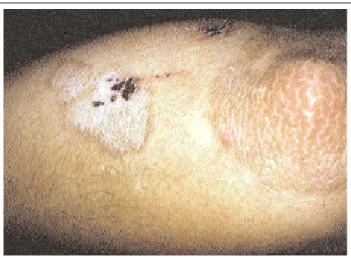
Ao exame dermatológico, apresentou placas eritêma-to-escamosas com hiperceratose exuberante, evidenciando sulcos irregulares de conformação cerebriforme, delimitadas nitidamente por um halo eritêmato-acastanhado, localizadas nos cotovelos e nos joelhos (Figuras 1 e 2). Também apre-sentava placas semelhantes, de contorno geográfico, nas faces extensoras das mãos e antebraços, mais pronunciadas à direita.
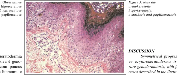
Foram realizadas duas biópsias de diferentes locais, que demonstraram achados semelhantes: hiperceratose ortoceratótica em trama de cesta, acantose discreta, papilo-matose exuberante, camada granulosa preservada, presença de ceratinócitos vacuolados, capilares dilatados e discreto infiltrado inespecífico de células mononucleares perivascu-lares na derme papilar (Figura 3).
Como tratamento, foram prescritos creme de tretinoí-na a 0,025% à noite e loção com lactato de amônia a 12% pela manhã. Após 60 dias, as lesões dos joelhos haviam regredido e nos cotovelos houve redução da hiperceratose, com a manutenção das lesões. Devido aos efeitos colaterais dos retinóides sistêmicos nessa faixa etária e à pouca quanti-dade de lesões, o uso da tretinoína tópica foi mantido.
Na ocasião da última consulta realizada, as lesões mantinham-se com pouca ceratose, leve eritema e limitadas aos cotovelos. A paciente abandonou o tratamento após o oitavo mês de acompanhamento clínico.
CASE REPORT
Female patient, nine years old, phototype IV, with a history of cutaneous lesions which appeared at 18 months of age with progression of the picture until she was about three of age. She did not complain of any loca-lized symptoms, her general state was good and she did not have any relevant morbid antecedents. There was no familial history of similar cases and the parents were not consanguineous.
Dermatological exam showed erythematous-scaly plaques and exuberant hyperkeratosis with irregular cere-briform furrows, clearly delimited by an erythematous to brownish-colored halo, located in the elbows and knees (Figures 1 and 2). She also presented similar plaques with geographical outline, in the extensor faces of the hands and forearms, which were more pronounced on the right side.
Two biopsies in different sites were performed, which demonstrated similar findings: orthokeratotic hyperkeratosis in a basket-weave pattern, discreet acan-thosis, exuberant papillomatosis, preserved granular layer, presence of vacuolated keratinocytes, dilated capillaries and a nonspecific discreet infiltrate of perivascular mono-nuclear cells in the papillary dermis (Figure 3).
For treatment, 0.025% tretinoin cream was prescri-bed to be applied at night in association with 12% ammo-nium lactate lotion in the morning. After 60 days, the lesions of the knees had regressed and there was a reduc-tion of the hyperkeratosis in the elbows, but with mainte-nance of the lesions. In view of the side effects of systemic retinoids in this age group and to the small quantity of lesions, the use of topical tretinoin was maintained.
At the time of the last consultation, the lesions still had a little keratosis, slight erythema and were limited to the elbows. The patient abandoned treatment after the eighth month of clinical follow-up.
Figu ra 1: Lesões n o cotovelo direito
Figu re 1: Lesion s in the right elbow
Figu ra 2: Lesões n o cotovelo esqu erdo
DISCUSSÃO
A eritroqueratodermia simétrica progressiva é geno-dermatose rara, com poucos casos descritos na literatura, e seu diagnóstico é firmado
muito mais em bases clínicas do que histológicas, uma vez que a última é inespecífica.2
Os primeiros relatos sobre a EQSPdatam de 1886, com Darier descrevendo um paciente com disqueratose congênita. Em 1908, Brocq e Dubreuilh estudaram o mesmo paciente de Darier e chamaram sua doença de "eri-troqueratodermia com placas simétricas".1,2,3 Em 1911,
Darier publicou o caso de um paciente com eritroquerato-dermia progressiva e simétrica verrucosa, como um raro distúrbio da ceratinização em que foram observadas pla-cas hiperceratótipla-cas sobre base eritematosa com distribui-ção simétrica nos joelhos, cotovelos, mãos e pés. Gottron, em 1923, denominou a mesma condição "eritroquerato-dermia simétrica progressiva", como é atualmente conhe-cida.1,2,3
O modo de herança da EQSPé autossômico domi-nante, com ocorrência de casos esporádicos que podem che-gar a 50% do total de pacientes diagnosticados com esta condição.2,3Acredita-se que o gene responsável pela doença
tenha penetrância incompleta, com expressividade variável, responsável por formas clínicas mais brandas, como o caso aqui descrito.1,2,3,5
A patogênese das eritroqueratodermias ainda é moti-vo de estudo. Análises com timidina tritiada demonstraram aumento da atividade mitótica na pele afetada dos pacientes com EQSP, tendo sido, no entanto, normal nos pacientes com EQV, sendo a fisiopatologia das duas condições dife-rente: enquanto na EQSP o defeito básico é o excesso de produção de células córneas, na EQVhá anomalias na coe-são das células da camada córnea.2
A microscopia ótica demonstra achados inespecífi-cos, podendo ocorrer em diferentes graus: acantose, hiper-ceratose em ortohiper-ceratose - eventualmente parahiper-ceratose focal - granular preservada com vacuolização perinuclear, ausência de atrofia suprapapilar, capilares dilatados e infil-trado linfo-histiocitário na derme papilar.2,7A análise
ultra-estrutural evidencia aumento do número e do tamanho das mitocôndrias do epitélio, que formam uma zona vacuoliza-da perinuclear na camavacuoliza-da granulosa. As células mais infe-riores da camada córnea possuem vacúolos lipídicos, que
DISCUSSION
Symmetrical progressi-ve erythrokeratoderma is a rare genodermatosis, with few cases described in the literatu-re, and its diagnosis is reached much more on a clinical than a histological basis, since the latter is nonspecific.2
The first reports of PSEK date from 1886, when Darier described a patient with congenital dyskeratosis. In 1908, Brocq and Dubreuilh studied the same patient as Darier and denominated the disease "erythrokeratoderma with symmetrical plaques".1,2,3In 1911, Darier published the
case of a patient with progressive erythrokeratoderma and symmetrical verrucosis, as a rare disturbance of the kerati-nization in that hyperkeratotic plaques were observed on an erythematous base with symmetrical distribution in the knees, elbows, hands and feet. In 1923, Gottron, denomina-ted the same condition "symmetrical progressive erythroke-ratoderma", as it is known today.1,2 3
PSEK is principally of autosomal dominant inheri-tance, with the occurrence of sporadic cases that can account for up to 50% of the total number of patients diag-nosed with this condition.2,3It is believed the gene
responsi-ble for the disease has incomplete penetrance, with varia-ble expressiveness, responsivaria-ble for milder forms as in the case described here.1,2,3,5
The pathogenesis of erythrokeratoderma is still a question for study. Analyses with tritiated thymidine have demonstrated an increased mitotic activity in the skin of patients affected by PSEK, however this was normal in patients with VEK, since the physiopathology of the two conditions is different: while in PSEK the basic defect is the excess of production of horny cells and in VEKthere are anomalies in the cohesion of the cells in the corneum stratum.2
Optical microscopy reveals nonspecific findings, which can occur in varying degrees: acanthosis, granular hyperkeratosis in orthokeratosis - occasionally focal para-keratosis - preserved with perinuclear vacuolization, absence of suprapapillary atrophy, dilated capillaries and lymphocytic and histiocytic (L&H) cells infiltrate in the papillary dermis.2,7 Ultrastructural analysis shows an
accumulation in the number and size of the mitochondria of the epithelium, which form a vacuolated perinuclear region within the granular layer. The lowermost cells of the
Figu ra 3: Observam-se hip erceratose ortoceratótica, acan tose e p ap ilomatose
Figu re 3: Note the orthok eratotic hyperk eratosis,
também podem ocorrer nas células espinhosas. Com o tra-tamento, essas alterações podem regredir, mesmo quando a melhora clínica não seja marcante.2
Ambos os sexos são acometidos de forma igual; o sur-gimento das lesões, mais comumente, ocorre nos primeiros meses de vida, porém já foram observados casos de início na idade escolar e puberdade, assim como desde o nascimen-to.2,3,4,6,7As lesões cutâneas instalam-se gradualmente nos
pri-meiros anos, tendem a permanecer estáveis quanto à forma, cor e localização até a puberdade, quando pode ocorrer remi-são espontânea.2,4,6
Clinicamente, são observadas placas de hipercerato-se sobre bahipercerato-se eritematosa, distribuídas de modo simétrico, principalmente nas extremidades, nádegas e face.1,2,4 Pode
ocorrer hiperceratose palmoplantar em aproximadamente 50% dos pacientes.2
Maldonado e cols.,4 ao descreverem 10 casos de
EQSP, fazem menção a variantes clínicas menores: uma hipocrômica, relatada por Saul,4e outra hipercrômica,
rela-tada por Kogoj.4
Em geral, os pacientes com EQSP não apresentam outros quadros clínicos relacionados, exceção feita àqueles agrupados sob a síndrome de Schnyder, que, além das lesões cutâneas típicas, apresentam surdez, miopatia ou atrofia muscular, neuropatia periférica e retardo mental. Outros achados são: ceratite, distrofia ungueal, infecções cutâneas recorrentes e retardo pôndero-estatural. Nesses pacientes, a análise da pele à microscopia eletrônica difere dos demais, por apresentar alterações no nível da junção dermoepidérmi-ca, o que sugere uma variante atípica da EQSP.8
O principal diagnóstico diferencial a ser lembrado é a EQV, descrita por Mendes da Costa em 1925. Nessa enti-dade, ocorrem áreas de eritema com expansão centrífuga, que podem variar rápida ou lentamente, associadas a placas ceratósicas persistentes; as lesões podem ser induzidas por alterações na temperatura ambiental, estresse emocional e pressão mecânica. Tendem a piorar durante a gestação. Cerca de 30% dos pacientes podem ter lesões ao nascimen-to, o que não ocorre na EQSP.2,5 A histopatologia é
seme-lhante nas duas condições, mas a microscopia eletrônica revela anomalias de coesão dos ceratinócitos da camada córnea, sem mitocôndrias edemaciadas ou aumentadas em número.
A terapêutica de escolha para as eritroqueratoder-mias são os retinóides orais.2,4,6O fator limitante para seu
uso são os efeitos colaterais, especialmente nas crianças. Podem ser usados, apresentando resultados variáveis, com remissões transitórias: coaltar, ácido salicílico, corticoeste-róides e retinóides tópicos.3-6A paciente aqui relatada
apre-sentou resposta clínica favorável com os retinóides tópicos, porém não a remissão total das lesões, o que vai ao encon-tro dos dados presentes na literatura. !
corneum stratum have lipid vacuoles, which can also occur in the spinous cells. On treatment, these alterations can regress, even without a marked clinical improvement.2
Both sexes are affected in a similar way; the onset of the lesions, most commonly, occurs in the first months of life, however cases have been observed at the beginning of school age and puberty, as well as at birth.2,3,4,6,7The
cuta-neous lesions develop gradually in the first few years, then tend to remain stable in terms of form, color and location until puberty, when spontaneous remission can occur.2,4,6
Clinically, hyperkeratose plaques are observed on an erythematous base, distributed in a symmetrical man-ner, mainly in the extremities, buttocks and face.1,2,4
Palmoplantar hyperkeratosis can occur in approximately 50% of the patients.2
Maldonado and cols.,4described 10 cases of PSEK
and made mention to lesser clinical variants: one hypoch-romic, reported by Saul4and the other hyperchromic,
des-cribed by Kogoj.4
In general, patients with PSEKdo not present other related clinical pictures, with the exception of those grou-ped under Schnyder's syndrome, which, besides the typical cutaneous lesions, present deafness, myopathy or muscular atrophy, peripheral neuropathy and mental retardation. Other findings include: keratitis, ungual dystrophy, recur-rent cutaneous infections and growth retardation. In these patients, analysis of the skin with electron microscopy dif-fers from the others, by presenting alterations at the level of the dermoepidermal junction, which suggests an atypi-cal variant of PSEK.8
The main differential diagnosis to be kept in mind is
VEK, described by Mendes da Costa in 1925. In this entity, there occurs areas of erythema with centrifugal expansion, which can vary rapidly or slowly, associated with persis-tent keratose plaques; these lesions can be induced by alte-rations in the ambient temperature, emotional stress and mechanical pressure. They tend to worsen during gesta-tion. Approximately 30% of the patients present the lesions at birth, which is not observed in PSEK.2,5Histopathologic
findings are similar in both conditions, but electron micros-copy reveals anomalies in the cohesion of keratinocytes in the corneum stratum, without however edematization of the mitochondria or an increase in their number.
The therapeutics of choice for erythrokeratoderma are oral retinoids.2,4,6 However, the side effects are a limiting
factor against their use, especially in children. Also used, but with variable results and transitory remissions are: coaltar, salicylic acid, corticosteroids and topical reti-noids.3-6 The patient reported here presented a favorable
clinical response to topical retinoids, however without total remission of the lesions, which is in agreement with the
ENDEREÇO PARA CORRESPONDÊNCIA: / MAILINGADDRESS:
Cristian e Dal Ma gro
SCN Qu ad ra 1 - Bl. E - Ed . Cen ral Pa rk - Sa la 710 Brasilia DF 70710-928
Tel.: (61) 327-2324
E-m ail: d a l.m a gro@a pis.com .br
REFERÊNCIAS / REFERENCES
1. Nico MMS, Neto CF, Oliveira ZNP. Eritroqueratodermia simétrica progressiva. An bras Dermatol 1995; 70(6):551-3. 2. Nazarro V, Blanchet-Bardon C. Progressive symmetric ery-throkeratodermia. Arch Dermatol 1986; 122(4):434-40.
3. Rodriguez-Pichardo A, Garcia-Bravo B, Sanchez-Pedreno P, Camacho-Martinez F. Progressive symmetric erythrokeratoder-mia. J Am Acad Dermatol 1998; 19(1 Pt 1): 129-30.
4. Ruiz-Maldonado R, Tamayo L, del Castilho V, Lozoya I. Erythrokeratodermia progressiva symmetrica: report of 10 cases. Dermatologica 1982; 164:133-41.
5. Gray LC, Davis LS, Guill MA. Progressive symmetric erythro-keratodermia. J Am Acad Dermatol 1996; 34(5 Pt 1):858-9. 6. Kudsi S, Naeyaert JM. Progressive symmetric erythrokerato-dermia of Darier Gottron. Dermatologica 1990; 180(3):196-7. 7. Griffiths WAD, Judge MR, Leigh IM. Disorders of keratiniza-tion. In: Champion RH, Burton JL, Burns DA, Breathnach SM,
eds. Textbook of Dermatology. Oxford: Blackwell Scientific Publications, 1998:1483-588.