Cryptococcoma mimicking a brain tumor in an
immunocompetent patient: case report of an extremely
rare presentation
Aline Lariessy Campos Paiva
I, Guilherme Brasileiro de Aguiar
II, Renan Maximilian Lovato
I, Arthus Vilar Deolindo Zanetti
I,
Alexandros Theodoros Panagopoulos
III, José Carlos Esteves Veiga
IVFaculdade de Ciências Médicas da Santa Casa de São Paulo (FCMSCSP), São Paulo (SP), Brazil
ABSTRACT
CONTEXT: Central nervous system (CNS) infectious diseases have high prevalence in developing coun-tries and their proper diagnosis and treatment are very important for public health planning. Cryptococcus neoformans is a fungus that may cause several CNS manifestations, especially in immunocompromised patients. Cryptococcal meningitis is the most common type of involvement. Mass-efect lesions are un-common: they are described as cryptococcomas and their prevalence is even lower among immunocom-petent patients. The aim here was to report an extremely rare case of cryptococcoma causing a mass efect and mimicking a brain tumor in an immunocompetent patient. The literature on CNS cryptococcal infec-tions was reviewed with emphasis on cryptococcomas. Clinical, surgical and radiological data on a female patient with this rare presentation of cryptococcoma mimicking a brain tumor are described.
CASE REPORT: A 54-year-old female patient presented to the emergency department with a rapid-onset progressive history of confusion and completely dependency for basic activities. Neuroimaging showed a left occipital lesion and neurosurgical treatment was proposed. From histopathological evaluation, a diagnosis of cryptococcoma was established. She received clinical support with antifungals, but despite optimal clinical treatment, her condition evolved to death.
CONCLUSIONS: Cryptococcal infections have several forms of presentation and, in immunocompetent patients, their manifestation may be even more diferent. Cryptococcoma is an extremely rare presenta-tion in which proper surgical and clinical treatment should be instituted as quickly as possible, but even so, there is a high mortality rate.
IMD. Resident, Discipline of Neurosurgery, Faculdade de Ciências Médicas da Santa Casa de São Paulo (FCMSCSP),
São Paulo (SP), Brazil.
IIMSc. Attending Neurosurgeon, Discipline of Neurosurgery, Faculdade de Ciências Médicas da Santa Casa de São Paulo (FCMSCSP), São Paulo (SP), Brazil.
IIIPhD. Attending Neurosurgeon, Discipline of Neurosurgery, Faculdade de Ciências Médicas da Santa Casa de São Paulo (FCMSCSP), São Paulo (SP), Brazil.
IVPhD. Full Professor and Head, Discipline of Neurosurgery, Faculdade de Ciências Médicas da Santa Casa de São Paulo (FCMSCSP), São Paulo (SP), Brazil.
KEY WORDS: Cryptococcosis. Brain neoplasms. Meningitis, cryptococcal. Immunocompetence.
INTRODUCTION
Cryptococcosis is the most common fungal infection of the central nervous system (CNS) and it occurs mainly among immunocompromised patients.1,2 Transmission occurs especially through inhalation of substances contained in the feces of pigeons and other birds.1 It is usu-ally considered to be a diferential diagnosis among immunocompromised patients who present diicult-to-treat long-term meningitis.1,3
he lungs are the primary sites of infection. From there, Cryptococcus spreads through a hematogenous route and can afect many organs such as the liver and spleen. When this organ-ism passes through the blood-brain barrier, it generally means that the host defenses are com-promised. his may be due to human immunodeiciency virus (HIV) infection or to chronic conditions such as renal and vascular diseases.1
In rarer cases, Cryptococcus neoformans infection may be manifested as neurocryptococ-coma, which is a granulomatous CNS lesion that may cause a mass efect.3 Few cases have been reported and the diferential diagnosis needs to include other neuroinfectious diseases and pri-mary or metastatic tumors.1,3 Dubey et al.4 reported only three cases of neurocryptococcoma over a 23-year period, over which 40 granulomatous brain lesions were considered. he treatments included antifungal medications and, in many cases, surgical removal of the lesions.
arterial hypertension. She presented to the emergency department with a complaint of confusion that evolved very quickly to com-pletely dependence for daily activities. Brain magnetic resonance imaging (MRI) showed mass-efect lesions in the let occipital lobe. She underwent neurosurgical intervention and meningitis treatment. Initially ater surgery, she showed some neurological improvement. However, despite optimal treatment, her condition evolved to death.
CASE REPORT
A 54-year-old female patient presented to the neurosurgical emergency department, brought by her family, who described a history of rapid and progressive mental confusion (starting around two months earlier, with signiicant worsening over the last two weeks), leading to complete dependence for basic daily activities such as baths and eating. he only signiicant condition in her medical history was hypertension. One relevant social fac-tor was that she had grown up on a farm and had had direct con-tact with several bird species including pigeons.
General and neurological physical examinations revealed that the patient was normotensive but in a poor general condition, was only able to obey simple orders, was restricted to bed (gait was not evaluated because the patient presented general weakness), seemed not to have any motor deicits and did not have any cranial nerves alterations or meningeal signs. It was diicult to perform a com-plete neurological examination because of her general condition. Based on her history and physical examination, she was categorized as having a score of 50 on the Karnofsky performance scale (KPS). An MRI scan (Figure 1) performed on the patient a few days before this evaluation was brought with her and this showed two let occipital lesions surrounded by edema. She did not have any other systemic impairment. It was decided to perform surgery to resect these lesions, which had a macroscopic appearance of a solid component with more gelatinous pseudocyst areas inside.
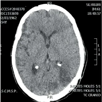
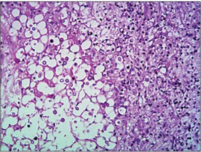
A quick check-up was performed, through laboratory tests, includ-ing a complete blood count before surgery, chest radiography and an electrocardiogram, and it did not show any abnormalities. Gross total removal was achieved and postoperative computed tomography (CT) scans were performed (Figure 2). In addition, samples from the lesion were sent for histopathological analysis (Figure 3). he result revealed that the lesion consisted of cryp-tococcoma. Because of this result, a more precise investigation was performed. he patient was found not to have either HIV or hepatitis (both tests were performed twice) or any other immu-nocompromised conditions. In addition, chest and abdominal CT scans were normal.
Ater the operation, the patient showed some neurological improvement and, ater some days, a lumbar puncture was per-formed to evaluate the presence of meningitis. he presence of cerebrospinal luid (CSF) infection was conirmed (large num-bers of specimens of Cryptococcus neoformans were observed in the samples, along with increased protein levels). Before the puncture, a brain CT scan was performed to rule out any signs of intracranial hypertension or any alteration that might make it impossible to perform this procedure. All punctures revealed increased intracranial pressure (ICP) that progressively wors-ened (the highest value was 29 cmH2O), but she did not pres-ent hydrocephalus.
Initially, ventriculoperitoneal shunt was not proposed, but when it was noticed that the ICP levels were progressively increasing and the procedure was indicated, she was seen to present a poor general
Figure 2. Axial computed tomography performed during the
postoperative period showing gross total resection.
Figure 1. A) Coronal T1-weighted magnetic resonance imaging (MRI)
showing enhanced left occipital lesions. B) Coronal T2 MRI showing extensive edema surrounding left occipital lesions.
condition with signiicant clinical impairment. Proper antifungal treatment consisting of amphotericin B (AmB) and luconazole was administered, in association with clinical support, because she presented renal failure due to use of many drugs and required hemodialysis. However, despite optimal clinical treatment in the intensive care unit (ICU), comprising antifungal drugs, broad-spectrum antibiotics against pneumonia, electrolyte replacement, hemodialysis and respiratory physiotherapy, the patient’s condi-tion evolved to death due to various complicacondi-tions, including the initial disease, the toxicity of the various medications and bron-choaspiration leading to sepsis.
DISCUSSION
Cryptococcosis is the most common fungal disease of the central nervous system1-4 and usually afects patients with a condition that reduces their immunological status. It therefore belongs to the group of opportunistic infections. It is most frequently asso-ciated with HIV infection3 but patients with chronic renal dis-ease, vascular conditions, hepatitis B or C, alcoholism, diabetes mellitus and oncological diseases are also typically more compro-mised than are immunocompetent patients, who rarely evolve with neurocryptococcosis.
Transmission occurs through inhalation of substances that are present in mammal and bird feces (pigeon feces have been described most frequently). hus, the lungs constitute the entrance for the dis-ease and pulmonary impairment is usually the irst manifestation.3 Ater the fungus enters the body, it can spread to several organs. he liver and spleen are generally afected3 and, depending on immu-nological status, the infection can cross the blood-brain barrier through fungal neurotropism and cause neurological impairment.
Central nervous system cryptococcal infections usually mani-fest as meningitis, meningoencephalitis, encephalitis or ventriculi-tis.5,6 hese patients usually present with raised intracranial pressure and hydrocephalus, and require shunt procedures. Moreover, spe-ciic medications are required to institute the proper treatment. Amphotericin B is the irst-line drug and lucytosine or lucon-azole are secondary agents.7 However, these drugs have severe side efects and, therefore, strict clinical follow-up is required. he main adverse efect is renal failure with electrolyte imbalance.
he World Health Organization (WHO) recommends two weeks of induction treatment with amphotericin B deoxycholate (AmBd) and lucytosine or luconazole, followed by eight weeks of consolidation treatment with oral luconazole.8 Toxicity analy-ses on AmB administered at a dose within the currently recom-mended dose range of 0.7 to 1 mg/kg/day for treatment durations of 5 to 14 days, alone or combined with a second antifungal have been conducted.7
Nevertheless, in rare cases, this chronic granulomatous process can lead to formation of a mass (cryptococcoma) that has a tumoral
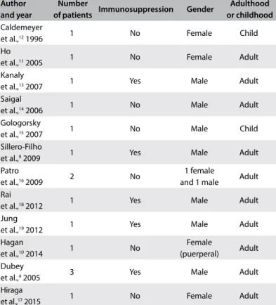
appearance.9 Metabolites released by Cryptococcus can inhibit the migration and function of leukocytes and promote survival and localized replication of the pathogen, thus facilitating chronic gran-ulomatous inlammation and cryptococcoma formation.6 here are a few reports of this condition, mainly in immuno-competent patients who usually did not develop this infection. A systematic review stored in the PubMed database was performed using the MESH terms cryptococcoma and neurocryptococcoma (Table 1). Twelve manuscripts described patients with cryptococcomas, but there were patients both with and without immunosuppressive conditions (Table 2).4,9-19
Surgical excision and debulking are indicated as adjunct thera-pies for lesions that are greater than or equal to 3 cm.11,20 he type of neurological manifestation and the indings from neuroimaging depend on the patient’s immunological status. Cryptococcomas are
Table 1. Search of the literature in medical databases for case reports
on cryptococcomas. The search was conducted on May 5, 2017
Database Search strategies Papers found Papers related MEDLINE (Via PubMed) “Brain Neoplasms”[Mesh] AND “Cryptococcosis”[Mesh]
AND Case Reports[ptyp]
28 12
Table 2. PubMed-indexed papers in English reporting on cerebral
cryptococcomas (MESH terms used: cryptococcoma; neurocryptococcoma)
Author and year
Number
of patients Immunosuppression Gender
Adulthood or childhood
Caldemeyer
et al.,12 1996 1 No Female Child
Ho
et al.,11 2005 1 No Female Adult
Kanaly
et al.,13 2007 1 Yes Male Adult
Saigal
et al.,14 2006 1 No Male Adult
Gologorsky
et al.,15 2007 1 No Male Child
Sillero-Filho
et al.,9 2009 1 Yes Male Adult
Patro
et al.,16 2009 2 No
1 female
and 1 male Adult Rai
et al.,18 2012 1 Yes Male Adult
Jung
et al.,19 2012 1 Yes Male Adult
Hagan
et al.,10 2014 1 No
Female
(puerperal) Adult Dubey
et al.,4 2005 3 Yes Male Adult
Hiraga
proportionally more likely to occur in immunocompetent patients than in immunosuppressed patients,11,21 but in terms of absolute numbers, this is still an extremely rare condition that is initially diicult to diagnose.21
Intraparenchymal cryptococcomas usually present with low signal intensity on T1-weighted MRI and high intensity on T2-weighted images. Solitary cryptococcomas are very rare lesions.22 However, despite these radiological hints, this is a very diicult diagnosis to make in non-immunocompromised patients without pulmonary impairment, before histopathological analysis has been conducted. At irst, without any precisely known clini-cal history, these images give rise to other diferential diagnoses, such as primary brain neoplasm (especially high-grade gliomas) and secondary lesions due to metastatic tumors. Other CNS infections such as tuberculosis are also diagnoses that need to be considered.
he patient of this paper was extensively investigated but did not have any immunosuppressive condition or pulmonary impair-ment. he only data that might have helped us to think of the diag-nosis of cryptococcoma was the patient’s previous contact with pigeons (which was not mentioned by the family until our team asked about this ater the histopathological diagnosis had been established). Because the patient had a neurological dysfunction that developed quickly, which made us think initially that the cause was an aggressive brain tumor, it was decided to perform surgi-cal removal. he histopathologisurgi-cal analysis (Figure 3) conirmed all the typical alterations relating to cryptococcosis, and proper treatment was quickly instituted. However, this is a condition with elevated mortality, which also led to death in her case.
CONCLUSIONS
Diagnosing the tumoral form of cryptococcosis (cryptococcoma) in immunocompetent patients is a challenge even in endemic regions. Primary and secondary brain tumors are usually the irst hypotheses in these cases. Proper investigation through anamnesis and imaging can lead to consideration of this diagnosis before the histopathological analysis has been conducted. A neurosurgical approach should be considered in cases of this type of mass lesion, to reduce the compressive factor and thus the intracranial pressure. Despite optimal treatment, this is a condition with high mortality.
REFERENCES
1. Awasthi M, Patankar T, Shah P, Castillo M. Cerebral cryptococcosis:
atypical appearances on CT. Br J Radiol. 2001;74(877):83-5.
2. Berkfield J, Enzenberger W, Lanfermann H. Cryptococcus
meningoencephalitis in AIDS: parenchymal and meningeal forms.
Neuroradiology. 1999;41(2):129-33.
3. Klock C, Cerski M, Goldani LZ. Histopathological aspects of
neurocryptococcosis in HIV-infected patients: autopsy report of 45
patients. Int J Surg Pathol. 2009;17(6):444-8.
4. Dubey A, Patwardhan RV, Sampth S, et al. Intracranial fungal granuloma:
analysis of 40 patients and review of the literature. Surg Neurol.
2005;63(3):254-60; discussion 260.
5. Dubbioso R, Pappatà S, Quarantelli M, et al. Atypical clinical and
radiological presentation of cryptococcal choroid plexitis in an
immunocompetent woman. J Neurol Sci. 2013;334(1-2):180-2.
6. Gavito-Higuera J, Mullins CB, Ramos-Duran L, et al. Fungal Infections
of the Central Nervous System: A Pictorial Review. J Clin Imaging Sci.
2016;6:24.
7. Bicanic T, Bottomley C, Loyse A, et al. Toxicity of Amphotericin
B Deoxycholate-Based Induction Therapy in Patients with
HIV-Associated Cryptococcal Meningitis. Antimicrob Agents Chemother.
2015;59(12):7224-31.
8. Alvarez-Uria G, Midde M, Pakam R, et al. Short-Course Induction
Treatment with Intrathecal Amphotericin B Lipid Emulsion for
HIV Infected Patients with Cryptococcal Meningitis. J Trop Med.
2015;2015:864271.
9. Sillero-Filho VJ, Souza ABM, Vaitsman RP, et al. Criptococoma cerebelar
simulando neoplasia metastática [Cerebellar cryptococcoma simulating
metastatic neoplasm]. Arq Neuropsiquiatr. 2009;67(2a):290-2.
10. Hagan JE, Ribeiro GS, Ko AI, et al. Puerperal brain cryptococcoma in
an HIV-negative woman successfully treated with fluconazole: a case
report. Rev Soc Bras Med Trop. 2014;47(2):254-6.
11. Ho TL, Lee HJ, Lee KW, Chen WL. Difusion-weighted and conventional
magnetic resonance imaging in cerebral cryptococcoma. Acta Radiol.
2005;46(4):411-4.
12. Caldemeyer KS, Mathews VP, Edwards-Brown MK, Smith RR. Central
nervous system cryptococcosis: parenchymal calciication and large
gelatinous pseudocysts. AJNR Am J Neuroradiol. 1996;18(1):107-9.
13. Kanaly CW, Selznick LA, Cummings TJ, Adamson DC. Cerebellar
cryptococcoma in a patient with undiagnosed sarcoidosis: case report.
Neurosurgery. 2007;60(3):E571; discussion E571.
14. Saigal G, Post MJ, Lolayekar S, Murtaza A. Unusual presentation of
central nervous system cryptococcal infection in an immunocompetent
patient. AJNR Am J Neuroradiol. 2005;26(10):2522-6.
15. Gologorsky Y, DeLaMora P, Souweidane MM, Greenield JP. Cerebellar
cryptococcoma in an immunocompetent child. Case report.
J Neurosurg. 2007;107(4 Suppl):314-7.
16. Patro SN, Kesavadas C, Thomas B, Kapilamoorthy TR, Gupta AK.
Uncommon presentation of intracranial cryptococcal infection
mimicking tuberculous infection in two immunocompetent patients.
Singapore Med J. 2009;50(4):e133-7.
17. Hiraga A, Yatomi M, Ozaki D, Kamitsukasa I, Kuwabara S. Cryptococcosis
mimicking lung cancer with brain metastasis. Clin Neurol Neurosurg.
2015;135:93-5.
18. Rai S, Marak RS, Jain S, Dhole TN. Posterior fossa midline cryptococcoma
in a patient with idiopathic CD4 lymphocytopenia. Indian J Med
Microbiol. 2012; 30(3):367-70.
19. Jung A, Korsukewitz C, Kuhlmann T, et al. Intracerebral mass lesion
diagnosed as cryptococcoma in a patient with sarcoidosis, a rare
opportunistic manifestation induced by immunosuppression with
corticosteroids. J Neurol. 2012;259(10):2147-50.
20. Perfect JR, Dismukes WE, Dromer F, et al. Clinical practice guidelines for
the management of cryptococcal disease: 2010 update by the infectious
diseases society of america. Clin Infect Dis. 2010;50(3):291-322.
21. Vender JR, Miller DM, Roth T, Nair S, Reboli AC. Intraventricular
cryptococcal cysts. AJNR Am J Neuroradiol. 1996;17(1):110-3.
22. Chen S, Chen X, Zhang Z, et al. MRI indings of cerebral cryptococcosis
in immunocompetent patients. J Med Imaging Radiat Oncol.
2011;55(1):52-7.
Sources of funding: None
Conlict of interest: None
Date of irst submission: February 12, 2017
Last received: April 9, 2017
Accepted: April 21, 2017
Address for correspondence:
Aline Lariessy Campos Paiva
Disciplina de Neurocirurgia da Faculdade de Ciências Médicas da Santa
Casa de São Paulo (FCMSCSP)
Rua Dr. Cesário Motta Júnior, 112
São Paulo (SP) — Brasil
CEP 01221-020
Tel./Fax. (+55 11) 2176-7000/93011-6370