Artrodese subtalar do pé pelo método de Ilizarov*
A
RTIGOO
RIGINALArtrodese subtalar of the foot for the Ilizarov* method
JOSÉ CARLOS BONGIOVANNI
1;
HILÁRIO BOATTO
2; EMÍLIO PALAZZO NETO
3; CARLOS LUIS ENGELEN
4; ANTONIO AUGUSTO COUTO DE MAGALHÃES
5;
PAULO ROBERTO MUSSI
6* * * *
* TTTTrabalho realizado no Departamento de Ortopedia e TTrabalho realizado no Departamento de Ortopedia e Trabalho realizado no Departamento de Ortopedia e Trabalho realizado no Departamento de Ortopedia e Trabalho realizado no Departamento de Ortopedia e Traumatologia da Universidade Fraumatologia da Universidade Fraumatologia da Universidade Fraumatologia da Universidade Fraumatologia da Universidade Federal de São Pederal de São Pederal de São Pederal de São Pederal de São Paulo - Escolaaulo - Escolaaulo - Escolaaulo - Escolaaulo - Escola P
PP P
Paulista de Medicina (Serviço do Profaulista de Medicina (Serviço do Profaulista de Medicina (Serviço do Profaulista de Medicina (Serviço do Profaulista de Medicina (Serviço do Prof. Dr. Dr. Dr. Dr. Dr. José Laredo Filho).. José Laredo Filho).. José Laredo Filho).. José Laredo Filho).. José Laredo Filho).
1 Doutor em Medicina, Professor Adjunto do Departamento de Ortopedia e Traumatologia, Chefe do Grupo de Fixadores Externos da Unifesp -Escola Paulista de Medicina.
2 Mestre em Ortopedia e Traumatologia, Assistente do Grupo de Fixadores Externos da Unifesp - Escola Paulista de Medicina. 3 Mestre emOrtopedia e Traumatologia, Assistente do Grupo de Fixadores Externos da Unifesp - Escola Paulista de Medicina.
4 Pós Graduando do Departamento de Ortopedia e Traumatologia, Assistente do Grupo de Fixadores Externos da Unifesp - Escola Paulista de Medicina.
5 Doutor em Medicina, Professor Adjunto do Departamento de Ortopedia e Traumatologia, Chefe do Grupo de Pé da Unifesp - Escola Paulista de Medicina.
6 Colaborador do Grupo de Fixadores Externos da Unifesp - Escola Paulista de Medicina.
RESUMO
RESUMO
RESUMO
RESUMO
RESUMO
Foram tratados 28 pacientes num total de 29 pés, Foram tratados 28 pacientes num total de 29 pés,Foram tratados 28 pacientes num total de 29 pés, Foram tratados 28 pacientes num total de 29 pés, Foram tratados 28 pacientes num total de 29 pés, com idade média de 25,8, predomínio do sexo masculino com idade média de 25,8, predomínio do sexo masculinocom idade média de 25,8, predomínio do sexo masculino com idade média de 25,8, predomínio do sexo masculino com idade média de 25,8, predomínio do sexo masculino 16 pacientes (57,2%), cor branca 25 pacientes (89,2%), 16 pacientes (57,2%), cor branca 25 pacientes (89,2%),16 pacientes (57,2%), cor branca 25 pacientes (89,2%), 16 pacientes (57,2%), cor branca 25 pacientes (89,2%), 16 pacientes (57,2%), cor branca 25 pacientes (89,2%), lado direito 16 pacientes (57,2%). Quanto à etiologia lado direito 16 pacientes (57,2%). Quanto à etiologialado direito 16 pacientes (57,2%). Quanto à etiologia lado direito 16 pacientes (57,2%). Quanto à etiologia lado direito 16 pacientes (57,2%). Quanto à etiologia houve um predomínio das seqüelas de fraturas de houve um predomínio das seqüelas de fraturas dehouve um predomínio das seqüelas de fraturas de houve um predomínio das seqüelas de fraturas de houve um predomínio das seqüelas de fraturas de calcâneo 11 pacientes (37,9%) e poliomielite 11 pacientes calcâneo 11 pacientes (37,9%) e poliomielite 11 pacientescalcâneo 11 pacientes (37,9%) e poliomielite 11 pacientes calcâneo 11 pacientes (37,9%) e poliomielite 11 pacientes calcâneo 11 pacientes (37,9%) e poliomielite 11 pacientes (37,9%), as outras causas foram necrose do talus 2 (37,9%), as outras causas foram necrose do talus 2(37,9%), as outras causas foram necrose do talus 2 (37,9%), as outras causas foram necrose do talus 2 (37,9%), as outras causas foram necrose do talus 2 pacientes (6,9%), barra óssea 2 pacientes (6,9%), artrose pacientes (6,9%), barra óssea 2 pacientes (6,9%), artrosepacientes (6,9%), barra óssea 2 pacientes (6,9%), artrose pacientes (6,9%), barra óssea 2 pacientes (6,9%), artrose pacientes (6,9%), barra óssea 2 pacientes (6,9%), artrose primária 2 pacientes (6,9%) e paralisia cerabral 1 paciente primária 2 pacientes (6,9%) e paralisia cerabral 1 pacienteprimária 2 pacientes (6,9%) e paralisia cerabral 1 paciente primária 2 pacientes (6,9%) e paralisia cerabral 1 paciente primária 2 pacientes (6,9%) e paralisia cerabral 1 paciente (3,5%). O tempo médio de consolidação foi de 3,5 (3,5%). O tempo médio de consolidação foi de 3,5(3,5%). O tempo médio de consolidação foi de 3,5 (3,5%). O tempo médio de consolidação foi de 3,5 (3,5%). O tempo médio de consolidação foi de 3,5 meses. Foram utilizados enxerto ósseo em 11 pacientes meses. Foram utilizados enxerto ósseo em 11 pacientesmeses. Foram utilizados enxerto ósseo em 11 pacientes meses. Foram utilizados enxerto ósseo em 11 pacientes meses. Foram utilizados enxerto ósseo em 11 pacientes (44%). Houve necessidade em 2 casos (6,9%) de se (44%). Houve necessidade em 2 casos (6,9%) de se(44%). Houve necessidade em 2 casos (6,9%) de se (44%). Houve necessidade em 2 casos (6,9%) de se (44%). Houve necessidade em 2 casos (6,9%) de se promover o alongamento do calcâneo para se aumentar promover o alongamento do calcâneo para se aumentarpromover o alongamento do calcâneo para se aumentar promover o alongamento do calcâneo para se aumentar promover o alongamento do calcâneo para se aumentar o pitch do mesmo. A montagem é simples e se baseia o pitch do mesmo. A montagem é simples e se baseiao pitch do mesmo. A montagem é simples e se baseia o pitch do mesmo. A montagem é simples e se baseia o pitch do mesmo. A montagem é simples e se baseia em 2 anéis com fixação transóssea através de 2 fios em 2 anéis com fixação transóssea através de 2 fiosem 2 anéis com fixação transóssea através de 2 fios em 2 anéis com fixação transóssea através de 2 fios em 2 anéis com fixação transóssea através de 2 fios olivados no talus e 2 no calcâneo. Como resultado foi olivados no talus e 2 no calcâneo. Como resultado foiolivados no talus e 2 no calcâneo. Como resultado foi olivados no talus e 2 no calcâneo. Como resultado foi olivados no talus e 2 no calcâneo. Como resultado foi obtido 100% de consolidação, ausência de dor e obtido 100% de consolidação, ausência de dor eobtido 100% de consolidação, ausência de dor e obtido 100% de consolidação, ausência de dor e obtido 100% de consolidação, ausência de dor e osteoporose e boa sensibilidade do pé. Das osteoporose e boa sensibilidade do pé. Dasosteoporose e boa sensibilidade do pé. Das osteoporose e boa sensibilidade do pé. Das osteoporose e boa sensibilidade do pé. Das complicações encontradas além das costumeiras ou complicações encontradas além das costumeiras oucomplicações encontradas além das costumeiras ou complicações encontradas além das costumeiras ou complicações encontradas além das costumeiras ou próprias do método 1 (3,5%) paciente apresentou próprias do método 1 (3,5%) paciente apresentoupróprias do método 1 (3,5%) paciente apresentou próprias do método 1 (3,5%) paciente apresentou próprias do método 1 (3,5%) paciente apresentou infecção profunda em 1 dos fios. O método mostrou-se infecção profunda em 1 dos fios. O método mostrou-seinfecção profunda em 1 dos fios. O método mostrou-se infecção profunda em 1 dos fios. O método mostrou-se infecção profunda em 1 dos fios. O método mostrou-se eficiente independente da etiologia com 100% de eficiente independente da etiologia com 100% deeficiente independente da etiologia com 100% de eficiente independente da etiologia com 100% de eficiente independente da etiologia com 100% de consolidação além de permitirem uma certa consolidação além de permitirem uma certaconsolidação além de permitirem uma certa consolidação além de permitirem uma certa consolidação além de permitirem uma certa independência ao paciente durante o tratamento. independência ao paciente durante o tratamento.independência ao paciente durante o tratamento. independência ao paciente durante o tratamento. independência ao paciente durante o tratamento.
SUMMARY
SUMMARY
SUMMARY
SUMMARY
SUMMARY
There had been heated 28 patients in a There had been heated 28 patients in a There had been heated 28 patients in a There had been heated 28 patients in a There had been heated 28 patients in a total of 29 feet, with na age average of 25,8, total of 29 feet, with na age average of 25,8, total of 29 feet, with na age average of 25,8, total of 29 feet, with na age average of 25,8, total of 29 feet, with na age average of 25,8, predominance in the quality of being male 16 patients predominance in the quality of being male 16 patients predominance in the quality of being male 16 patients predominance in the quality of being male 16 patients predominance in the quality of being male 16 patients (57,2%), white
(57,2%), white (57,2%), white (57,2%), white (57,2%), white
color 25 patients (89,2%), right side 16 patients color 25 patients (89,2%), right side 16 patients color 25 patients (89,2%), right side 16 patients color 25 patients (89,2%), right side 16 patients color 25 patients (89,2%), right side 16 patients (57,2%). As the ethiology there were a predominancy of (57,2%). As the ethiology there were a predominancy of (57,2%). As the ethiology there were a predominancy of (57,2%). As the ethiology there were a predominancy of (57,2%). As the ethiology there were a predominancy of the calcaneum fracture´s consequence 11 patients the calcaneum fracture´s consequence 11 patients the calcaneum fracture´s consequence 11 patients the calcaneum fracture´s consequence 11 patients the calcaneum fracture´s consequence 11 patients (37,9%) and poliomyelitis 11 patients (6,9%), bone bar 2 (37,9%) and poliomyelitis 11 patients (6,9%), bone bar 2 (37,9%) and poliomyelitis 11 patients (6,9%), bone bar 2 (37,9%) and poliomyelitis 11 patients (6,9%), bone bar 2 (37,9%) and poliomyelitis 11 patients (6,9%), bone bar 2 patients (6,9%), primary arthrosis 02 patients (6,9%) abd patients (6,9%), primary arthrosis 02 patients (6,9%) abd patients (6,9%), primary arthrosis 02 patients (6,9%) abd patients (6,9%), primary arthrosis 02 patients (6,9%) abd patients (6,9%), primary arthrosis 02 patients (6,9%) abd cerebral palsy 1 patient (3,5%). The average time of cerebral palsy 1 patient (3,5%). The average time of cerebral palsy 1 patient (3,5%). The average time of cerebral palsy 1 patient (3,5%). The average time of cerebral palsy 1 patient (3,5%). The average time of consolidation was 3,5 months. There had been used bone consolidation was 3,5 months. There had been used bone consolidation was 3,5 months. There had been used bone consolidation was 3,5 months. There had been used bone consolidation was 3,5 months. There had been used bone graft in 11 patients (44%). There were the necessity to graft in 11 patients (44%). There were the necessity to graft in 11 patients (44%). There were the necessity to graft in 11 patients (44%). There were the necessity to graft in 11 patients (44%). There were the necessity to promote the calcaneum extension to raise the pitch of promote the calcaneum extension to raise the pitch of promote the calcaneum extension to raise the pitch of promote the calcaneum extension to raise the pitch of promote the calcaneum extension to raise the pitch of itself, in 2 cases (6,9%). The mounting is simple and it itself, in 2 cases (6,9%). The mounting is simple and it itself, in 2 cases (6,9%). The mounting is simple and it itself, in 2 cases (6,9%). The mounting is simple and it itself, in 2 cases (6,9%). The mounting is simple and it relies on 2 rings transbone settlement through 2 strings relies on 2 rings transbone settlement through 2 strings relies on 2 rings transbone settlement through 2 strings relies on 2 rings transbone settlement through 2 strings relies on 2 rings transbone settlement through 2 strings fixed in the talus and 2 in the calcaneum. As the result fixed in the talus and 2 in the calcaneum. As the result fixed in the talus and 2 in the calcaneum. As the result fixed in the talus and 2 in the calcaneum. As the result fixed in the talus and 2 in the calcaneum. As the result was obtained in 100% of consolidation, pain´s absence was obtained in 100% of consolidation, pain´s absence was obtained in 100% of consolidation, pain´s absence was obtained in 100% of consolidation, pain´s absence was obtained in 100% of consolidation, pain´s absence and osteopethosis and feet´s great sensibility
and osteopethosis and feet´s great sensibility and osteopethosis and feet´s great sensibility and osteopethosis and feet´s great sensibility
and osteopethosis and feet´s great sensibility. In the. In the. In the. In the. In the complications there were found, besides the usual or complications there were found, besides the usual or complications there were found, besides the usual or complications there were found, besides the usual or complications there were found, besides the usual or method itself 1 (3,5%) patient. Showed deep infection in method itself 1 (3,5%) patient. Showed deep infection in method itself 1 (3,5%) patient. Showed deep infection in method itself 1 (3,5%) patient. Showed deep infection in method itself 1 (3,5%) patient. Showed deep infection in 1 of the strings. The method has showed efficient, 1 of the strings. The method has showed efficient, 1 of the strings. The method has showed efficient, 1 of the strings. The method has showed efficient, 1 of the strings. The method has showed efficient, independent of the ethiology with 100% of consolidation independent of the ethiology with 100% of consolidation independent of the ethiology with 100% of consolidation independent of the ethiology with 100% of consolidation independent of the ethiology with 100% of consolidation beyond to permit a certain independancy of the patient beyond to permit a certain independancy of the patient beyond to permit a certain independancy of the patient beyond to permit a certain independancy of the patient beyond to permit a certain independancy of the patient during the treatment.
INTRODUÇÃO
INTRODUÇÃO
INTRODUÇÃO
INTRODUÇÃO
INTRODUÇÃO
Inúmeros são os métodos existentes para se promover a Artrodese Subtalar do Pé que vai desde procedimentos mais simples como o utilizado pela técnica da trefina (Magalhães e Col.)21 até procedimentos mais complexos em que há necessidade de colocação de enxerto e osteossíntese interna.
Os autores mostram uma nova opção de tratamento nos casos em que existe a necessidade da artrodese subtalar ou da artrodese tríplice através da fixação externa, com o desenvolvimento de uma montagem pessoal simplificada, mas que dá grande estabilidade e compressão no foco da artrodese, além de dar grande comodidade ao paciente permitindo que o mesmo deambule com carga no pós-operatório imediato.
MA
MA
MA
MA
MATERIAL E MÉTODO
TERIAL E MÉTODO
TERIAL E MÉTODO
TERIAL E MÉTODO
TERIAL E MÉTODO
A casuística consta de 28 pacientes, com um total de 29 pés tratados no Departamento de Ortopedia e Traumatologia da Universidade Federal de São Paulo - Escola Paulista de Medicina, no período de 1989 a 1997. A idade mínima foi de 15 anos e a máxima de 54 anos com uma média de 25,8 anos. Quanto ao sexo 16 pacientes (57,2%) eram masculino e 12 (42,8%) feminino; a cor branca apresentou 25 pacientes (89,3%) e não branca 03 pacientes (10,7%). Quanto ao lado, 16 pés (55,2%) eram do direito e 13 (44,8%) do esquerdo. O tempo mínimo de tratamento foi de 2,8 meses e o máximo foi de 6,5 meses, com uma média de 3,8 meses. O tempo de seguimento mínimo foi de 1 ano e o máximo de 8 anos com uma média de 4,3 anos. Quanto à etiologia houve predomínio das seqüelas de fraturas do calcâneo em 11 pés (37,9%), seqüela de poliomielite em 11 pés (37,9%) sendo as outras causas necrose do talus em 2 pés (6,9%), barra óssea 2 pés (6,9%), artrose primária 2 pés (6,9%) e paralisia cerebral 1 pé (3,5%).
Todos os pacientes apresentavam dor incapacitante ao apoio e à marcha e algum desvio em varo e valgo do retro pé no pré operatório.
Um dos pacientes com necrose do talus e osteossíntese com parafuso, apresentava concomitantemente infecção (Staphilococus aureus) que atingia a tíbio társica.
A metodologia utilizada foi a idealização de uma montagem simplificada a 2 semi-anéis acoplados à placas de conexão, um fixado no talus com 2 fios olivados cruzados e um semi-anel fixado no calcâneo também com 2 fios olivados.
Quando houve necessidade de se artrodesar a médiotársica (talo-navicular e calcâneo-cubóidea) um 3° fio era acoplado no
INTRODUCTION
INTRODUCTION
INTRODUCTION
INTRODUCTION
INTRODUCTION
Countless they are the existent methods to promote Artrodese Subtalar of the Foot that is going from simpler procedures as used him/it by the technique of the trefina (Magalhães and Col. )21 even more complex procedures in than there are need of graft placement and osteossíntese it interns.
The authors show a new treatment option in the cases in that the need of the artrodese subtalar exists or of the artrodese triple through the fixation expresses, with the development of a simplified personal assembly, but that gives great stability and compression in the focus of the artrodese, besides giving great comfort to the patient allowing
MA
MA
MA
MA
MATERIAL AND METHOD
TERIAL AND METHOD
TERIAL AND METHOD
TERIAL AND METHOD
TERIAL AND METHOD
The casuistry consists of 28 patient, with a total of 29 feet treated in the Department of Orthopedics and Traumatologia of the Federal University of São Paulo - School From São Paulo of Medicine, in the period from 1989 to 1997. The minimum age was of 15 years and the 54 year-old maxim with a 25,8 year-old average. With relationship to the sex 16 patient (57,2%) they were masculine and 12 (42,8%) feminine; the white color presented 25 patient (89,3%) and not white 03 patient (10,7%). With relationship to the side, 16 feet (55,2%) they were of the right and 13 (44,8%) of the left. The minimum time of treatment was of 2,8 months and the maximum was of 6,5 months, with an average of 3,8 months. The time of minimum following was of 1 year and the 8 year-old maximum with a 4,3 year-old average. With relationship to the aetiology there was prevalence of the sequels of fractures of the calcâneo in 11 feet (37,9%), poliomyelitis sequel in 11 feet (37,9%) being the other causes necrosis of the talus in 2 feet (6,9%), it obstructs bony 2 feet (6,9%), artrose primary 2 feet (6,9%) and paralysis cerebral 1 foot (3,5%).
All the patients presented pain incapacitante to the support and the march and some deviation in I beat and valgo of the retro foot in the operative pré.
One of the patients with necrosis of the talus and osteossíntese with screw, presented concomitantemente infection (Staphilococus aureus) that reached the tíbio társica.
sistema em forma arqueada para que houvesse possibilidade de compressão dessas articulações ou as mesmas foram fixados com agrafes de Blount. Nos casos em que não havia uma importante lesão articular, promovia-se a abertura prévia da articulação, com cruentização da mesma e compressão através do fixador.
Em 13 pés (44,8%) houve a necessidade de utilização de enxerto ósseo, retirado do ilíaco contra lateral, para preenchimento do espaço articular. Em 02 casos (6,9%) houve a necessidade de se promover o alongamento posterior do calcâneo para se aumentar o pitch do mesmo.
Carga parcial com muletas foi apresentado no P.O imediato e após 2 a 3 semanas, após a retirada dos pontos e ter cedido o processo inflamatório provocado pelo ato cirúrgico carga total foi liberada. Uma orientação fisioterápica foi dada e realizada no pós operatório.
When there was need of if artrodesar the médiotársica (shaft-navicular and calcâneo-cubóidea) a 3° thread was coupled in the system in arched form so that there was possibility of compression of those articulations or the same ones were fastened with agrafes of Blount. In the cases in that there was not an important lesion to articulate, the previous opening of the articulation was promoted, with cruentização of the same and compression through the fixador. In 13 feet (44,8%) there was the need of use of bony graft, retired of the ilíaco against lateral, for completion of the space to articulate. In 02 cases (6,9%) there was the need to promote the subsequent prolongation of the calcâneo to increase the pitch of the same.
Partial load with crutches was presented in immediate P.O and after 2 to 3 weeks, after the retreat of the points and to have given up the inflammatory process provoked by the act surgical total load it was liberated. An orientation fisioterápica was given and accomplished in the operative powders.
Fig. 1a - Rx initial (Sequel of calcâneo Fracture) 1b - front Rx with fixador
1c - profile Rx with fixador
a
b
c
ILLUSTRATIONS
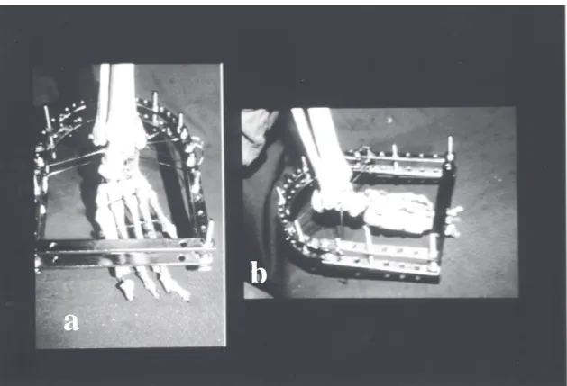
Fig. 2a - Assembly of seen Fixador of front 2b - Assembly fixador seen lateral
RESUL
RESUL
RESUL
RESUL
RESULT
T
T
T
TADOS
ADOS
ADOS
ADOS
ADOS
Foi obtido consolidação nos 29 pés (100%). Em todos os casos houve desaparecimento da dor. Em 24 casos (82,7%) houve correção total do desvio do retro pé sendo que em 3 pés (10,3%) permaneceu desvio , 2 em varo e 1 em valgo que não ultrapassou 5° e 1 paciente permaneceu com varo menor que 5°.
O caso que apresentou necrose do talus, curou após artrodese da tíbio-társica.
RESUL
RESUL
RESUL
RESUL
RESULTS
TS
TS
TS
TS
It was obtained consolidation in the 29 feet (100%). In all the cases there was disappearance of the pain. In 24 cases (82,7%) there was total correction of the deviation of the retro foot and in 3 feet (10,3%) deviation stayed, 2 in I beat and 1 in valgo that didn't surpass 5° and 1 patient stayed with I beat smaller than 5°. The case that presented necrosis of the talus, cured after artrodese of the tíbio-társica.
a
COMPLICAÇÕES
COMPLICAÇÕES
COMPLICAÇÕES
COMPLICAÇÕES
COMPLICAÇÕES
Além das complicações menores comuns ao método como a dor, edema, infecção superficial, um dos pacientes apresentou infecção profunda em um dos fios havendo necessidade da troca do mesmo com cura do processo.
A paciente que apresentava necrose do talus com osteossíntese por parafuso (seqüela de fratura-luxação) apresentou regressão do quadro após a retirada do material de síntese, houve reagudização do processo durante o período de evolução havendo necessidade de se promover a artrodese da tíbio társica com cura total do processo infeccioso.
COMPLICA
COMPLICA
COMPLICA
COMPLICA
COMPLICATIONS
TIONS
TIONS
TIONS
TIONS
Besides the smaller complications common to the method as the pain, edema, superficial infection, one of the patients presented deep infection in one of the threads having need of the change of the same with cure of the process.
The patient that presented necrosis of the talus with osteossíntese for screw (fracture-dislocation sequel) it presented regression of the picture after the retreat of the synthesis material, there was reagudização of the process during the evolution period having need to promote the artrodese of the tíbio társica with total cure of the infectious process.
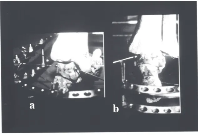
Fig 3a - Seen subsequent assembly fixador. Larger
arrow shows sub-talar open before the compression. Arrow minor shows threads olivados.
3b - Seen subsequent assembly of the fixador. Smaller arrow shows sub-talar closed after compression enter the rings. Smaller arrows show threads olivados.
ILLUSTRATIONS
ILLUSTRATIONS
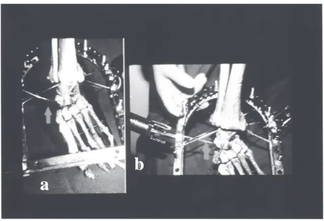
Fig. 4a - Seen previous it shows passage of the 3° thread in the navicular and cubóide. Arrow shows shaft-navicular open before the positioning of the thread. 4b - Seen previous showing the tencionamento of the 3° thread. Arrow showing closing of the shaft-navicular and calcâneo-cubóidea.
RESUL
RESUL
RESUL
RESUL
RESULT
T
T
T
TADOS
ADOS
ADOS
ADOS
ADOS
- Houve consolidação em 100% das artrodeses.
- Desaparecimento da dor incapacitente em todos os casos. - Correção de deformidade do retro pé em 82,7% sendo que em 10,3% dos casos, as deformidades que permaneceram não eram de fundamental importância.
RESUL
RESUL
RESUL
RESUL
RESULTS
TS
TS
TS
TS
- There was consolidation in 100% of the artrodeses. - Disappearance of the pain incapacitente in all the cases. - Correction of deformity of the retro foot in 82,7% and in 10,3% of the cases, the deformities that stayed they were not of fundamental importance.
a
Fig. 5a,5b - Patient during treatment. Stooping and in foot with total load.
CONCLUSÕES
CONCLUSÕES
CONCLUSÕES
CONCLUSÕES
CONCLUSÕES
O método mostrou-se eficiente independentemente da etiologia com 100% da consolidação, desaparecimento do sintoma doloroso e correções das deformidades além de dar opção nos casos em que há um grande afundamento do calcâneo com elevação da tuberosidade posterior do mesmo de se promover o abaixamento com alongamento posterior e para baixo, aumentando-se dessa maneira o pitch do calcâneo.
Pela simplicidade da montagem possibilita um conforto ao paciente no pós operatório imediato permitindo carga total precocemente.
CONCLUSIONS
CONCLUSIONS
CONCLUSIONS
CONCLUSIONS
CONCLUSIONS
The method was shown efficient independently of the aetiology with 100% of the consolidation, disappearance of the painful symptom and corrections of the deformities besides giving option in the cases in that there is a great sinking of the calcâneo with elevation of the subsequent tuberosidade of the same of promoting the abaixamento with subsequent prolongation and down, increasing of that sorts out the pitch of the calcâneo.
For the simplicity of the assembly it makes possible a comfort to the patient in the immediate operative powders allowing load total precocemente.
ILLUSTRATIONS
ILLUSTRATIONS
Fig. 6a - final Rx without the fixador. Artrodese consolidated subtalar.
6b - final Patient.
a
b
REFERÊNCIAS
REFERÊNCIAS
REFERÊNCIAS
REFERÊNCIAS
REFERÊNCIAS
1. ASAMI GROUP - Joint ContracturesJoint ContracturesJoint ContracturesJoint Contractures. Operative Principles ofJoint Contractures Ilizarov. Ed. Medi Surgical Vídeo. Cap. 33: 436-440, 1991.
2. ALBEE, F. H. - Orthopedic and Reconstruction Surgery, Industrial and Civilian, Philadelphia London, Saunders, 1917 appud SOREN, A -Safe inlay of bone graft in arthrodesis. Clin. Orthop.58Clin. Orthop.58Clin. Orthop.58Clin. Orthop.58Clin. Orthop.58: 147 - 152, 1968.
3. BATALHA, E.S.C. - New method of intra-articular arthrodesis by transposition of local cancellous bone. A preliminary report. Am.J. Am.J. Am.J. Am.J. Am.J. Surg., 85
Surg., 85 Surg., 85 Surg., 85
Surg., 85-92, 1950.
4. BIANCHI-MAIOCCHI, A; BENEDETTI,G.B.; CATAGNI, M.A;CATTANEO,R.; TENTORI,L.; VILLA, A; L´osteossinteseL´osteossinteseL´osteossinteseL´osteossinteseL´osteossintese T
T T T
Transsosea Secondo G.A Ilizarovranssosea Secondo G.A Ilizarovranssosea Secondo G.A Ilizarovranssosea Secondo G.A Ilizarovranssosea Secondo G.A Ilizarov. Aspetti Clinici Dell´impiego Dell´apparato di Compressione-Distrazione di G.A Ilizarov in Traumatologia. Ed. Medi Surgical Video, Milano - Itália. Cap.2, pag. 157-77, 1983.
5. BLAIR, H.C. - Comminute fractures and fracture dislocations of the body of the astragalus. Operative treatment. Am.J.Surg., 59Am.J.Surg., 59Am.J.Surg., 59Am.J.Surg., 59Am.J.Surg., 59:37-43, 1943.
6. CAMPBELL, P. - Arthrodesis of the ankle with modified distraction-compressiom and bone grafting. J. Bone Joint surg., 72-A:J. Bone Joint surg., 72-A:J. Bone Joint surg., 72-A:J. Bone Joint surg., 72-A:J. Bone Joint surg., 72-A:551-556, 1990.
7. CANTALAMESSA, C. - Arthrodesi della tíbio-társica per esiti di fratture del collo piede. Revisione della casística e controllo a distanza. Clin. Orthop., 268:
Clin. Orthop., 268: Clin. Orthop., 268: Clin. Orthop., 268:
Clin. Orthop., 268: 49-55, 1991.
8. CATAGNI, M.A.; FFFratture e PFFratture e Pratture e Pratture e Pseudoartrosi - Tratture e Pseudoartrosi - Tseudoartrosi - Tseudoartrosi - Tseudoartrosi - Trattamento conrattamento conrattamento conrattamento conrattamento con fissatore esterno
fissatore esterno fissatore esterno fissatore esterno
fissatore esterno circolare di Ilizarovcircolare di Ilizarovcircolare di Ilizarovcircolare di Ilizarovcircolare di Ilizarov; Studio CA, 1997.
9. CATAGNI, M.A.; MALZEV, V.; KIRIENKO, A.; Advances in IlizarovAdvances in IlizarovAdvances in IlizarovAdvances in IlizarovAdvances in Ilizarov Apparatus Assembly
Apparatus Assembly Apparatus Assembly Apparatus Assembly
-ILLUSTRATIONS
Fig. 7a - final Picture of the patient's foot. 7b - final Picture of the foot seen for behind.
a
b
Lengthening Deformity Correction; Ed. A. Bianchi-Maiochi, 155p., 1997.
10. DENNIS, D.A; CLAYTON, M.L.; WONG, D.A; MACK, R.P.; SUSMAN, M.H. - Internal fixation compression arthrodesis of the ankle. Clin. Clin. Clin. Clin. Clin. Orthop., 253:
Orthop., 253: Orthop., 253: Orthop., 253:
Orthop., 253: 212-220, 1990.
11. FONTANILLS, C.F.; LOPEZ, A; ROQUE, D.F.; CAMPOS, F.R. -Artrodesis de tobillo por compresion externa. RerRerRerRer. Cuba.Ortop.Rer. Cuba.Ortop.. Cuba.Ortop.. Cuba.Ortop.. Cuba.Ortop. T
T T T
Traumatol., 1:raumatol., 1:raumatol., 1:raumatol., 1:raumatol., 1: 17-21, 1987.
12. GOLYAKHOVSKY, V.: Dobradiças. Manual de TécnicasManual de TécnicasManual de TécnicasManual de TécnicasManual de Técnicas Operatórias do Método de Ilizarov
Operatórias do Método de Ilizarov Operatórias do Método de Ilizarov Operatórias do Método de Ilizarov
Operatórias do Método de Ilizarov, 6:, 6:, 6:, 6:, 6: 123-130, 1996.
13. GOLYAKHOVSKY, V.: Correção das contraturas articulares. ManualManualManualManualManual de Técnicas Operatórias do Método de Ilizarov
de Técnicas Operatórias do Método de Ilizarov de Técnicas Operatórias do Método de Ilizarov de Técnicas Operatórias do Método de Ilizarov
de Técnicas Operatórias do Método de Ilizarov, 6:, 6:, 6:, 6: 130-, 6: 139, 1996.
14. GRUEN, G.S. & MEARS, D.C. - Arthrodesis of the ankle and subtalar joints, Clin. Orthop., 268 Clin. Orthop., 268 Clin. Orthop., 268 Clin. Orthop., 268: 15-20, 1991. Clin. Orthop., 268
15. HAWKINS, B.J.; LANGERMAN, R.J., ANGER, D.M.; CALHOUN, J.H. - The Ilizarov technique in ankle fusion. Clin. Orthop., 303:Clin. Orthop., 303:Clin. Orthop., 303:Clin. Orthop., 303:Clin. Orthop., 303: 217 - 225, 1994.
16. ILIZAROV, G.A. - The Apparatus: Components and Biome-The Apparatus: Components and Biome-The Apparatus: Components and Biome-The Apparatus: Components and Biome-The Apparatus: Components and Biome-chanical Principles of Aplication
chanical Principles of Aplication chanical Principles of Aplication chanical Principles of Aplication
chanical Principles of Aplication. Transosseus Osteosynthe-sis. Ed. Springer - Verlag, 329-68, 1992.
17. JAHSS, M. - Disorders of the foot & ankle Disorders of the foot & ankle Disorders of the foot & ankle Disorders of the foot & ankle Disorders of the foot & ankle, 2 ed. Toronto, W.B. Saunders Company, 1991,
18. JOHNSON, J.E. & JOHNSON, K.A - Dowel arthrodesis for degen-erative arthrites of the tarsometatarsal (Lisfranc) joints. Foot & Ankle Foot & Ankle Foot & Ankle Foot & Ankle Foot & Ankle int., 6:
int., 6: int., 6: int., 6:
19. JOHNSON, K.A - Arthrodesis of the foot and ankle. In: Surgery ofSurgery ofSurgery ofSurgery ofSurgery of the foot and ankle
the foot and ankle the foot and ankle the foot and ankle
the foot and ankle. New York, Raven Press, 1989, v.6, p. 151-208.
JOHNSON, E.E.; WELTMER, J.; LIAN, G.J.; CRACCHIOLLO, III, A -Ilizarov ankle arthrodesis. Clin. Orthop., 280:Clin. Orthop., 280:Clin. Orthop., 280:Clin. Orthop., 280:Clin. Orthop., 280: 160-169 , 1992.
21. MAGALHÃES, A C. - Artrodese do T Artrodese do T Artrodese do T Artrodese do Tornozelo pelo Método Artrodese do Tornozelo pelo Métodoornozelo pelo Métodoornozelo pelo Métodoornozelo pelo Método das T
das T das T das T
das Trefinas: Estudo de 30 Prefinas: Estudo de 30 Prefinas: Estudo de 30 Prefinas: Estudo de 30 Prefinas: Estudo de 30 Pacientes.acientes.acientes.acientes.acientes. Tese apresentada à Universidade Federal de São Paulo - Escola Paulista de Medicina para obtenção do Título de Doutor em Medicina, São Paulo, 1995.
22. MEAD, N.C. - Fusion of the ankle joint. A description of a simple, effective surgical technic. Clin. Orthop., 23: Clin. Orthop., 23: Clin. Orthop., 23: Clin. Orthop., 23: Clin. Orthop., 23: 211-15, 1953.
23. MOECKEL, B.H.; PATTERSON, B.M.; INGLIS, A E.; SCULCO, T.P. - Ankle Arthrodesis. A comparison of internal and external fixation. Clin. Orthop., 268:
Clin. Orthop., 268: Clin. Orthop., 268: Clin. Orthop., 268:
Clin. Orthop., 268: 78-83, 1991.
24. MYERSON, M.S. & QUILL, G. - Ankle arthrodesis. A compariosn of na arthroscopic and na open method of treatment. Clin. Orthop.,Clin. Orthop.,Clin. Orthop.,Clin. Orthop.,Clin. Orthop., 268: 84
268: 84 268: 84 268: 84
268: 84-95, 1991.
25. NERY, C.A S.; BRUSCHINI, S.; MAGALHÃES, A A C.; SODRÉ, H.; ,IZUSAKI, J.M. - Uso de trefinas: Um método simples e útil para artrodeses do tornozelo e pé. RerRerRerRerRer. Bras. Ortop. 27:. Bras. Ortop. 27:. Bras. Ortop. 27:. Bras. Ortop. 27:. Bras. Ortop. 27: 157 - 161, 1992.
26. PAPA, J. & GIRARD, P. - Salvage, with arthrodesis, in intractable diabetic neuropathic arthropathy of the foot and ankle J. Bone J. Bone J. Bone J. Bone J. Bone Joint Surg., 75-A:
Joint Surg., 75-A: Joint Surg., 75-A: Joint Surg., 75-A:
Joint Surg., 75-A: 1056-1066, 1993.
27. PETERSON, H.A - Dowel graft technique for triple arthrodesis in talocalcaneal coalition - report of a case with 12-year follow-up. Foot & Ankle int. vol (9).:
Foot & Ankle int. vol (9).: Foot & Ankle int. vol (9).: Foot & Ankle int. vol (9).:
Foot & Ankle int. vol (9).: 201-203, 1989.
28 REAGNAULD, B. The foot. PThe foot. PThe foot. PThe foot. PThe foot. Pathology , aetiologyathology , aetiologyathology , aetiologyathology , aetiologyathology , aetiology, semiol-, semiol-, semiol-, semiol-, semiol-ogy
ogy ogy ogy
ogy, clinical investigation and therapy, clinical investigation and therapy, clinical investigation and therapy, clinical investigation and therapy, clinical investigation and therapy... Berlim, Springer-Verlag, 1986.
29. SHEVTSOV, V.I. (et al) : Osteossíntese Transóssea pelo Método do Centro de Reabilitação Ortopédica do Acadêmico Gavriil Abramovich Ilizarov. Defeitos Ósseos de Membros InferioresDefeitos Ósseos de Membros InferioresDefeitos Ósseos de Membros InferioresDefeitos Ósseos de Membros InferioresDefeitos Ósseos de Membros Inferiores, 287-301, 1996.
30. STAPLE, S.O - Posterior arthrodesis of the ankle and subtalar joint. J. Bone Joint Surg., 38-A:
J. Bone Joint Surg., 38-A: J. Bone Joint Surg., 38-A: J. Bone Joint Surg., 38-A:
J. Bone Joint Surg., 38-A: 50, 1956.
31. STEWART, M. - Arthrodesis. In: Campbell ´s Operative Ortho-Campbell ´s Operative Ortho-Campbell ´s Operative Ortho-Campbell ´s Operative Ortho-Campbell ´s Operative Ortho-paedics
paedics paedics paedics