Vimar Is a Novel Regulator of Mitochondrial
Fission through Miro
Lianggong Ding1,2, Ye Lei1,2, Yanping Han2, Yuhong Li2, Xunming Ji2*, Lei Liu2*
1State Key Laboratory of Membrane Biology, School of Life Sciences, Peking University, Beijing, China,
2Aging and Disease lab of Xuanwu Hospital and Beijing Institute for Brain Disorders, Capital Medical University, Youanmen, Beijing, China
*[email protected](XJ);[email protected](LL)
Abstract
As fundamental processes in mitochondrial dynamics, mitochondrial fusion, fission and transport are regulated by several core components, including Miro. As an atypical Rho-like small GTPase with high molecular mass, the exchange of GDP/GTP in Miro may require assistance from a guanine nucleotide exchange factor (GEF). However, the GEF for Miro has not been identified. While studying mitochondrial morphology inDrosophila, we inci-dentally observed that the loss ofvimar, a gene encoding an atypical GEF, enhanced mito-chondrial fission under normal physiological conditions. Because Vimar could
co-immunoprecipitate with Miroin vitro, we speculated that Vimar might be the GEF of Miro. In support of this hypothesis, a loss-of-function (LOF)vimarmutant rescued mitochondrial enlargement induced by a gain-of-function (GOF)Mirotransgene; whereas a GOFvimar
transgene enhancedMirofunction. In addition,vimarlost its effect under the expression of a constitutively GTP-bound or GDP-bound Miro mutant background. These results indicate a genetic dependence of vimar on Miro. Moreover, we found that mitochondrial fission played a functional role in high-calcium induced necrosis, and a LOFvimarmutant rescued the mitochondrial fission defect and cell death. This result can also be explained by vimar’s function through Miro, because Miro’s effect on mitochondrial morphology is altered upon binding with calcium. In addition, aPINK1mutant, which induced mitochondrial enlarge-ment and had been considered as aDrosophilamodel of Parkinson’s disease (PD), caused fly muscle defects, and the loss ofvimarcould rescue these defects. Furthermore, we found that the mammalian homolog of Vimar, RAP1GDS1, played a similar role in regulat-ing mitochondrial morphology, suggestregulat-ing a functional conservation of this GEF member. The Miro/Vimar complex may be a promising drug target for diseases in which mitochon-drial fission and fusion are dysfunctional.
Author Summary
Mitochondrial dynamics including fusion, fission and transport are essential for energy supply in eukaryotic cells; and defects in mitochondrial dynamics often result in prema-ture aging and diseases such as Parkinson's disease (PD). In mitochondrial transport a11111
OPEN ACCESS
Citation:Ding L, Lei Y, Han Y, Li Y, Ji X, Liu L (2016) Vimar Is a Novel Regulator of Mitochondrial Fission through Miro. PLoS Genet 12(10): e1006359. doi:10.1371/journal.pgen.1006359
Editor:Nils-Go¨ran Larsson, Max Planck Institute for Biology of Ageing, GERMANY
Received:April 1, 2016
Accepted:September 12, 2016
Published:October 7, 2016
Copyright:©2016 Ding et al. This is an open access article distributed under the terms of the
Creative Commons Attribution License, which permits unrestricted use, distribution, and reproduction in any medium, provided the original author and source are credited.
Data Availability Statement:All relevant data are within the paper and its Supporting Information files.
Funding:This work is supported by the Chinese Ministry of Science and Technology (Grant No.
2013CB530700;http://www.most.gov.cn/) to LL
machinery, the Miro/Milton complex loads mitochondria onto microtubule through kine-sin motor proteins; and regulates mitochondrial fusion and fission through unknown mechanisms. As a small GTPase, the exchange of GTP/GDP in Miro requires a specific guanine nucleotide exchange factor (GEF). However, the GEF for Miro has not been iden-tified. In this study, we identified Vimar as a new regulator of mitochondrial dynamics in
Drosophila. We found that loss ofvimarpromoted mitochondrial shortening; and this
function was mediated through Miro. As a GEF, Vimar partially localized on mitochon-dria and could physically interact with Miro. In the pathophysiological conditions, includ-ing aPink1mutant to model PD and a calcium-overload induced stress to model neuronal necrosis inDrosophila, loss ofvimarsuppressed both aberrant mitochondrial fusion and fragmentation in PD and necrosis, respectively. As the mammalian homolog of Vimar, RAP1GDS1 function was similar to Vimar. Therefore, Vimar/ RAP1GDS1 may be a great drug target to deal with diseases caused by defective mitochondrial dynamics.
Introduction
Mitochondrial fission, fusion and transport play important roles for the function of this organ-elle [1,2]. The balance between fusion and fission controls mitochondrial morphology, which is mediated by series of large dynamin-related GTPases [3]. Among these GTPases, mitofu-sin1/mitofusin2 (MFN1/MFN2) and optic atrophy protein1 (OPA1) are the core components that are responsible for mitochondrial fusion [4–7], whereas dynamin-related protein 1 (Drp1) is the core component that is responsible for mitochondrial fission [8,9]. In addition to these GTPases in dynamin-related family, mitochondrial Rho (Miro), an atypical member of the Rho small GTPase family, has a well-known function of transporting the mitochondria along microtubules [10,11]. Miro also regulates mitochondrial morphology via inhibition of fission under physiological Ca2+conditions, although the mechanism is not that clear [12–16]. Large GTPases such as dynamin-like GTPase family members hydrolyze GTP and exchange GTP and GDP without the assistance from other regulators [17,18]. However, members of the small GTPase family often require other proteins to help release their tightly bound GDP or enhance their low GTPase activities. These proteins are referred to as guanine nucleotide exchange factors (GEFs) and GTPase activating proteins (GAPs), respectively [19]. To date, most small GTPases require unique GEFs or GAPs [19].
An understanding of the regulation of mitochondrial dynamics may help us to address many human diseases. For instance, mutations in OPA1 or MFN2 result in dominant optic atrophy or Charcot-Marie-Tooth neuropathy type 2A [20,21]. Abnormal mitochondrial fis-sion also promotes aging and cell death [22,23]. In necroptosis, the formation of the necro-some promotes mitochondrial fission through dephosphorylation of Drp1 [24]. In neuronal excitotoxicity, calcium ions are overloaded, resulting in reduced levels of the MFN2 protein, which enhances mitochondrial fission and leads to neuronal necrosis [25,26]. In addition, other components such as Miro may participate in this process [26]. Miro has two EF hand motifs that bind calcium; thus, Miro can couple calcium increase with reduced mitochondrial motility to meet the locally increased energy demands [16,27]. Interestingly, Miro also pro-motes fission in the presence of excess calcium, which is distinct from its inhibitory role in fis-sion under normal calcium concentrations [16]. It is unclear whether Miro plays a functional role in neuronal necrosis [26].
The mitochondrial morphology represents a transient balance between mitochondrial fusion and fission [28]. Using a systematic genetic screen in yeast covering approximately 88%
of genes, 117 genes that regulate mitochondrial morphology were identified [29]. Similarly, a screen of 719 genes that are predicted to encode mitochondrial proteins in worms demon-strated that more than 80% of these genes regulate mitochondrial morphology [30]. Although many genes may regulate mitochondrial morphology, their relationships to the core mitochon-drial fusion and fission components are unclear.
In studying mitochondrial morphology, we accidently discovered that the loss ofvimar (vis-ceral mesodermal armadillo-repeats), which encodes an atypical GEF [31–33], promoted mito-chondrial fission inDrosophilaflight muscle cells. Furthermore, we found that vimar was capable of interacting with Miroin vitro. Genetically, vimar required normal GDP- or GTP-bound activity of Miro to affect mitochondrial morphology, suggesting vimar is likely the Miro GEF. In addition, we found that the Miro/vimar complex suppressed mitochondrial fission during necrosis and mitochondrial fusion inPINK1mutant model of Parkinson’s disease (PD), making vimar a potential drug target.
Results
Vimar is a novel regulator of mitochondrial morphology in
Drosophila
under normal conditions
To identify novel regulators of mitochondrial morphology, we studied the flight muscle in
Dro-sophilaadults, because they have a stereotypic distribution of mitochondria in the longitudinal
myofibers [34]. To visualize the mitochondria in the muscle cells, a muscle-specific promoter, Mhc-Gal4, was used to drive a mitochondria-targeted GFP (UAS-mitoGFP); the progenies were referred to asMhc>mitoGFP. The mitochondrial morphology was clearly observed (Fig 1Aa and 1Af). Using these flies, we accidently observed that mitochondrial fission was
enhanced when avimar(visceral mesodermal armadillo-repeats) RNAi was expressed (Fig 1Ab and 1Af). To further confirm the loss-of-function (LOF) effect ofvimar, we testedvimark16722, a P-element mutant with the mobile element inserted into the 5’-UTR region of thevimar gene. Again, we observed the trend of enhanced mitochondrial fission in the heterozygous
vimark16722mutant (Fig 1Ac and 1Af). Because the homozygousvimark16722mutant was
embryonic lethal, we selected a deficient mutant (Df(2R) ED1612) covering thevimarlocus and generated a trans-heterozygousvimar(vimark16722/Df) mutant to further test the effect of vimar. In these flies, the mitochondria exhibited a stronger fission morphology compared to the heterozygous mutant (Fig 1Ad and 1Af). These results indicate thatvimarplays a dominant role in regulating mitochondrial morphology in a dosage-dependent manner. To confirm the mitochondrial defect was generated from loss ofvimar, we tried to rescuevimark16722/Dfby
tubulin-Gal4/UAS-vimar(tubulin-Gal4is a ubiquitously expressed promoter). The result
showed that the shortened mitochondria in thevimark16722/Dfmutant were rescued byvimar overexpression (S1A Fig); while overexpression ofvimaralone did not affect the mitochondrial morphology (Fig 1Ae and 1Af), suggesting that the levels of the vimar protein may be saturated under normal physiological condition. Using a polyclonal antibody of vimar, we confirmed that the protein levels of vimar were reduced invimark16722andvimarRNAi, and increased in
thevimaroverexpression line (S1B and S1C Fig).
To examine mitochondrial distribution, we studiedDrosophilalarval oenocytes because of their stereotypical location and morphology. The wild type mitochondria, labeled with
UAS-mitoGFPdriven by an oenocytes-specificpromoter,PromE(800)-Gal4, were evenly distributed
Fig 1.Drosophila vimaraffects mitochondrial morphology under normal conditions.(A)a-e, Live imaging of the mitochondrial morphology in the flight muscle of adult flies. The mitochondria are labeled withUAS-mitoGFPdriven by
(Fig 1Bb–1Bd). Interestingly, knocking downvimarby RNAi showed a similar distribution pattern (Fig 1Be). For mitochondrial morphology, loss ofKhc,Milton,Miroandvimarresulted in mitochondrial shortening (Fig 1Ba'–1Be' and 1Bf). Similarly, mitochondria in the eye disc of
GMR>mitoGFP/vimar RNAiwas also shortened (Fig 1C). These results suggest that vimar
reg-ulates mitochondrial morphology in different cell types, such as muscle, oenocyte and eye disc. Transport of mitochondria along the axon can be quantified inDrosophilaneuronsin vivo [36]. As a positive control, theCCAP-Gal4>Miro RNAiline (CCAP-Gal4is a promoter label-ing a slabel-ingle axon within a neuron bundle) displayed reduced flux of mobile mitochondria in both anterograde (soma to synapse) and retrograde (synapse to soma) transport (Fig 1D). The
CCAP-Gal4>vimar RNAiline showed a similar result (Fig 1D). RNAi of bothvimarandMiro
resulted in a similar reduction of mitochondrial transport asMiroRNAi alone (Fig 1D), sug-gesting vimar and Miro may function in the same pathway.
To test vimar subcellular localization, proteins from the thoraces of adult flies (Mhc> Mi-toGFP) were extracted and separated into cytosolic and mitochondrial fractions. The Western blot data showed that endogenous vimar was present in both cytosol and mitochondria (Fig 1E). Apart from mitochondria, to test whether Vimar can distribute in other subcellular com-partments, we fractionized organelles of ER, lysosome and Golgi apparatus, and found that Vimar was also enriched in the ER fraction, as well as in the cytosol (S2A Fig). This result is consistent with reports suggesting that Miro protein is localized and function in the site of mitochondria-ER junction [37,38].
Vimar functions through Miro to regulate mitochondrial morphology
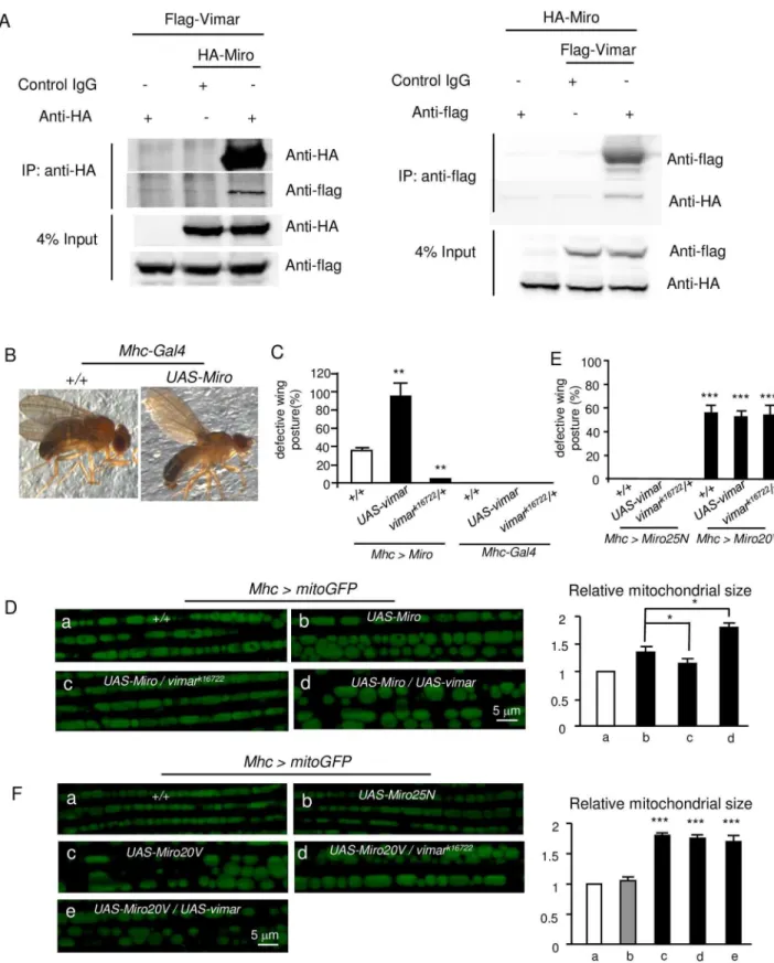
We asked whether vimar regulates mitochondrial morphology through controlling the GTP/ GDP exchange of Miro, because Miro is a well-known small GTPase that regulates mitochon-drial transport and morphology [10,14].First, we evaluated their physical interactions. The Flag-tagged Vimar (Vimar-Flag) and HA-tagged Miro (Miro-HA) were ectopically expressed in the HEK293T cells. By co-immuno-precipitation (co-IP) assays with anti-HA and anti-Flag antibodies, Miro and Vimar could pull down with each other (Fig 2A). This result suggests that Miro and Vimar can bind with each other, at least under the overexpression conditions.
Next, we tested their genetic interactions. The wing posture defects underline dysfunctional flight muscles that control wing position and movement [39]. It has been reported that overex-pression of Miro induces mitochondrial enlargement [13,15,16]. Consistently, we observed this mitochondrial change in the Miro overexpression condition. Meanwhile, the wing posture defects ofMho>Miroflies increased progressively after eclosion and reached the maximum to
bar represents the control, the gray bar represents no statistical different from the control, and the black bar represents significantly different from the control.*for p<0.05;**for p<0.01;***for p<0.001. (B) Mitochondrial distribution and morphology in larval oenocytes.a, The mitochondria are labeled withPromE(800)>mitoGFP.b-e, The effects ofKhc,
Milton,Miroandvimar RNAiare shown. The dotted red lines denote the cell boundaries, which were determined by the mitoGFP background.a’-e’, Enlarged view of the white box labeled area in the upper panel.f, To quantify the
mitochondrial length, the averaged mitochondrial length of the control (+/+) is set as 1, and the relative ratios of the other genotypes to the control are shown. Mitochondrial length of five oenocytes was quantified per genotype and shown as means±SD. (C) Live imaging of mitochondria in eye disc after knocking downvimarbyGMR>mitoGFP. Three eye discs were analyzed for each genotype. (D) Effect ofvimaron mitochondrial transport. The mitochondria are labeled with mitoGFP (CCAP>mitoGFP), and their movements in the axons were recorded and transformed into kymographs. Mitochondria motion in ten axons from five larvae was analyzed for each genotype. The quantification is shown on the bar graph. (E) Subcellular vimar protein distribution by protein fractionation. The proteins from adult thoraces (Mhc>MitoGFP) were separated into cytosolic and crude mitochondrial fractions. The vimar protein enrichment was analyzed by
immunobloting with the anti-vimar antibody. The mitoGFP protein was detected by the anti-GFP antibody; andβ-actin is a cytosolic protein.
approximately 30% at the seventh day after eclosion (Fig 2B and 2C). Interestingly,vimark16722 almost completely abolished the wing defect induced by theMirooverexpression; whilevimar overexpression greatly enhanced the wing posture defect. As controls, vimar overexpression alone or vimar mutant (vimark16722) had no wing posture defect (Fig 2C). For the mitochon-drial morphology in theMhc>mitoGFPflies,Mirooverexpression resulted in aberrant mito-chondrial size enlargements (Fig 2Da and 2Db), and these defects could be rescued by the heterozygousvimark16722mutant (Fig 2Dc). Moreover,vimaroverexpression further enhanced mitochondrial size increase under theMirooverexpression background (Fig 2Dd). These results suggest thatvimarmay genetically interact withMiro.
We cannot test effect of GOFvimarunder theMiro RNAibackground, becauseMiro RNAi did not induce the wing posture defects in the flight muscles. To further test Miro/vimar inter-action, we generated transgenes of constitutively GDP-bound or GTP-bound mutant of Miro. The rational is that GOF or LOFvimarshould not affect these mutant phenotypes if vimar functions as a Miro GEF. Based on a previous report [40], the amino acid substitutions of A20V (Miro20V) and T25N (Miro25N) should render Miro constitutively GTP-bound and GDP-bound, respectively. As expected, a Miro25N overexpression in the flight muscle
(Mhc>Miro25N) did not affect the wing posture (Fig 2E) or mitochondria morphology (Fig
2Fa and 2Fb). In contrast, a strong wing posture defect (Fig 2E) and enlarged mitochondria size (Fig 2Fc) were observed in the Miro20V overexpression line (Mhc>Miro20V). Impor-tantly, GOF or LOFvimarfailed to affect the defects in the Miro20V overexpression line (Fig 2E, 2Fd and 2Fe). We also examined vimar effect on mitochondrial transport in the GOF
Mir-oWT,Miro20VandMiro25Nbackground. However, we found that almost no mitochondria
were distributed in the axons in the GOF MiroWT or Miro20V background. This data is con-sistent with previous reports indicating GOF Miro strongly increased mitochondrial length and reduced transportation [41,42]. We could not examine their mitochondrial transports. In contrast, mitochondrial transport was unaltered under GOF vimar background or combined with Miro25N expression (S2B Fig). Together, these results suggest that vimar requires the nor-mal GTP/GDP binding activity of Miro for its function.
To test whether the vimar/Miro interaction depends on the GTPase activity of Miro, we co-transfected vimar-Flag with inactive (Miro25N) and active (Miro20V) form ofDrosophila HA-Miro in the HEK293T cells. The co-IP results showed that the vimar/Miro interaction was unaffected by these Miro mutants (S2C Fig). This result suggests that Miro/vimar interaction is not regulated by the GTPase activity of Miro. For mitochondrial distribution of vimar under the LOFMirobackground (Mhc>mitoGFP/Miro RNAi), we observed that the mitochondrial fraction of vimar was unaltered (S3A Fig). This result indicates that vimar may attach with mitochondria by itself or with other partners.
It has been reported that mitochondrial shortening caused by Miro loss required the func-tion of Drp1 [16]. Therefore, we could expect that loss of Drp1 might rescue the mitochondrial shortening in the muscle ofMiroRNAi background. Indeed, it is the case (S3B Fig). Regarding the interaction between Drp1 and Vimar, our data showed that loss of Drp1 also rescued the mitochondrial shortening of the Vimar mutant (S3B Fig). This result indicates that Miro/ Vimar complex is likely to regulate mitochondrial fission through Drp1.
in theMhc-Gal4background. Trial N = 3, with 100–150 flies examined in each experiment. (D) Live imaging of the mitochondrial morphology in the fly flight muscle. The genotype of each fly muscle is labeled on the micrograph. Five thoraces were quantified for each genotype. (E) Quantification of defective wing posture in the Miro20V and Miro25N overexpression background. Trial N = 3, with 100–150 flies examined in each experiment. (F) Live imaging of the mitochondrial morphology in the fly flight muscle in the indicated genotypes. Five thoraces were quantified for each genotype.
To study whether Miro/vimar affected the Drp1 recruitment to mitochondria underMiro
RNAiorvimar RNAibackgrounds, we used a transgene with a 9.35 kb genomic DNA insertion,
which contains an endogenous Drp1 gene labeled by a HA tag (Flag-FlAsH-HA-Drp1) [43,44]. The result showed that the mitochondria fraction of Drp1 monomer was unaltered in these RNAi conditions (S3C and S3D Fig). This result indicates that loss of Miro/vimar may not affect the recruitment of Drp1 to mitochondria, and how Miro/vimar affects Drp1 function is unclear.
Vimar promotes mitochondrial fission in response to high calcium
concentrations
Miro plays distinct roles in regulating mitochondrial morphology under normal and high cal-cium conditions [16]. In normal conditions, Miro increases mitochondrial size through inhibi-tion of Drp1 funcinhibi-tion [13,15,16]; however, it promotes mitochondrial fission in high calcium conditions by increasing Drp1 activity, such as in depolarized neurons [16,35]. If vimar func-tions through Miro, we expect that vimar may promote mitochondrial fission in high calcium conditions.
To test Miro/vimar response at high calcium state, We had previously established a fly model to study the high calcium-induced cellular response, and accomplished calcium over-load by expressing a leaky cation channel, the glutamate receptor 1 Lurcher mutant (GluR1Lc) [45,46]. This fly model (simplified as theAGmodel) containedAppl-Gal4(a neuron-specific promoter),UAS-GluR1Lcandtub-Gal80ts(an inhibitor of Gal4 at 18°C, which lost its function at 30°C). Thus, theAGflies were normal at 18°C, and calcium overload was induced upon a shift to 30°C [45]. Following the time progression after the GluR1Lcinduction, calcium accu-mulates and neuronal necrosis increases gradually in theAGflies [45].
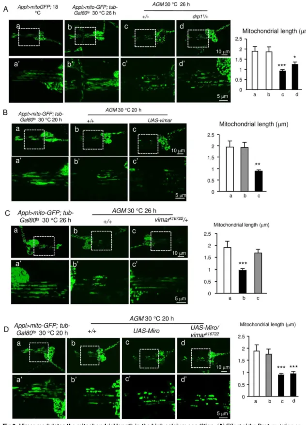
It is well known that mitochondrial fragmentation occurs upon calcium overloaded [47]. To recapitulate this phenomenon and observe mitochondrial morphology by live cell imaging, we addedUAS-mitoGFPto theAGflies (simplified as theAGMmodel). After theAGMlarval flies were raised at 30°C to induce calcium influx for 20 hours, mitochondrial fragmentation in the chordotonal neurons showed subtle fission compared to control; while at the 26 hour, the mitochondria in theAGMdendrites underwent dramatic fragmentation (Fig 3A–3D). For the rescue effect of a given genetic manipulation, we showed the 26 hour time point (to rescue the more severe defects); and for the enhancer effect of a given genetic manipulation, we showed the 20 hour time point (to enhance a less defective phenotype). As a positive control, a LOF mutant of Drp1,drp11, which possessed an A186V amino acid substitution at the Dynamin-GTPase domain [44], strongly suppressed the mitochondrial fission defect (Fig 3Ac and 3Ad). These results suggest that theAGMmodel can be adopted to study the mitochondrial morphol-ogy in high calcium conditions.
Because Miro promotes mitochondrial fission in the high calcium conditions [16], we expected that vimar might enhance Miro function under high calcium concentrations; and the
LOFvimarmight rescue the mitochondrial fission defect in theAGMflies. Indeed, GOFvimar
Fig 3. Vimar modulates the mitochondrial length in the high calcium condition.(A) Effect of the Drp1 mutation on mitochondrial fission in high calcium conditions.aandb, Mitochondrial dendrites in the larval chordotonal neurons in the control flies (Appl>mitoGFPat 18˚C andAppl>mitoGFP,tub-Gal80tsat 30˚C).candd, Mitochondrial morphology in the
AGMandAGM/drp11flies.a’-d’are the enlarged view from the boxed area ina-d, and the mitochondrial lengths ina’-d’
The loss of
vimar
suppressed both necrotic cell death and muscle
defects in a
Drosophila
PD model
Mitochondrial fission may enhance calcium overload-induced necrotic cell death in neuron cultures [47]. However, there is still insufficient genetic evidence to demonstrate that mito-chondrial fission plays a causal role in neuronal necrosis [48]. To study this question, we previ-ously showed that we could quantify necrosis in theAGGflies (theAGflies containing
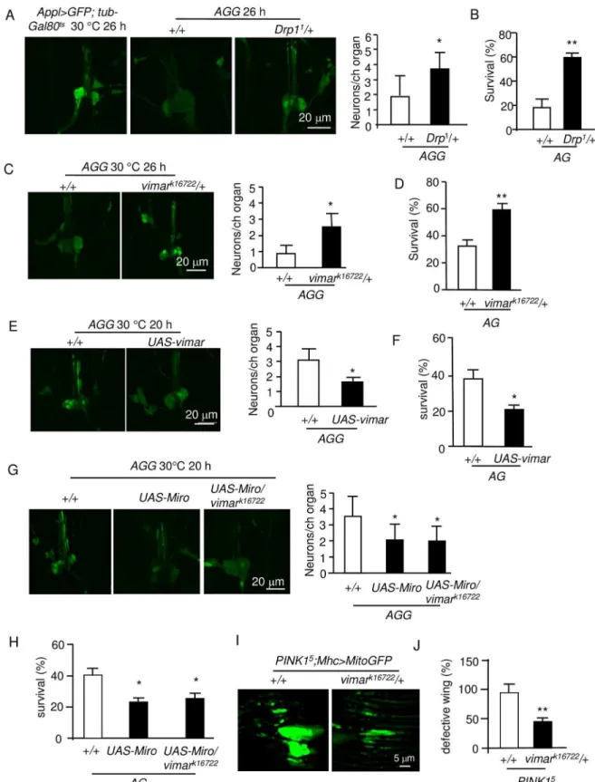
UAS-GFP) at single cell resolution [45]. The result showed thatDrp11could rescue necrosis in
the chordotonal neurons (Fig 4A). In addition, the function of these neurons could be assessed at the behavioral level by quantifying adult fly death [45];Drp11rescued the lethality of theAG flies (Fig 4B). Strikingly,vimark16722exhibited a rescue effect in theAGGflies at both the cellu-lar and behavioral levels (Fig 4C and 4D). In contrast, the GOFvimartransgene had the oppo-site effect (Fig 4E and 4F). This result is consistent with the suppression of mitochondrial fission in this mutant. Furthermore, the GOFMirotransgene enhanced necrosis; however,
vimark16722did not rescue the GOFMirophenotype (Fig 4G and 4H), similar to its effect on
mitochondria. These results indicate that Miro has the dominant role in the Miro/vimar com-plex and that the Miro/vimar comcom-plex plays a functional role in neuronal necrosis.
In thePINK1mutant of the Parkinson's disease (PD)Drosophilamodel, mitochondrial fusion is enhanced, and the LOFMiromutant could suppress this mitochondrial defect [13]. Therefore, we speculate that the LOFvimarmutant might rescue the defective mitochondrial fusion in thePINK1mutant. To test this hypothesis, we studied aPINK1mutant,PINK15[49]. In thePINK15flies, the mitochondria are abnormally elongated and fused (Fig 4I), and the fly wing posture is defective (Fig 4J). Strikingly,vimark16722could rescue both the mitochondrial morphology and wing posture defects (Fig 4I and 4J). Together, these results indicate that LOF of the Miro/vimar complex suppressed both mitochondrial fragmentation during necrosis and
PINK1mutant ofDrosophilaPD model.
Furthermore, we found thatvimark16722andUAS-vimarhad no effect on classical apoptosis induced by Hid expression [50] (S5 Fig), suggesting that vimar may specifically affect PD and necrosis, but does not regulate apoptosis.
The mammalian homolog of vimar (RAP1GDS1) plays a similar role in
mitochondrial morphology and cell death
A protein sequence comparison showed thatDrosophilavimar shares great similarity with the mammalian protein RAP1GDS1 (S6 Fig); however, it is not clear whether vimar is a functional homolog of RAP1GDS1 [51]. Here, we further investigated the role of RAP1GDS1 in mito-chondrial morphology. First, we used a lentivirus to transfect a RAP1GDS1 shRNA into HEK293T cells and established a stable cell line. As expected, the protein level of the
RAP1GDS1 was significantly reduced in the shRNA line (S7A Fig). Then, this shRNA line was transiently transfected with a mitochondrial reporter, mitoDsRed. We found that the mito-chondrial length showed a trend of reduction in the RAP1GDS1 shRNA cells (Fig 5A). Next, we studied the effect of RAP1GDS1 on necrosis. Necrotic cell death was induced by a calcium ionophore (A23187), which causes calcium overloading and necrosis [52]. As expected, the cal-cium ionophore induced mitochondrial fragmentation, and the RAP1GDS1 shRNA rescued the mitochondrial defect (Fig 5B). To quantify the cell death, we measured cellular ATP level
on the mitochondrial length after induction ofAGMexpression for 26 hours. 10 to 16 chordotonal organs were examined for each genotype. (C) Effect ofvimarmutant (vimark16722) on the mitochondrial fragmentation of theAGMflies. 10 to 16 chordotonal organs were examined for each genotype. (D) Effect ofMirooverexpression and thevimarmutant on the mitochondrial fragmentation of theAGMflies. 10 to 16 chordotonal neurons were examined for each genotype.
Fig 4. Vimar suppresses neuronal necrosis and muscle degeneration induced by thePink1mutant.(A) Effect of the
and performed propidium iodide (PI) staining [53]. The result showed that RAP1GDS1 shRNA rescued necrosis in both assays (Fig 5C and 5D). Moreover, we tested the RAP1GDS1 shRNA in another human cell line, the SH-SY5Y neuroblastoma cells. Similar to the HEK293T cells, the RAP1GDS1 shRNA protected the SH-SY5Y cells from calcium overload (S7B and S7C Fig). In addition, we examined the effect of a Miro-1 siRNA on calcium ionophore induced necrosis. The result showed that it also rescued the cell death (Fig 5E and 5F, and the Miro-1 siRNA effect is shown inS7D Fig). Furthermore, the HA-tagged Miro1 and the Flag-tagged RAP1GDS1 could co-immunoprecipitatein vitro(Fig 5G). Together, these results indi-cate that the function of the Miro1/RAP1GDS1 complex in regulating mitochondrial morphol-ogy and necrosis is conserved with theDrosophilaMiro/vimar complex.
Discussion
Vimar is a novel regulator of mitochondrial morphology
Mitochondrial function can be assessed by the enzymatic activity of citrate synthase (CS), the first enzyme in the Krebs cycle that converts acetyl-CoA and oxaloacetate to citrate [54]. In culturedDrosophilaS2 cells,vimarknock down by RNAi resulted in reduced CS activity [54], indicating that vimar may positively regulate mitochondrial function. Because mitochondrial fission has generally been associated with reduced mitochondrial respiration [55], the decreased CS activity may be a result of mitochondrial fission. Consis-tent with this notion, our results demonstrated that the LOF ofvimarpromoted mitochon-drial fission. In addition, a GOFvimartransgene had a minimal effect on mitochondrial morphology, indicating that vimar activity might be saturated under normal physiological conditions.
Vimar functions through Miro to regulate mitochondrial morphology
Because Vimar has been predicted to be a GEF, we hypothesized that vimar may regulate mito-chondrial morphology by affecting a small GTPase, which requires a GEF to help with the GTP/GDP exchange process [19]. Interestingly, Miro is one such small GTPase that is known to play important roles in mitochondrial fission and transport [10,14,16]. We propose that vimar and Miro may function as a complex. First, a fraction of the vimar protein was localized to the mitochondria, possibly indicating a functional role on mitochondria. Interestingly, the mitochondrial localization of vimar seems not dependent on Miro, because LOF Miro did not affect the mitochondrial fraction of vimar. This indicates that vimar may directly bind with mitochondria or through other scaffolding proteins. Second, vimar and Miro could physically interact with each other, at leastin vitro. Their interaction seems not affected by the GTPase activity of Miro, because the constitutively GDP- or GTP-bound Miro mutants did not affect their interactions. Third, vimar genetically interacted with Miro. This included the result dem-onstrating that the LOFvimarmutant reduced the effect of Miro on mitochondrial fissionof the cell loss. For all quantification of neuronal necrosis, trial N = 5, with 10–15 flies were examined in each trial in this figure. (B) Effect of theDrp1mutant on the survival of theAGadult flies. For all quantification ofAGlethality, trial N = 3, with 100–150 flies were examined for each trial. (C) Effect ofvimarmutant on neuronal necrosis. (D) Effect of thevimarmutant on the survival of theAGflies. (E) Effect ofvimaroverexpression on neuronal necrosis. (F) Effect ofvimaroverexpression on the survival of theAGflies. (G) Effect ofMirooverexpression on neuronal necrosis. The result showed thatMirooverexpression enhanced neuronal necrosis; and thevimarmutant had no rescue effect on this defect. (H) Effect ofMirooverexpression on the survival of theAGflies. (I) Effect of thevimarmutant (vimark16722) onPINK1mutant induced mitochondrial defect. The live image showed the mitochondrial morphology in thePINK1mutant (PINK15) and under thevimarmutant background. Ten thoraces were analyzed for each genotype. (J) Effect of thevimarmutant (vimark16722) on the wing posture defect of thePINK1 mutant (PINK15). Trial N = 3, with 100–150 flies were examined in each trial.
inhibition and the GOF vimar transgene had the opposite effect. Moreover, in the constitutive GFP-bound or GDP-bound Miro mutants, the effect of the GOF or LOFvimarwas abolished. Therefore, vimar requires the normal GDP/GTP binding activity of Miro to function. It is also known that Miro1 overexpression increase mitochondrial size partially by suppression of the Drp1 function [15,16]. Consistently, increased mitochondrial fission in the LOF of Miro or vimar was abolished by loss of Drp1, suggesting the Miro/vimar complex depends on Drp1 to regulate mitochondrial morphology.
The Miro/vimar complex may regulate PD and neuronal necrosis
through mitochondrial fusion and fission
Familial PD caused by mutations inPINK1orParkinresults in a series of mitochondrial dys-functions, particularly the failure to eliminate damaged mitochondria through mitophagy [56,
57]. In thesePINK1orParkinmutants, the key proteins involved in mitochondrial fusion and fission, such as Marf/Mitofusin and Miro, accumulate [13,58]. In thePINK1mutant flies, the flight muscle is damaged, resulting in wing posture defects [59]. Similarly, we observed that Miro overexpression in the flight muscle resulted in a strong wing posture defect. This result may explain the wing posture defect in thePINK1mutant, in which the levels of the Miro pro-tein are increased [13]. Our result demonstrated that the LOF ofvimarcould rescue the wing defect in thePINK1mutant, consistent with the hypothesis that vimar functions through Miro.
When the intracellular calcium level is high, Miro switches from promoting mitochon-drial fission inhibition to enhancing mitochonmitochon-drial fission [16]. The mechanism for this switch is unclear, although alterations of Drp1 function could be one possibility [16]. Inter-estingly, Gem1, the yeast homolog of Miro GTPase, has been reported to function as a nega-tive regulator for ER-mitochondria contacts, where Drp1 aggregates and cleaves
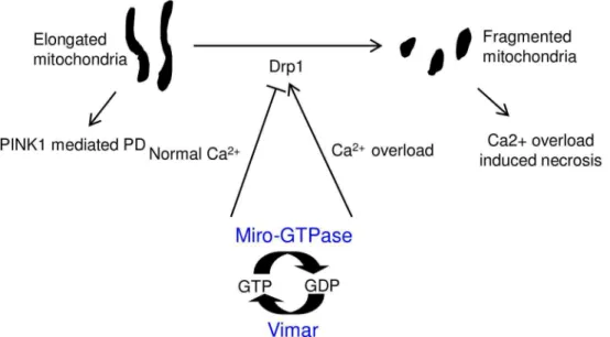
mitochondria into smaller units [37]. This may serve as the mechanism for Miro to regulate mitochondrial morphology via Drp1. In addition to affect mitochondrial fission, Miro also regulates mitochondrial transport in a calcium dependent manner. For mitochondrial trans-port, Miro forms protein complexes with Milton, a kinesin adaptor, and with motor pro-teins, such as kinesin and dynein [35]. In high calcium conditions, Miro alters its binding patterns and results in reduced transport activity [27,60,61]. Based on these reports, we pro-posed that the Miro/vimar complex acted together to affect mitochondrial morphology: at normal condition, Miro/vimar inhibits fission via Drp1; at high calcium state, Ca2+bound Miro switches its function to promote fission. Indeed, vimar responds to the calcium change in the same way as Miro (Fig 6). In addition, our data demonstrated that knocking down
control shRNA). Trial N = 3, with 100 cells were quantified in each trial. (B) Effect of RAP1GDS1 knocking down on the mitochondrial fragmentation under calcium overload stress. The HEK293T cells were treated with 20μM calcium ionophore (A23187) for 4 hours. The result showed that RAP1GDS1 shRNA reduced fragmented mitochondria upon calcium ionophore treatment. Trial N = 3, with 100 cells were quantified in each trial. (C) Effect of the RAP1GDS1 shRNA on calcium ionophore-induced necrosis. The HEK293T control and RAP1GDS1 shRNA stable cell lines were treated with 20μM A23187 for 14 hours. Then, the cell death was quantified by the ATP assay. The result indicated that less cell death occurred in the RAP1GDS1 shRNA expressing cells. Trial N = 3. (D) Effect of the RAP1GDS1 shRNA on calcium ionophore-induced necrosis. The PI and DAPI staining patterns are shown. The red signals indicate the PI-positive cells and the blue channel indicates the DAPI staining. Trial N = 3. (E) Effect of theMiro1siRNA on calcium ionophore induced necrosis determined by the ATP assay. TheMiro1siRNA was transiently transfected in HEK293T cells for 48 hours. Trial N = 3. (F) Effect of theMiro1siRNA on calcium ionophore induced necrosis determined by the PI staining assay. The PI and DAPI staining patterns are shown. The same result was observed as inE. Trial N = 3. (G) Co-Immunoprecipitation of RAP1GDS1 and Miro1. The proteins were collected from the HEK293T cells that expressed Flag-tagged RAP1GDS1 (Flag-RAP1GDS1) and HA-tagged Miro1 (HA-Miro1). The control IgG is shown as a negative control. The total protein input is shown as the protein loading control. Trial N = 3.
RAP1GDS1 and Miro1 increased mitochondrial fission and could rescue calcium overload induced necrosis, similar to the loss of vimar or Miro inDrosophila. These data support the hypothesis that RAP1GDS1 is the mammalian homolog of vimar, supporting a previous pre-diction [51].
Mitochondrial fission plays important role in apoptosis by promoting mitochondrial outer-membrane permeabilization (MOMP) to release cytochrome c from the mitochondria [62]. The use of the Drp1 inhibitor mdivi to block fission has been shown to be an effective treat-ment for stroke [47], and the function of mitochondrial fission on necrotic cell death has been well documented [24,26,48]. The uncertainty lies in the lack of genetic evidence and down-stream mechanism of mitochondrial fission in necrosis [48]. Our data demonstrated that mito-chondrial fragmentation occurred in necrotic neurons, and the LOFDrp1andvimarmutants both suppressed neuronal necrosis.
Much evidence suggests that the mitochondrial fusion and fission defects are directly linked to many human diseases [22], and strategies that target the Miro/vimar complex may affect a broad spectrum of diseases. For instance, mutations in the fragile X mental retardation 1 (FMR1) gene, which result from expansion of trinucleotide repeat in the 50untranslated region,
often cause enhanced mitochondrial fission and mental retardation syndrome [63]. Likewise, aberrant mitochondrial fusion was observed in aDrosophilaAlzheimer's disease model induced by the ectopic expression of a human tau mutant (tauR406W) [43]. In this case, the tau mutant may promote excessive actin stabilization to decrease Drp1 recruitment to the mito-chondria, which results in excessive mitochondrial fusion and neurodegeneration [43,64]. Due to the dual function of the Miro/vimar complex in high-Ca2+induced necrosis andPINK1 mutant induced PD, a drug to target this complex may benefit both disease states. As a modula-tor, it may be safer to target vimar/ RAP1GDS1.
Fig 6. A schematic model of Miro/vimar function on mitochondrial morphology.In normal calcium conditions, the Miro/vimar complex promotes mitochondrial fission inhibition, and their GOF results in elongated mitochondria. Increased mitochondrial fusion is known to occur in thePINK1mutant flies, and this defect can be rescued by LOF Miro/vimar. In the high calcium state, the Miro/vimar complex promotes mitochondrial fragmentation, which accelerates neuronal necrosis. Regardless of the intracellular calcium level, vimar enhances the function of Miro, because vimar is likely the GEF to promote Miro’s GTP/GDP exchange.
Supporting Information
S1 Fig. (A)a-c, Live imaging of the mitochondrial morphology in the flight muscle of adult flies. The mitochondria are labeled withUAS-mitoGFPdriven byTubulin-Gal4(Tubulin>mitoGFP). The genotype is indicated on each micrograph.d, The averaged mitochondrial size of the control (+/+) is set as 1, and the relative ratios of the other genotypes to the control are shown. Five tho-races from each genotype were quantified. Bar graphs throughout all figures are means ± SD. The white bar represents the control, the gray bar represents no statistical different from the con-trol, and the black bar represents significantly different from the control.for p<0.05;for p<0.01;for p<0.001.(B) and (C) Vimar protein level in the adult thoraces. The Western blot shows immunobloting with a vimar antibody, with the genotype listed on each lane.β-actin is shown as the protein loading control. The quantified data is shown as means ± SD. Trial N = 3. (PDF)
S2 Fig. (A) Vimar distribution in other subcellular compartments. The fly homogenate was sep-arated into cytosol, lysosome, Golgi apparatus and ER. Vimar protein level was determined by immunobloting using a vimar antibody. Calnexin, Lamp1, GM130 andβ-actin are markers for ER, lysosome, Golgi apparatus and cytoplasm, respectively. (B) Effect ofvimaroverexpression on mitochondrial transport. The mitochondria are labeled with mitoGFP (CCAP>mitoGFP), and their movements in the axons were recorded and transformed into kymographs. Overex-pression ofMiro25N,vimaror both of them had no effects towards mitochondria transport. Mitochondria motion in ten axons from five larvae was analyzed for each genotype. (C) Effect of
DrosophilaMiro mutants on its interaction with vimarin vitro. The HA-tagged Miro, Miro20V
(a constitutive GTP-bound mutant) and Miro25N (a constitutive GDP-bound mutant) was indi-vidually co-transfected with Flag-tagged vimar. The co-IP experiment showed that GTP or GDP state of Miro did not affect its interaction with vimar. Trial N = 3. IgG is shown as a negative control. The total protein input is shown as the protein loading control.
(PDF)
S3 Fig. (A) Effect of LOF Miro on the mitochondrial localization of vimar. In theMhc>
mi-toGFP/Miro RNAiflies, the mitochondrial fraction of vimar was unaltered as the control
Mho>mitoGFPflies. Anti-ATP5A is shown as the mitochondria marker and actin as cytosolic
marker. Trial N = 2. (B) Live image of mitochondria in flight muscle.Miro RNAiandvimar RNAiresulted in shortened mitochondria, which could be blocked byDrp1 RNAi. Five thoraces from each genotype were quantified. (C) and (D) Drp1 recruitment to mitochondria inMiro
RNAiorvimar RNAibackground. Thoracic homogenate was separated into cytosol and
mito-chondria and Drp1 protein level in different fractions was immunoblotted. Anti-ATP5A is shown as the mitochondria marker and actin as cytosolic marker. Trial N = 2.
(PDF)
S4 Fig. Vimar protein distribution in mitochondria fraction under high calcium stress.The AGfly heads were homogenized and separated in cytoplasmic fraction and crude mitochon-dria. Vimar level was labeled by a vimar antibody. Anti-ATP5A is shown as the mitochondria marker and actin as cytosolic marker. Trial N = 2.
(PDF)
S5 Fig. Effect of LOF and GOFvimaron apoptosis.The apoptotic flies (GMR-Gal4;
GMR-Hid) showed smaller eye size. Addition ofUAS-P35, a known apoptosis inhibitor, is
shown as a positive control, which rescued the smaller eye size defect. However,vimark16722or
UAS-vimarshowed no effect on the eye size defect.
S6 Fig. Protein sequence comparison between vimar (635 a.a) and RAP1GDS1 (608 a.a). The alignment is generated from CLUSTAL alignment algorithm.
(PDF)
S7 Fig. (A) Effect of the RAP1GDS1 shRNA on the level of the RAP1GDS1 protein in the sta-ble HEK293T cells.β-actin was used as the loading control. (B) Effect RAP1GDS1 shRNA on necrosis in the stable SH-SY5Y cells. The cells were treated with 20 μM A23187 for 1 hour. The bright field images of the cells showed less cell death in the RAP1GDS1 shRNA cells upon cal-cium ionophore treatment. Trial N = 4. (C) Quantification of necrosis by the ATP assay. The stable SH-SY5Y cell lines were treated with 20 μM A23187 for 6 hours. The result showed that less cell death occurred in the RAP1GDS1 shRNA cells than the control (scrambled shRNA) cells. Trial N = 3. (D) The efficiency of theMiro1siRNA onMirotranscripts in 293T cells. The transcript level ofMiro1was determined by qRT-PCR. The result showed thatMiro1siRNA significantly knocked downMiro1transcripts. Trial N = 3.
(PDF)
Author Contributions
Conceptualization:LD XJ LL.
Data curation:DL YH LL.
Formal analysis:LD YeL.
Investigation:LD YeL YH YuL.
Methodology:YeL YH YuL.
Project administration:LD LL.
Resources:YeL YuL.
Supervision:LL.
Writing – original draft:LD XJ LL.
Writing – review & editing:XJ.
References
1. Sheng ZH, Cai Q. Mitochondrial transport in neurons: impact on synaptic homeostasis and neurode-generation. Nat Rev Neurosci. 13(2):77–93. Epub 2012/01/06. doi: nrn3156 [pii] doi:10.1038/nrn3156
PMID:22218207.
2. Detmer SA, Chan DC. Functions and dysfunctions of mitochondrial dynamics. Nat Rev Mol Cell Biol. 2007; 8(11):870–9. doi:10.1038/nrm2275PMID:17928812.
3. Chan DC. Mitochondrial fusion and fission in mammals. Annu Rev Cell Dev Biol. 2006; 22:79–99. doi:
10.1146/annurev.cellbio.22.010305.104638PMID:16704336.
4. Westermann B. Mitochondrial fusion and fission in cell life and death. Nat Rev Mol Cell Biol. 2010; 11 (12):872–84. doi:10.1038/nrm3013PMID:21102612.
5. Alexander C, Votruba M, Pesch UE, Thiselton DL, Mayer S, Moore A, et al. OPA1, encoding a dyna-min-related GTPase, is mutated in autosomal dominant optic atrophy linked to chromosome 3q28. Nat Genet. 2000; 26(2):211–5. doi:10.1038/79944PMID:11017080.
6. Chen H, Detmer SA, Ewald AJ, Griffin EE, Fraser SE, Chan DC. Mitofusins Mfn1 and Mfn2 coordi-nately regulate mitochondrial fusion and are essential for embryonic development. J Cell Biol. 2003; 160(2):189–200. doi:10.1083/jcb.200211046PMID:12527753; PubMed Central PMCID:
7. Cipolat S, Martins de Brito O, Dal Zilio B, Scorrano L. OPA1 requires mitofusin 1 to promote mitochon-drial fusion. Proc Natl Acad Sci U S A. 2004; 101(45):15927–32. Epub 2004/10/29. doi: 0407043101 [pii] doi:10.1073/pnas.0407043101PMID:15509649; PubMed Central PMCID: PMC528769.
8. Bleazard W, McCaffery JM, King EJ, Bale S, Mozdy A, Tieu Q, et al. The dynamin-related GTPase Dnm1 regulates mitochondrial fission in yeast. Nat Cell Biol. 1999; 1(5):298–304. doi:10.1038/13014
PMID:10559943; PubMed Central PMCID: PMC3739991.
9. Otera H, Wang C, Cleland MM, Setoguchi K, Yokota S, Youle RJ, et al. Mff is an essential factor for mitochondrial recruitment of Drp1 during mitochondrial fission in mammalian cells. J Cell Biol. 2010; 191(6):1141–58. doi:10.1083/jcb.201007152PMID:21149567; PubMed Central PMCID:
PMC3002033.
10. Guo X, Macleod GT, Wellington A, Hu F, Panchumarthi S, Schoenfield M, et al. The GTPase dMiro is required for axonal transport of mitochondria to Drosophila synapses. Neuron. 2005; 47(3):379–93. doi:10.1016/j.neuron.2005.06.027PMID:16055062.
11. Glater EE, Megeath LJ, Stowers RS, Schwarz TL. Axonal transport of mitochondria requires milton to recruit kinesin heavy chain and is light chain independent. J Cell Biol. 2006; 173(4):545–57. doi:10. 1083/jcb.200601067PMID:16717129; PubMed Central PMCID: PMC2063864.
12. Frederick RL, McCaffery JM, Cunningham KW, Okamoto K, Shaw JM. Yeast Miro GTPase, Gem1p, regulates mitochondrial morphology via a novel pathway. J Cell Biol. 2004; 167(1):87–98. doi:10. 1083/jcb.200405100PMID:15479738; PubMed Central PMCID: PMC2172521.
13. Liu S, Sawada T, Lee S, Yu W, Silverio G, Alapatt P, et al. Parkinson’s disease-associated kinase PINK1 regulates Miro protein level and axonal transport of mitochondria. PLoS Genet. 8(3):e1002537. Epub 2012/03/08. doi:10.1371/journal.pgen.1002537PGENETICS-D-11-02331 [pii]. PMID:
22396657; PubMed Central PMCID: PMC3291531.
14. Fransson A, Ruusala A, Aspenstrom P. Atypical Rho GTPases have roles in mitochondrial homeosta-sis and apoptohomeosta-sis. J Biol Chem. 2003; 278(8):6495–502. doi:10.1074/jbc.M208609200PMID:
12482879.
15. Fransson S, Ruusala A, Aspenstrom P. The atypical Rho GTPases Miro-1 and Miro-2 have essential roles in mitochondrial trafficking. Biochem Biophys Res Commun. 2006; 344(2):500–10. doi:10.1016/ j.bbrc.2006.03.163PMID:16630562.
16. Saotome M, Safiulina D, Szabadkai G, Das S, Fransson A, Aspenstrom P, et al. Bidirectional Ca2 +-dependent control of mitochondrial dynamics by the Miro GTPase. Proc Natl Acad Sci U S A. 2008; 105(52):20728–33. doi:10.1073/pnas.0808953105PMID:19098100; PubMed Central PMCID: PMC2634948.
17. Ferguson SM, De Camilli P. Dynamin, a membrane-remodelling GTPase. Nat Rev Mol Cell Biol. 2012; 13(2):75–88. doi:10.1038/nrm3266PMID:22233676; PubMed Central PMCID: PMC3519936.
18. Gasper R, Meyer S, Gotthardt K, Sirajuddin M, Wittinghofer A. It takes two to tango: regulation of G proteins by dimerization. Nat Rev Mol Cell Biol. 2009; 10(6):423–9. doi:10.1038/nrm2689PMID:
19424291.
19. Cherfils J, Zeghouf M. Regulation of small GTPases by GEFs, GAPs, and GDIs. Physiological reviews. 2013; 93(1):269–309. doi:10.1152/physrev.00003.2012PMID:23303910.
20. Delettre C, Lenaers G, Griffoin JM, Gigarel N, Lorenzo C, Belenguer P, et al. Nuclear gene OPA1, encoding a mitochondrial dynamin-related protein, is mutated in dominant optic atrophy. Nat Genet. 2000; 26(2):207–10. doi:10.1038/79936PMID:11017079.
21. Kijima K, Numakura C, Izumino H, Umetsu K, Nezu A, Shiiki T, et al. Mitochondrial GTPase mitofusin 2 mutation in Charcot-Marie-Tooth neuropathy type 2A. Hum Genet. 2005; 116(1–2):23–7. doi:10.1007/ s00439-004-1199-2PMID:15549395.
22. Itoh K, Nakamura K, Iijima M, Sesaki H. Mitochondrial dynamics in neurodegeneration. Trends in cell biology. 2013; 23(2):64–71. doi:10.1016/j.tcb.2012.10.006PMID:23159640; PubMed Central PMCID: PMC3558617.
23. Knott AB, Bossy-Wetzel E. Impairing the mitochondrial fission and fusion balance: a new mechanism of neurodegeneration. Annals of the New York Academy of Sciences. 2008; 1147:283–92. doi:10. 1196/annals.1427.030PMID:19076450; PubMed Central PMCID: PMC2605288.
24. Wang Z, Jiang H, Chen S, Du F, Wang X. The mitochondrial phosphatase PGAM5 functions at the convergence point of multiple necrotic death pathways. Cell. 2012; 148(1–2):228–43. doi:10.1016/j. cell.2011.11.030PMID:22265414.
26. Wang W, Zhang F, Li L, Tang F, Siedlak SL, Fujioka H, et al. MFN2 couples glutamate excitotoxicity and mitochondrial dysfunction in motor neurons. J Biol Chem. 2015; 290(1):168–82. doi:10.1074/jbc. M114.617167PMID:25416777; PubMed Central PMCID: PMC4281719.
27. Wang X, Schwarz TL. The mechanism of Ca2+ -dependent regulation of kinesin-mediated mitochon-drial motility. Cell. 2009; 136(1):163–74. doi:10.1016/j.cell.2008.11.046PMID:19135897; PubMed Central PMCID: PMC2768392.
28. Frazier AE, Kiu C, Stojanovski D, Hoogenraad NJ, Ryan MT. Mitochondrial morphology and distribu-tion in mammalian cells. Biological chemistry. 2006; 387(12):1551–8. doi:10.1515/BC.2006.193
PMID:17132100.
29. Altmann K, Westermann B. Role of essential genes in mitochondrial morphogenesis in Saccharomy-ces cerevisiae. Molecular biology of the cell. 2005; 16(11):5410–7. doi:10.1091/mbc.E05-07-0678
PMID:16135527; PubMed Central PMCID: PMC1266436.
30. Ichishita R, Tanaka K, Sugiura Y, Sayano T, Mihara K, Oka T. An RNAi screen for mitochondrial pro-teins required to maintain the morphology of the organelle in Caenorhabditis elegans. J Biochem. 2008; 143(4):449–54. doi:10.1093/jb/mvm245PMID:18174190.
31. Kikuchi A, Kaibuchi K, Hori Y, Nonaka H, Sakoda T, Kawamura M, et al. Molecular cloning of the human cDNA for a stimulatory GDP/GTP exchange protein for c-Ki-ras p21 and smg p21. Oncogene. 1992; 7(2):289–93. PMID:1549351.
32. Kaibuchi K, Mizuno T, Fujioka H, Yamamoto T, Kishi K, Fukumoto Y, et al. Molecular cloning of the cDNA for stimulatory GDP/GTP exchange protein for smg p21s (ras p21-like small GTP-binding pro-teins) and characterization of stimulatory GDP/GTP exchange protein. Molecular and cellular biology. 1991; 11(5):2873–80. doi:10.1128/MCB.11.5.2873PMID:1901951; PubMed Central PMCID: PMC360075.
33. Yamamoto T, Kaibuchi K, Mizuno T, Hiroyoshi M, Shirataki H, Takai Y. Purification and characteriza-tion from bovine brain cytosol of proteins that regulate the GDP/GTP exchange reaccharacteriza-tion of smg p21s, ras p21-like GTP-binding proteins. The Journal of biological chemistry. 1990; 265(27):16626–34. PMID:2118909.
34. Greene JC, Whitworth AJ, Kuo I, Andrews LA, Feany MB, Pallanck LJ. Mitochondrial pathology and apoptotic muscle degeneration in Drosophila parkin mutants. Proc Natl Acad Sci U S A. 2003; 100 (7):4078–83. doi:10.1073/pnas.0737556100PMID:12642658; PubMed Central PMCID: PMC153051.
35. Birsa N, Norkett R, Higgs N, Lopez-Domenech G, Kittler JT. Mitochondrial trafficking in neurons and the role of the Miro family of GTPase proteins. Biochemical Society transactions. 2013; 41(6):1525– 31. doi:10.1042/BST20130234PMID:24256248.
36. Wang X, Schwarz TL. Imaging axonal transport of mitochondria. Methods in enzymology. 2009; 457:319–33. doi:10.1016/S0076-6879(09)05018-6PMID:19426876; PubMed Central PMCID: PMC2996865.
37. Murley A, Lackner LL, Osman C, West M, Voeltz GK, Walter P, et al. ER-associated mitochondrial divi-sion links the distribution of mitochondria and mitochondrial DNA in yeast. eLife. 2013; 2:e00422. doi:
10.7554/eLife.00422PMID:23682313; PubMed Central PMCID: PMC3654481.
38. Kornmann B, Osman C, Walter P. The conserved GTPase Gem1 regulates endoplasmic reticulum-mitochondria connections. Proceedings of the National Academy of Sciences of the United States of America. 2011; 108(34):14151–6. doi:10.1073/pnas.1111314108PMID:21825164; PubMed Central PMCID: PMC3161550.
39. Beall CJ, Fyrberg E. Muscle abnormalities in Drosophila melanogaster heldup mutants are caused by missing or aberrant troponin-I isoforms. J Cell Biol. 1991; 114(5):941–51. doi:10.1083/jcb.114.5.941
PMID:1908472; PubMed Central PMCID: PMC2289106.
40. Babic M, Russo GJ, Wellington AJ, Sangston RM, Gonzalez M, Zinsmaier KE. Miro’s N-terminal GTPase domain is required for transport of mitochondria into axons and dendrites. J Neurosci. 2015; 35(14):5754–71. doi:10.1523/JNEUROSCI.1035-14.2015PMID:25855186; PubMed Central PMCID: PMC4388930.
41. Liu S, Sawada T, Lee S, Yu W, Silverio G, Alapatt P, et al. Parkinson’s disease-associated kinase PINK1 regulates Miro protein level and axonal transport of mitochondria. PLoS genetics. 2012; 8(3): e1002537. doi:10.1371/journal.pgen.1002537PMID:22396657; PubMed Central PMCID: PMC3291531.
43. DuBoff B, Gotz J, Feany MB. Tau promotes neurodegeneration via DRP1 mislocalization in vivo. Neu-ron. 2012; 75(4):618–32. doi:10.1016/j.neuron.2012.06.026PMID:22920254; PubMed Central PMCID: PMC3428596.
44. Verstreken P, Ly CV, Venken KJ, Koh TW, Zhou Y, Bellen HJ. Synaptic mitochondria are critical for mobilization of reserve pool vesicles at Drosophila neuromuscular junctions. Neuron. 2005; 47 (3):365–78. doi:10.1016/j.neuron.2005.06.018PMID:16055061.
45. Liu K, Ding L, Li Y, Yang H, Zhao C, Lei Y, et al. Neuronal necrosis is regulated by a conserved chro-matin-modifying cascade. Proc Natl Acad Sci U S A. 2014; 111(38):13960–5. doi:10.1073/pnas. 1413644111PMID:25201987.
46. Kohda K, Wang Y, Yuzaki M. Mutation of a glutamate receptor motif reveals its role in gating and delta2 receptor channel properties. Nat Neurosci. 2000; 3(4):315–22. doi:10.1038/73877PMID:
10725919.
47. Grohm J, Kim SW, Mamrak U, Tobaben S, Cassidy-Stone A, Nunnari J, et al. Inhibition of Drp1 pro-vides neuroprotection in vitro and in vivo. Cell Death Differ. 2012; 19(9):1446–58. doi:10.1038/cdd. 2012.18PMID:22388349; PubMed Central PMCID: PMC3422469.
48. Jahani-Asl A, Germain M, Slack RS. Mitochondria: joining forces to thwart cell death. Biochimica et biophysica acta. 2010; 1802(1):162–6. doi:10.1016/j.bbadis.2009.09.006PMID:19747972.
49. Clark IE, Dodson MW, Jiang C, Cao JH, Huh JR, Seol JH, et al. Drosophila pink1 is required for mito-chondrial function and interacts genetically with parkin. Nature. 2006; 441(7097):1162–6. doi:10. 1038/nature04779PMID:16672981.
50. Bergmann A, Agapite J, McCall K, Steller H. The Drosophila gene hid is a direct molecular target of Ras-dependent survival signaling. Cell. 1998; 95(3):331–41. doi:10.1016/S0092-8674(00)81765-1
PMID:9814704.
51. Lo PC, Frasch M. bagpipe-Dependent expression of vimar, a novel Armadillo-repeats gene, in Dro-sophila visceral mesoderm. Mech Dev. 1998; 72(1–2):65–75. doi:10.1016/S0925-4773(98)00016-1
PMID:9533953.
52. Karch J, Kwong JQ, Burr AR, Sargent MA, Elrod JW, Peixoto PM, et al. Bax and Bak function as the outer membrane component of the mitochondrial permeability pore in regulating necrotic cell death in mice. eLife. 2:e00772. Epub 2013/08/31. doi:10.7554/eLife.0077200772 [pii]. PMID:23991283; PubMed Central PMCID: PMC3755340.
53. Degterev A, Huang Z, Boyce M, Li Y, Jagtap P, Mizushima N, et al. Chemical inhibitor of nonapoptotic cell death with therapeutic potential for ischemic brain injury. Nature chemical biology. 2005; 1(2):112– 9. doi:10.1038/nchembio711PMID:16408008.
54. Chen J, Shi X, Padmanabhan R, Wang Q, Wu Z, Stevenson SC, et al. Identification of novel modula-tors of mitochondrial function by a genome-wide RNAi screen in Drosophila melanogaster. Genome Res. 2008; 18(1):123–36. Epub 2007/11/29. doi: gr.6940108 [pii] doi:10.1101/gr.6940108PMID:
18042644; PubMed Central PMCID: PMC2134776.
55. Westermann B. Bioenergetic role of mitochondrial fusion and fission. Biochimica et biophysica acta. 2012; 1817(10):1833–8. doi:10.1016/j.bbabio.2012.02.033PMID:22409868.
56. Narendra DP, Jin SM, Tanaka A, Suen DF, Gautier CA, Shen J, et al. PINK1 is selectively stabilized on impaired mitochondria to activate Parkin. PLoS biology. 2010; 8(1):e1000298. doi:10.1371/journal. pbio.1000298PMID:20126261; PubMed Central PMCID: PMC2811155.
57. Matsuda N, Sato S, Shiba K, Okatsu K, Saisho K, Gautier CA, et al. PINK1 stabilized by mitochondrial depolarization recruits Parkin to damaged mitochondria and activates latent Parkin for mitophagy. The Journal of cell biology. 2010; 189(2):211–21. doi:10.1083/jcb.200910140PMID:20404107; PubMed Central PMCID: PMC2856912.
58. Ziviani E, Tao RN, Whitworth AJ. Drosophila parkin requires PINK1 for mitochondrial translocation and ubiquitinates mitofusin. Proc Natl Acad Sci U S A. 2010; 107(11):5018–23. doi:10.1073/pnas. 0913485107PMID:20194754; PubMed Central PMCID: PMC2841909.
59. Park J, Lee SB, Lee S, Kim Y, Song S, Kim S, et al. Mitochondrial dysfunction in Drosophila PINK1 mutants is complemented by parkin. Nature. 2006; 441(7097):1157–61. doi:10.1038/nature04788
PMID:16672980.
60. Macaskill AF, Rinholm JE, Twelvetrees AE, Arancibia-Carcamo IL, Muir J, Fransson A, et al. Miro1 is a calcium sensor for glutamate receptor-dependent localization of mitochondria at synapses. Neuron. 2009; 61(4):541–55. doi:10.1016/j.neuron.2009.01.030PMID:19249275; PubMed Central PMCID: PMC2670979.
61. Chen Y, Sheng ZH. Kinesin-1-syntaphilin coupling mediates activity-dependent regulation of axonal mitochondrial transport. The Journal of cell biology. 2013; 202(2):351–64. doi:10.1083/jcb.201302040
62. Cassidy-Stone A, Chipuk JE, Ingerman E, Song C, Yoo C, Kuwana T, et al. Chemical inhibition of the mitochondrial division dynamin reveals its role in Bax/Bak-dependent mitochondrial outer membrane permeabilization. Dev Cell. 2008; 14(2):193–204. doi:10.1016/j.devcel.2007.11.019PMID:18267088; PubMed Central PMCID: PMC2267902.
63. Ross-Inta C, Omanska-Klusek A, Wong S, Barrow C, Garcia-Arocena D, Iwahashi C, et al. Evidence of mitochondrial dysfunction in fragile X-associated tremor/ataxia syndrome. The Biochemical journal. 2010; 429(3):545–52. doi:10.1042/BJ20091960PMID:20513237; PubMed Central PMCID: PMC4011071.