and Dysfunction in Neuroblastoma Cells
So Jung Park1, Doo Sin Jo1, Ji Hyun Shin1, Eun Sung Kim1, Yoon Kyung Jo1, Eun Sun Choi1, Hae Mi Seo1,2,
Sung Hyun Kim3, Jung Jin Hwang4, Dong-Gyu Jo5, Jae-Young Koh6, Dong-Hyung Cho1*
1Department of East-West Medical Science, Graduate School of East-West Medical Science, Kyung Hee University, Yongin, South Korea,2Department of Genetic Engineering, Kyung Hee University, Yongin, South Korea,3School of Medicine, Kyung Hee University, Seoul, South Korea,4Asan Institute for Life Science, Institute for Innovative Cancer Research, University of Ulsan College of Medicine, Asan Medical Center, Seoul, South Korea,5School of Pharmacy, Sungkyunkwan University, Suwon, South Korea,6Department of Neurology, University of Ulsan College of Medicine, Asan Medical Center, Seoul, South Korea
Abstract
To date, several regulatory proteins involved in mitochondrial dynamics have been identified. However, the precise mechanism coordinating these complex processes remains unclear. Mitochondrial chaperones regulate mitochondrial function and structure. Chaperonin 10 (Cpn10) interacts with heat shock protein 60 (HSP60) and functions as a co-chaperone. In this study, we found that down-regulation of Cpn10 highly promoted mitochondrial fragmentation in SK-N-MC and SH-SY5Y neuroblastoma cells. Both genetic and chemical inhibition of Drp1 suppressed the mitochondrial fragmentation induced by Cpn10 reduction. Reactive oxygen species (ROS) generation in 3-NP-treated cells was markedly enhanced by Cpn10 knock down. Depletion of Cpn10 synergistically increased cell death in response to 3-NP treatment. Furthermore, inhibition of Drp1 recovered Cpn10-mediated mitochondrial dysfunction in 3-NP-treated cells. Moreover, an ROS scavenger suppressed cell death mediated by Cpn10 knockdown in 3-NP-treated cells. Taken together, these results showed that down-regulation of Cpn10 increased mitochondrial fragmentation and potentiated 3-NP-mediated mitochondrial dysfunction in neuroblastoma cells.
Citation:Park SJ, Jo DS, Shin JH, Kim ES, Jo YK, et al. (2014) Suppression of Cpn10 Increases Mitochondrial Fission and Dysfunction in Neuroblastoma Cells. PLoS ONE 9(11): e112130. doi:10.1371/journal.pone.0112130
Editor:Thiruma V. Arumugam, National University of Singapore, Singapore ReceivedJuly 7, 2014;AcceptedOctober 12, 2014;PublishedNovember 12, 2014
Copyright:ß2014 Park et al. This is an open-access article distributed under the terms of the Creative Commons Attribution License, which permits unrestricted use, distribution, and reproduction in any medium, provided the original author and source are credited.
Data Availability:The authors confirm that all data underlying the findings are fully available without restriction. All relevant data are within the paper. Funding:This research was supported by a grant of the Korean Health 21 R&D Project and Korea-UK Collaborative Alzheimer’s Disease Research Project, Ministry of Health & Welfare, Republic of Korea (A092042, A120196). The funders had no role in study design, data collection and analysis, decision to publish, or preparation of the manuscript.
Competing Interests:The authors have declared that no competing interests exist. * Email: [email protected]
Introduction
Mitochondria are constantly undergoing division and fusion in normal cells. The balance of mitochondrial dynamics influences mitochondrial morphology, distribution and function [1]. Several GTPase proteins including dynamin-related protein1 (Drp1), optic dominant atrophy 1 (Opa1), and mitofusin 1/22 (Mfn1/2) have been identified as regulators of mitochondrial dynamics [2–5]. Mfn1/2 are involved in outer mitochondrial membrane (OMM) fusion with adjacent mitochondria [6]. Opa1 is responsible for inner mitochondrial membrane fusion, and maintains both mitochondrial DNA and cristae morphogenesis [7]. In contrast, mitochondrial fission relies on the Drp1 protein. Drp1 shares common mechanism with its homolog, dynamin GTPase, which is involved in endocytosis [8]. Upon stimulation of mitochondrial fission, Drp1 is translocated from the cytosol into the OMM and interacts with its receptors such as mitochondrial fission 1 (Fis1) and mitochondrial fission factor (MFF). This binding initiates the mitochondrial fission process [9]. Mitochondrial fusion functions as a cell protection mechanism, whereas massive mitochondrial fission potentiates cell death [10]. Owing to their high energy demands, mitochondrial function is particularly important in neuronal cells. Disruption of mitochondria dynamics can affect a
wide range of neuronal activities such as synaptic transmission, axonal/dendritic transport, and neuronal calcium homeostasis [11]. Therefore, an imbalance in mitochondrial dynamics can contribute to neurodegenerative diseases including Huntington’s disease (HD), Alzheimer’s disease (AD) and Parkinson’s disease (PD) [12]. HD, an autosomal dominant disorder is a genetic disease caused by mutation in the Huntingtin (Htt) gene. Mutant
Htt leads to excessive mitochondrial fission and neuronal dysfunction [13]. 3-nitropropionic acid (3-NP), a mitochondrial oxidative phosphorylation complex II inhibitor, triggers a move-ment disorder similar in many respects to HD. 3-NP induces abnormal mitochondrial fission, prolonged energy impairments and subsequent neuronal injury [14].
(Cpn10)/heat shock 10 kDa protein 1(HSPE1) interacts with heat shock protein 60 (HSP60), and functions as a co-chaperone. The Cpn10-HSP60 complex regulates the folding of proteins imported into mitochondria [19]. Therefore, Cpn10 is important for the maintenance of normal mitochondrial structure and activity [20,21]. Cpn10 is a multifunctional protein. The overexpression of Cpn10 has been reported alongside various tumors such as in prostate and lymphomas [22,23]. Overexpression of Cpn10 modulates also apoptosis by increasing anti-apoptotic Bcl-2 proteins and decreasing the pro-apoptotic Bax protein [24]. In addition, Cpn10 is involved in the Ras GTPase pathway, bone marrow cell differentiation, and the IGF-1R signaling pathway [25,26]. However, the underlying mechanism for Cpn10 regula-tion in mitochondria-mediated neuro-toxicity remains unclear.
In this study, we found that down-regulation of Cpn10 greatly increased mitochondrial fragmentation, and potentiated 3-NP-mediated mitochondrial dysfunction in neuroblastoma cells.
Materials and Methods
Cell culture and stable transfection
SK-N-MC and SH-SY5Y neuroblastoma cells were obtained from the American Type Culture Collection (ATCC). Wild type mouse embryo fibroblast (MEF) and Drp1 deficient MEF cells were generally provided by Dr. Katsuyoshi Mihara (Kyushu University, Japan) [27]. All cells were cultured at 37uC in a 5% CO2 incubator and maintained in Dulbecco’s modified Eagle’s medium (DMEM) containing 1% penicillin/streptomycin as well as 10% fetal bovine serum (Invitrogen, Carlsbad, CA). To generate stable cell line (SK/mito-YFP), SK-N-MC cells were transfected with pmito-YFP using Lipofectamine 2000 according to manufacturer’s protocol (Invitrogen, Carlsbad, CA). The cells were selected by growth in selection medium containing Geneticin (1 mg/ml) for 10 days. After single cell dropping, the stable clone was selected under a fluorescence microscope.
Reagents
The YFP-fused mito-tracker plasmid (pmito-YFP) was previ-ously described [28]. 3-nitropropionic acid (3-NP) and N-acetylcysteine (NAC) were purchased from Sigma (St. Louis, MO USA). Mdivi-1 (3-(2,4-Dichloro-5-methoxyphenyl)-2,3-dihy-dro-2-thioxo-4(1H)-quinazolinone) was purchased from Enzo life sciences (Farmingdale, NY, USA). A mitoTracker probe was purchased from Invitrogen (Carlsbad, CA). The validated siRNA targeting for Cpn10 (#1, 59 -CAAAGUAGUUCUAGAUGAC-39), (#2, 59-GCGUGAAAGUUGGAGAUAA-39) negative scram-bled siRNA (59-CCUACGCCACCAAUUUCGU-39) were pur-chased form Dharmacon (Thermo Scientific). And previously validated Drp1 siRNA (59-GAGGUUAUUGAACGACUCA-39) and Opa1 siRNA (59-CUGGAAAGACUAGUGUGUU-39) were synthesized from Bioneer (Daejeon, Korea).
Western blotting
For Western blotting, all lysates were prepared with protein sample buffer (62.5 mM Tris-HCl, pH 6.8, 25% glycerol, 2% SDS, 5%b-mercaptoethanol, 0.01% Bromophenol blue) (BioRad, Hercules, CA). Then the samples were separated by SDS-PAGE and transferred to PVDF membrane (BioRad). After blocking with 4% skim milk in TBST (25 mM Tris, 3 mM 140 mM NaCl, 0.05% Tween-20), the membranes were incubated over-night with specific primary antibodies at 4uC. Anti-Drp1 antibody was from BD (San Jose, CA); anti-Cpn10 antibody was from BD (San Jose, CA); anti-Actin antibody was from Millipore (Temecula, CA). For
protein detection, the membranes were incubated with HRP-conjugated secondary antibodies (Pierce, Rockford, IL).
ROS measurement
Intracellular ROS levels were assayed using a fluorescent dye, 29,79-dichlorofluorescein diacetate (DCFH-DA) (Invitrogen, Carls-bad, CA), which is converted to the highly fluorescent 29,79 -dichlorofluorescein (DCF) in the presence of oxidant. Briefly, cells plated in 96-well plate were transfected with siRNA. With or without further treatment of 3-NP for 8 hr, and the cells were incubated with DCFH-DA (20 uM) in serum free medium for 30 min (excitation/emission wave length 358/485) (Victor X3, Perkinelmer). Relative ROS level was presented as the change in fluorescence of drug treated sample compared with that of control sample.
Measurement of cellular total ATP level
SK-N-MC cells were transfected with Cpn10 siRNA for 5 days. The cells were further treated with or without 3-NP for 8 hr, and then cellular total ATP level was detected with an ATP bioluminescence detection kit according to the manufacturer’s protocol (Promega, Madison, WI).
Measurement of mitochondria membrane potential Mitochondrial membrane potential was measured with a unique fluorescent cationic dye, JC-1 (5,59,6,69 -tetrachloro-1,19,3,39-tetraethylbenzimidazolylcarbocyanine iodide, BD, San Jose, CA) that detects loss of signal of mitochondrial membrane potential. The fluorescence intensity was monitored using plate reader (PerkinElmer, Waltham, MA) at excitation and emission wavelength of 485 nm and 535 nm for monomeric form as well as 535 nm and 590 nm for JC1-aggregates form
Apoptotic cell death analysis
Apoptotic cell death was determined by using an Annexin V-FITC/PI Apoptosis Detection Kit according to the manufacturer’s protocol (BD Pharmingen, San Diego, CA). Briefly, cells transfected with Cpn10 siRNA was exposed to 3-NP for 24 hr. Then, the cells were stained with Annexin V-FITC and propidium iodide (BD Pharmingen, San Jose, CA). After staining, cell death was analyzed by using flow cytometer (BD, San Jose, CA).
Statistical analysis
Data were obtained from least three independent experiments, and presented as means 6 S.E.M. Statistical evaluation of the results was performed with one-way ANOVA. Data were considered significant at a value of *p,0.02.
Results
Down-regulation of Cpn10 induces mitochondrial fragmentation in neuroblastoma cells
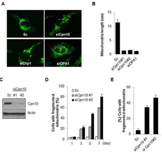
similar to Opa1 knock downs cells (Figure 1A–1D). We further confirmed the Cpn10 knock down effect in other neuroblastoma cells. Consistent with previous results, reduced expression of Cpn10 markedly increased mitochondrial fragmentation in SH-SY5Y cells (Figure 1E). These results suggest that down-regulation of Cpn10 promotes mitochondrial fission in neuroblastoma cells.
Inhibition of Drp1 suppresses Cpn10 knock down-mediated mitochondria fission and dysfunction
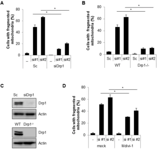
Drp1 is a key player in the machinery of mitochondrial fission. To investigate the effects of Drp1 on siCpn10-mediated mito-chondrial fission, Cpn10 siRNAs were transfected with or without Drp1 siRNA, into SK/mito-YFP cells. The results showed that a loss of Drp1 expression strongly suppressed Cpn10 knock down-mediated mitochondrial fission (Figure 2A). These effects were further confirmed in Drp1-deficient cells. Either wild type (WT) or Drp1-deficient (Drp1-/-) mouse embryonic fibroblast (MEF) cells were transfected with siCpn10, and mitochondrial morphology was monitored. Consistently, siCpn10-induced mitochondrial fragmentation was completely blocked in Drp1-/-MEF cells when
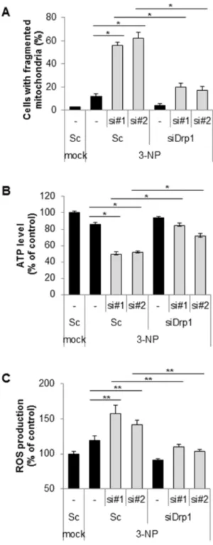
compared with control cells (Figure 2B). The inhibition of Drp1 expression in siDrp1 transfected cells and Drp1-/- cells was addressed using Western blot analysis (Figure 2C). Finally, we enhanced the effect of Drp1 with a selective chemical inhibitor of Drp1, Mdivi-1 [30]. Similar with Drp1 down-regulation experi-ment, Mdivi-1 treatment significantly reduced mitochondria fragmentation in Cpn10 known down cells (Figure 2D). Taken together, these results suggested that Drp1 plays a crucial role in regulating mitochondrial fragmentation in Cpn10 knock down cells. Mitochondrial functions are also influenced by mitochondrial dynamics. Since excessive mitochondrial fragmentation increases mitochondrial dysfunctions, we next examined the effect of Cpn10 knock down on mitochondrial function. Both mitochondrial membrane potential and total cellular ATP level were measured in Cpn10 knockdown cells. As shown in Figure 3, loss of Cpn10 significantly reduced cellular ATP level as well as mitochondrial membrane potential in SK-N-MC cells (Figure 3A, 3B). In addition, depletion of Cpn10 slightly increased ROS production (Figure 3C), suggesting that down-regulation of Cpn10 associated with mitochondrial dysfunctions in neuroblstoma cells.
Figure 1. Down-regulation of Cpn10 induces mitochondrial fragmentation in neuroblastoma cells.(A, B) SK-N-MC cells stably expressing mito-YFP (SK/mito-YFP) were transfected with either a control scrambled siRNA (Sc) or a specific siRNA against Cpn10 for 5 days. Then mitochondrial morphology (A) and mitochondrial length (B) were examined with a fluorescence microscope. Both Drp1 siRNA (siDrp1) and Opa1 siRNA (siOpa1) were used as positive controls. (C) The reduced expression of Cpn10 by siRNA was confirmed by Western blotting. (D) SK/mito-YFP cells were transfected with scrambled siRNA or Cpn10 siRNAs (si#1, si#2). And the mitochondrial fragmentation was observed by a fluorescence microscopy at the indicated time points. (E) SH-SY5Y cells were transfected with either a control scrambled siRNA (Sc) or specific siRNAs against Cpn10 (siCPN10#1, #2). After 5 days, the cells were stained with Mito-tracker (100 nM), and the cells containing fragmented mitochondria were counted using a fluorescence microscope. Data are represented as the mean6SEM. (n.3).
Figure 2. Inhibition of Drp1 suppresses mitochondria fragmentation induced by loss of Cpn10.(A) Drp1 siRNA was co-transfected with either scrambled siRNA (Sc) or Cpn10 siRNA (#1,#2) in SK/mito-YFP cells. 5 days later, the cells with fragmented mitochondria were counted under a fluorescence microscopy. (B) Wild type MEF (WT) and Drp1 deficient MEF (Drp1-/-) cells were transfected with either a control scrambled siRNA (Sc) or specific siRNAs against Cpn10 (siCPN10 #1,#2). After 5 days, the cells were labeled with a fluorescence MitoTracker (100 nM) to observe mitochondrial morphology. The cells with fragmented mitochondria were counted under a fluorescence microscopy. (C) The reduced expression of Drp1 in Drp1 siRNA transfected cells and in Drp1 knock out MEF cells was confirmed by Western blotting. (D) SK/mito-YFP cells transfected with scrambled siRNA (Sc) or Cpn10 siRNA (si#1, si#2) were treated with a Drp1 inhibitor, Mdivi-1 (20mM). The cells with fragmented mitochondria were counted under a fluorescence microscope. Data are represented as the mean6SEM. (n.3) and were considered significant at a value of *p,0.02.
doi:10.1371/journal.pone.0112130.g002
Figure 3. Down-regulation of Cpn10 promotes mitochondrial dysfunction in SK-N-MC cells.(A-C), SK-N-MC cells were transfected with a control scrambled (Sc) or Cpn10 siRNA (si#1, si#2). After 5 days, the alteration of mitochondrial membrane potential was monitored by the MitoProbe JC-1 assay (A). The cellular total ATP level was examined by an ATP bioluminescence detection assay (B). The Intracellular ROS level was measured by a DCFH-DA fluorescence ROS detection assay (C). Data are represented as the mean6SEM (n.3), and were considered significant at a value of *p,0.05.
Down-regulation of chaperonin10 promotes 3-NP-mediated mitochondrial dysfunction
Chaperone family proteins can prevent the aggregation of polyglutamine (polyQ), which is linked with Huntington’s disease (HD)-like pathology and symptoms. 3-nitropropionic acid (3-NP) serves as an experimental model of HD and induces abnormal mitochondria morphology and neurotoxicity [14,31]. Thus, we further investigated the effect of Cpn10 knock down on 3-NP-mediated mitochondrial dysfunction. As shown in Figure 3A, down-regulation of Cpn10 synergistically enhanced mitochondrial fragmentation in 3-NP treated cells when compared with control cells (Figure 4A). In accordance with the previous results, the Cpn10 knock down–mediated mitochondrial fragmentation was highly suppressed by Drp1 inhibition in 3NP-treated cells (Figure 4A). Interestingly, the reduced ATP level and increased ROS production by siCpn10 were much more impaired by 3-NP treatment (Figure 3B and 3C). However, both ATP reduction and ROS induction were reinstated by inhibition of Drp1 in 3-NP-treated cells (Figure 4B and 4C). Collectively, these results suggest that Cpn10 knock down exacerbates 3-NP-mediated mitochon-drial dysfunction in neuroblastoma cells.
Down-regulation of Cpn10 potentiates 3-NP-mediated neurotoxicity
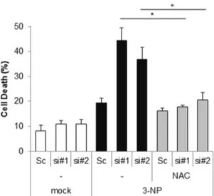
Excessive mitochondrial fragmentation leads to mitochondrial dysfunction and subsequent cell death, therefore, we additionally examined the effect of Cpn10 knock down on 3-NP-induced cell death. SK-N-MC cells transfected with siCpn10 were treated with 3-NP. As shown in Figure 5, depletion of Cpn10 synergistically increased cell death in response to 3-NP in SK-N-MC cells. Interestingly, the cell death was remarkably suppressed by treatment with an ROS scavenger, N-acetylcystein (NAC) (Figure 5). These results suggest that down-regulation of Cpn10 potentiates 3-NP-induced neurotoxicity via excessive ROS pro-duction (Figure 5).
Discussion
Altered mitochondrial dynamics are associated with mitochon-drial dysfunctions in neurodegenerative diseases. In this study, we showed that down-regulation of Cpn10 promotes mitochondrial fission and potentiates neurotoxin-induced mitochondrial dysfunc-tion and cell death. Cpn10 plays many roles in mitochondrial homeostasis. Cpn10 is considered a cooperating partner of HSP60 in protein folding processes [32]. The HSP60-Cpn10 protein complex accelerates the folding of polypeptides imported into mitochondria and reduces aggregation of unfolded inactive polypeptides. It has been reported that expression of mitochon-drial HSP proteins is up-regulated to protect against cellular damage following global brain ischemia [33]. Overexpression of Cpn10 and HSP60 suppresses cytotoxicity by inhibiting mito-chondrial depolarization and modulating mitomito-chondrial Bcl-2 family proteins in cardiomyocytes [34]. Ectopic expression of Cpn10 increases Bcl-xL protein levels and restores the mitochon-drial membrane potential as well as reducing caspase activation in doxorubicin-treated cells [24]. In accordance with results, we have found that loss of Cpn10 promotes mitochondrial dysfunction and potentiates cytotoxicity in neuroblastoma cells. Down-regulation of Cpn10 synergistically increased mitochondrial fragmentation and dysfunction following 3-NP or 6-hydroxyl dopamine treat-ment (Figure 3 and data not shown). Our data presented here further emphasize the importance of Cpn10 in mitochondria. Cpn10 is overexpressed during carcinogenesis of the large bowel and uterine exocervix [35]. In addition, Cpn10 is up-regulated by
Figure 4. Suppression of Cpn10 exacerbates 3-NP-mediated mitochondrial dysfunction in SK-N-MC cells. (A) SK-N-MC cells were transfected with either scrambled siRNA or Cpn10 siRNA (si#1, si#2) with or without Drp1 siRNA (siDrp1) for 5 days. The cells were further exposed to 3-NP (10 mM) for 8 hr, then cells with fragmented mitochondria were counted with a fluorescence microscope. (B, C) SK-N-MC cells were transfected with either scrambled siRNA or Cpn10 siRNA (si#1, si#2) with or without Drp1 siRNA (siDrp1) for 5 days, then cells were further exposed to 3-NP (10 mM) for 8 hr. The total cellular ATP level was measured by an ATP bioluminescence detection assay (B). The intracellular ROS level was determined by a DCFH-DA fluorescence ROS detection assay (C). Data are represented as the mean 6SEM (n.3), and were considered significant at a value of *p,0.02, **p,0.05.
neuronal vesicular cell trafficking and neuronal synaptic plasticity [36]. Therefore, further studies on the role of Cpn10 in regulating expression in physiology, and patho-physiological conditions will be helpful in understanding its functions and these processes.
Recently our group reported that loss of mitochondrial chaperone proteins could efficiently induce mitochondrial fission and dysfunction in neuronal cells [18]. Despite the known role of Cpn10 in mitochondria, the molecular mechanism underlying mitochondrial fragmentation by Cpn10 knock down is still
unknown. Loss of Cpn10 induces mitochondrial fission in a Drp1-dependent manner (Figure 2). Drp1 is a large GTPase protein and several post-translational modifications such as phosphorylation, S-nitrosylation, ubiquitination and O-Glcnacyla-tion of Drp1 protein modulate its GTPase activity [37–41]. Among them, phosphorylation is thought to be an important mechanism of Drp1 regulation. The phosphorylation at Serine 616 on Drp1 by many kinases such as CDK1, ERK1/2 and
PKC-denhances the fission activity of Drp1 in different conditions [42– 44]. Phosphorylation of Drp1 at another site (Serine 637) by PKA and CamK1-ahas shown the opposite effect on Drp1 activation. Phosphorylation of Drp1 by PKA inhibits Drp1 activity, while phosphorylation by CamK1-a increases Drp1 activity [45–46]. More recently, Chou et al have reported that GSK3b also mediates the phosphorylation of Drp1 at Serine 693. The phosphorylation of Drp1 by GSK3b inhibits Drp1 function and elongates mitochondria in response to oxidative stress [47]. It has been suggested that Cpn10 could regulate cellular signaling pathways. Cpn10 is found in secretory granules as well as the mitochondrial matrix. Erythropoietin treatment highly promotes the expression and secretion of Cpn10 in endothelial cells [48]. Interestingly, Cpn10 treatment increases phosphorylation of GSK3b and induces cell differentiation [49]. Therefore, the possibility of GSK3b-mediated regulation of mitochondrial dynamics by Cpn10 ought to be elucidated.
In conclusion, we have demonstrated that down-regulation of Cpn10 leads to mitochondrial dysfunctions through mitochondrial fragmentation. In addition, this down-regulation potentiates neurotoxin-mediated neurotoxicity.
Author Contributions
Conceived and designed the experiments: DHC. Performed the experi-ments: SJP DSJ JHS ESK YKJ ESC HMS. Analyzed the data: SJP DHC. Contributed reagents/materials/analysis tools: SHK JJH DGJ JK. Wrote the paper: DHC SJP.
References
1. Detmer SA, Chan DC (2007) Functions and dysfunctions of mitochondrial dynamics. Nat Rev Mol Cell Biol 8: 870–879.
2. Chen H, Chan DC (2005) Emerging functions of mammalian mitochondrial fusion and fission. Hum Mol Genet 14: R283–R289.
3. Cipolat S, Martins de Brito O, Dal Zilio B, Scorrano L (2004) OPA1 requires mitofusin 1 to promote mitochondrial fusion. Proc Natl Acad Sci USA 101: 15927–15932.
4. Kageyama Y, Zhang Z, Sesaki H (2011) Mitochondrial division: molecular machinery and physiological functions. Curr Opin Cell Biol 23: 427–434. 5. Tamura Y, Itoh K, Sesaki H (2011) SnapShot: Mitochondrial Dynamics. Cell
145: 1158.
6. Ishihara N, Eura Y, Mihara K (2004) Mitofusin 1 and 2 play distinct roles in mitochondrial fusion reactions via GTPase activity. J Cell Sci 117: 6535–6546. 7. Olichon A, Baricault L, Gas N, Guillou E, Valette A, et al. (2003) Loss of OPA1 perturbates the mitochondrial inner membrane structure and integrity leading to cytochrome c release and apoptosis. J Biol Chem 278: 7743–7746.
8. Smirnova E, Griparic L, Shurland DL, van der Bliek AM (2001) Dynamin-related protein Drp1 is required for mitochondrial division in mammalian cells. Mol Biol Cell 12: 2245–2256.
9. Loso´n OC, Song Z, Chen H, Chan DC (2013) Fis1, Mff, MiD49, and MiD51 mediate Drp1 recruitment in mitochondrial fission. Mol Biol Cell 24: 659–667. 10. Cho DH, Nakamura T, Lipton SA (2010) Mitochondrial dynamics in cell death
and neurodegeneration. Cell Mol. Life Sci 67: 3435–3447.
11. Obashi K, Okabe S (2013) Regulation of mitochondrial dynamics and distribution by synapse position and neuronal activity in the axon. Eur J Neur-osci 38: 2350–2363.
12. Wilson TJ, Slupe AM, Strack S (2013) Cell signaling and mitochondrial dynamics: Implications for neuronal function and neurodegenerative disease. Neurobiol Dis 51: 13–26.
13. Song W, Chen J, Petrilli A, Liot G, Klinglmayr E, et al. (2011) Mutant huntingtin binds the mitochondrial fission GTPase dynamin-related protein-1 and increases its enzymatic activity. Nat Med 17: 377–382.
14. Liot G, Bossy B, Lubitz S, Kushnareva Y, Sejbuk N, et al. (2009) Complex II inhibition by 3-NP causes mitochondrial fragmentation and neuronal cell death via an NMDA- and ROS-dependent pathway. Cell Death Differ 16: 899–909. 15. Czarnecka AM, Campanella C, Zummo G, Cappello F (2006) Mitochondrial chaperones in cancer: from molecular biology to clinical diagnostics. Cancer Biol Ther 5: 714–720.
16. Elwi AN, Lee B, Meijndert HC, Braun JE, Kim SW (2012) Mitochondrial chaperone DnaJA3 induces Drp1-dependent mitochondrial fragmentation. Int J Biochem Cell Biol 44: 1366–1376.
17. Merkwirth C, Dargazanli S, Tatsuta T, Geimer S, Lo¨wer B, et al. (2008) Prohibitins control cell proliferation and apoptosis by regulating OPA1-dependent cristae morphogenesis in mitochondria. Genes Dev 22: 476–488. 18. Park SJ, Shin JH, Jeong JI, Song JH, Jo YK, et al. (2014) Down-regulation of
mortalin exacerbates Ab-mediated mitochondrial fragmentation and dysfunc-tion. J Biol Chem 289: 2195–2204.
19. Levy-Rimler G, Viitanen P, Weiss C, Sharkia R, Greenberg A, et al. (2001) The effect of nucleotides and mitochondrial chaperonin 10 on the structure and chaperone activity of mitochondrial chaperonin 60. Eur J Biochem 268: 3465– 3472.
20. Gupta RS (1995) Evolution of the chaperonin families (Hsp60, Hsp10 and Tcp-1) of proteins and the origin of eukaryotic cells. Mol Microbiol 15: 1–11. 21. Lau S, Patnaik N, Sayen MR, Mestril R (1997) Simultaneous Overexpression of
Two Stress Proteins in Rat Cardiomyocytes and Myogenic Cells Confers Protection Against Ischemia-Induced Injury. Circulation 96: 2287–2294. 22. Cappello F, Rappa F, David S, Anzalone R, Zummo G (2003)
Immunohisto-chemical evaluation of PCNA, p53, HSP60, HSP10 and MUC-2 presence and expression in prostate carcinogenesis. Anticancer Res 23: 1325–1231. 23. Ghobrial IM, McCormick DJ, Kaufmann SH, Leontovich AA, Loegering DA,
et al. (2005) Proteomic analysis of mantle-cell lymphoma by protein microarray. Blood 105: 3722–3730.
Figure 5. Loss of Cpn10 potentiates 3-NP-mediated cell death in SK-N-MC cells. (A) SK/mito-YFP cells were transfected with scrambled siRNA (Sc) or Cpn10 siRNA (si#1, si#2). After 5 days, the cells were further incubated with 3-NP (10 mM) in the presence or absence of a ROS inhibitor, NAC (1 mM) for 24 hr. Then cells were stained with Annexin V and propidium iodide to measure cell viability by a flow cytometric analysis. Data are represented as the mean6SEM (n.3), and were considered significant at a value of *p,0.02.
24. Shan YX, Liu TJ, Su HF, Samsamshariat A, Mestril R, et al. (2003) Hsp10 and Hsp60 modulate Bcl-2 family and mitochondria apoptosis signaling induced by doxorubicin in cardiac muscle cells. J Mol Cell Cardiol 35: 1135–1143. 25. Lin KM, Hollander JM, Kao VY, Lin B, Macpherson L, et al. (2004) Myocyte
protection by 10 kD heat shock protein (Hsp10) involves the mobile loop and attenuation of the Ras GTP-ase pathway. FASEB J 18: 1004–1006. 26. Cappello F, Tripodo C, Farina F, Franco V, Zummo G (2004) HSP10 selective
preference for myeloid and megakaryocytic precursors in normal human bone marrow. Eur J Histochem 48: 261–265.
27. Ishihara N, Nomura M, Jofuku A, Kato H, Suzuki SO, et al. (2009) Mitochondrial fission factor Drp1 is essential for embryonic development and synapse formation in mice. Nat Cell Biol 11: 958–966.
28. Yoon YS, Yoon DS, Lim IK, Yoon SH, Chung HY, et al. (2006) Formation of elongated giant mitochondria in DFO-induced cellular senescence: involvement of enhanced fusion process through modulation of Fis1. J Cell Physiol 209: 468– 480.
29. Wang X, Petrie TG, Liu Y, Liu J, Fujioka H, et al. (2012) Parkinson’s disease-associated DJ-1 mutations impair mitochondrial dynamics and cause mitochon-drial dysfunction. J Neurochem 121: 830–839.
30. Tanaka A, Youle RJ (2008) A chemical inhibitor of DRP1 uncouples mitochondrial fission and apoptosis. Mol Cell 29: 409–410.
31. Solesio ME, Saez-Atienzar S, Jordan J, Galindo MF (2013) 3-Nitropropionic acid induces autophagy by forming mitochondrial permeability transition pores rather than activating the mitochondrial fission pathway. Br J Pharmacol 168: 63–75.
32. Hartl FU, Bracher A, Hayer-Hartl M (2011) Molecular chaperones in protein folding and proteostasis. Nature 475: 324–332.
33. Ray PS, Martin JL, Swanson EA, Otani H, Dillmann WH, et al. (2001) Transgene overexpression of alphaB crystallin confers simultaneous protection against cardiomyocyte apoptosis and necrosis during myocardial ischemia and reperfusion. FASEB J 15: 393–402.
34. Lin KM, Lin B, Lian IY, Mestril R, Scheffler IE, et al. (2001) Combined and individual mitochondrial HSP60 and HSP10 expression in cardiac myocytes protects mitochondrial function and prevents apoptotic cell deaths induced by simulated ischemia-reoxygenation. Circulation 103: 1787–1792.
35. Cappello F, Bellafiore M, David S, Anzalone R, Zummo G (2003) Ten kilodalton heat shock protein (HSP10) is overexpressed during carcinogenesis of large bowel and uterine exocervix. Cancer Lett 196: 35–41.
36. Khawaja X, Xu J, Liang JJ, Barrett JE (2004) Proteomic analysis of protein changes developing in rat hippocampus after chronic antidepressant treatment: Implications for depressive disorders and future therapies. J Neurosci Res 75: 451–460.
37. Cho DH, Nakamura T, Fang J, Cieplak P, Godzik A, et al. (2009) S-nitrosylation of Drp1 mediates beta-amyloid-related mitochondrial fission and neuronal injury. Science 324: 102–105.
38. Gawlowski T, Suarez J, Scott B, Torres-Gonzalez M, Wang H, et al. (2012) Modulation of dynamin-related protein 1 (DRP1) function by increased O-linked-b-N-acetylglucosamine modification (O-GlcNAc) in cardiac myocytes. J Biol Chem 287: 30024–30034.
39. Chen Y, Dorn GW (2013) PINK1-phosphorylated mitofusin 2 is a Parkin receptor for culling damaged mitochondria. Science 340: 471–475.
40. Cribbs JT, Strack S (2007) Reversible phosphorylation of Drp1 by cyclic AMP-dependent protein kinase and calcineurin regulates mitochondrial fission and cell death. EMBO Rep 8: 939–944.
41. Karbowski M, Youle RJ (2011) Regulating mitochondrial outer membrane proteins by ubiquitination and proteasomal degradation. Curr Opin Cell Biol 23: 476–482.
42. Taguchi N, Ishihara N, Jofuku A, Oka T, Mihara K (2007) Mitotic phosphorylation of dynamin-related GTPase Drp1 participates in mitochondrial fission. J Biol Chem 282: 11521–11529.
43. Yu T, Jhun BS, Yoon Y (2011) High-glucose stimulation increases reactive oxygen species production through the calcium and mitogen-activated protein kinase-mediated activation of mitochondrial fission. Antioxid Redox Signal 14: 425–437.
44. Qi X, Disatnik MH, Shen N, Sobel RA, Mochly-Rosen D (2011) Aberrant mitochondrial fission in neurons induced by protein kinase C{delta} under oxidative stress conditions in vivo. Mol Biol Cell 22: 256–265.
45. Chang CR, Blackstone C (2007) Cyclic AMP-dependent protein kinase phosphorylation of Drp1 regulates its GTPase activity and mitochondrial morphology. J Biol Chem 282: 21583–21587.
46. Han XJ, Lu YF, Li SA, Kaitsuka T, Sato Y, et al. (2008) CaM kinase I alpha-induced phosphorylation of Drp1 regulates mitochondrial morphology. J Cell Biol 182: 573–585.
47. Chou CH, Lin CC, Yang MC, Wei CC, Liao HD, et al. (2012) GSK3beta-mediated Drp1 phosphorylation induced elongated mitochondrial morphology against oxidative stress. PLoS One 7: e49112.
48. Sadacharan SK, Cavanagh AC, Gupta RS (2001) Immunoelectron microscopy provides evidence for the presence of mitochondrial heat shock 10-kDa protein (chaperonin 10) in red blood cells and a variety of secretory granules. Histochem Cell Biol 116: 507–517.