Ce ntro m e re do m ain o rganizatio n
and histo ne m o dificatio ns
Karolinska Institute, Sodertorn University College, Huddinge, Sweden P. Bjerling and K. Ekwall
Abstract
Centromere function requires the proper coordination of several
sub-functions, such as kinetochore assembly, sister chromatid cohesion,
binding of kinetochore microtubules, orientation of sister
kineto-chores to opposite spindle poles, and their movement towards the
spindle poles. Centromere structure appears to be organized in
differ-ent, separable domains in order to accomplish these functions. Despite
the conserved nature of centromere functions, the molecular genetic
definition of the DNA sequences that form a centromere in the yeasts
Saccharomyces cerevisiae
and
Schizosaccharomyces pombe
, in the
fruit fly
Drosophila melanogaster
, and in humans has revealed little
conservation at the level of centromere DNA sequences. Also at the
protein level few centromere proteins are conserved in all of these four
organisms and many are unique to the different organisms. The recent
analysis of the centromere structure in the yeast
S. pombe
by electron
microscopy and detailed immunofluorescence microscopy of
Droso-phila
centromeres have brought to light striking similarities at the
overall structural level between these centromeres and the human
centromere. The structural organization of the centromere is generally
multilayered with a heterochromatin domain and a central core/inner
plate region, which harbors the outer plate structures of the
kineto-chore. It is becoming increasingly clear that the key factors for
assembly and function of the centromere structure are the specialized
histones and modified histones which are present in the centromeric
heterochromatin and in the chromatin of the central core. Thus, despite
the differences in the DNA sequences and the proteins that define a
centromere, there is an overall structural similarity between
cen-tromeres in evolutionarily diverse eukaryotes.
Co rre spo nde nce K. Ekwall Karolinska Institute Sodertorn University College Box 4101
S-141 04 Huddinge Sweden
Fax: + 46-8-585-88-510 E-mail: karl.ekwall@ cbt.ki.se
Presented at the XXX Annual Meeting of the Brazilian Society of Biochemistry and Molecular Biology, Caxambu, MG, Brazil, May 19-22, 2001.
Research supported by Cancerfonden (grant # 4284-B01-03XAB) and the Swedish Medical Research Council (grant # K00-31X-12562-03A) to K. Ekwall.
Received January 14, 2002 Accepted February 19, 2002
Ke y wo rds
·Centromere ·Kinetochore ·Histone acetylation ·Electron microscopy
Intro ductio n
The centromere is the region of the
chro-mosome that is responsible for its segregation
at mitosis and meiosis. Centromeres are
there-fore essential for genetic stability and there are
several examples where defects in centromere
function are associated with aneuploidy
(aber-rant chromosome number) causing birth
characterization of
Drosophila
and
Schizosac-charomyces pombe
centromeres (3,4). We also
address the exciting mechanistic insights
re-garding the involvement of histone
modifica-tions in centromere assembly and function.
Ce ntro m e ric D NA se que nce s
A survey of centromere DNA sequences
from yeast,
Drosophila
and humans is
pre-sented in Table 1. In human cells the
centro-meric DNA consists of AT-rich
a
-satellite
repeats (5).
However,
this DNA sequence is
not absolutely required for centromere
as-sembly and function
since so-called
neo-centromeres can sometimes be formed in
regions of the chromosome which lack
a
-satellite DNA (6,7). In
Drosophila
, the
cen-tromere has been assigned on a Dp1187
minichromosome to a 420-kb region rich in
repetitive DNA sequences (8). Also in
Dro-sophila
neocentromeres can in some cases
be formed on other centromere sequences
(9). Thus, both in human and
Drosophila
cells there is plasticity with respect to the
DNA sequences that can form a centromere.
In fission yeast (
S. pombe
) the centromere
DNA encompasses 40- to 100-kb regions of
the chromosome and has a symmetric
organ-ization. The minimal DNA sequence
require-ment for an
S. pombe
centromere is a central
core sequence (
CC/cnt
) flanked by a portion
of repeated arrays that consist of inner (
imr/
B
) and outer (
otr/K + L
) sequences (10-12).
In budding yeast, centromeres are relatively
small and 120 bp of DNA is sufficient for a
fully functional centromere (13). Thus, at
least in humans and
Drosophila
the exact
DNA sequences are not important for the
formation of a functional centromere and in
general there is no conservation between
species with regard to the DNA sequences
that can form a centromere.
Ce ntro me re pro te ins
A survey of centromere proteins from
two yeasts,
Drosophila
and humans is
pre-sented in Table 1. Several centromere
pro-teins have been defined in humans through
the binding of autoantibodies from human
patients with scleroderma. These
immuno-globulins react with several distinct
cen-tromere proteins (CENP) (14,15). In
Droso-phila
many centromere proteins have been
defined: MEI-S332, POLO, PROD, ROD,
ZW10, BUB1, BUB3, CENP-meta, HP1 and
CID (DmCENP-A) (16-24). These proteins
are part of different subdomains of the
Dro-sophila
centromere: the pericentric
hetero-chromatin, the inner plate and the outer
kine-tochore (3). In
S. pombe
several centromere
proteins have also been identified: Swi6,
Chp1, Cnp1, Mis6, Mis12, Ndc80, Nuf2,
Spc24, Alp14/Dis1 and Mtc1 (25-34).
Chro-matin immunoprecipitation crosslinking
ex-periments have demonstrated that these
pro-teins are also part of different centromeric
subdomains. For example, Cnp1 (
S. pombe
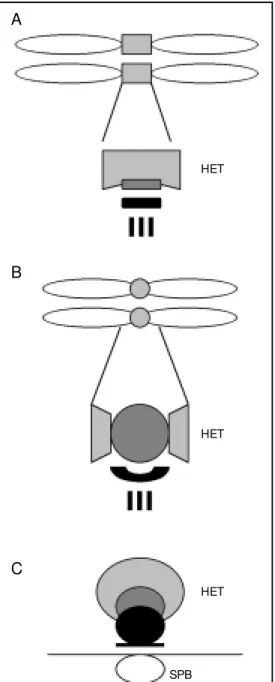
pro-Table 1. A survey of centromere DNA sequences and proteins in humans, Drosophila, fission yeast and budding yeast.
Centromere Human Drosophila Fission yeast Budding yeast
DNA sequences Tandem arrays of 171 bp Simple satellites and A 15-kb central core w ith Three conserved regions
(references - M onomer a-satellite repeats single complete unique sequences CDE-I TCACATGAT, CDE-II
see text) transposable elements (cnt and imr) flanked 80-90 bp >90% A+T, and
by 20-100 kb or CDE-III TGATTTCCGAA
(otr/K+L) repeats
M inimal DNA <500 kb of DXZ1 a-satellites 420 kb is sufficient for 7 kb of central core + 2 kb of 120 bp of DNA sufficient
sequence are sufficient for centromere centromere function the otr/K repeat sufficient for function (9). Deletion of
requirement for function on a Dp1187 for function CDE-I or CDE-II reduces
function minichromosome centromere function but
(references - point mutations in the
see text) central CCG in CDE-III
completely inactivate the centromere
Centromere proteins (references - see text)
Heterochromatin CENP-B Cph1 (CENP-B homologue 1)
INCENP
M 31 (mouse) HP1 Sw i6, Chp1
SUV39H1 (mouse) Su(var)3-9* Clr4*
PROD M EI-S322
Inner plate/ CENP-A CID (DmCENP-A) Cnp1 (SpCENP-A) Cse4 (ScCENP-A)
central core CENP-C
M is6
Cph2 (CENP-B homologue 2)
Outer kinetochore HEC, Nuf2R Ndc80, Nuf2, Spc24 Ndc80, Nuf2, Spc24,
Spc25
CENP-E CENP-meta
CENP-F
hBUB1 BUB1 Bub1
BUB3
Dam1 Spc19 Spc34 ZW10
POLO ROD
XM AP215* Alp14/Dis1 Stu2
M tc1
CLIP170* Bik1
BimC kinesins* Cin8
Aurora B Ipl1
Homologous proteins are underlined and placed on the same horizontal line. * Localization at centromere not determined.
teins are missing. The
S. cerevisiae
cen-tromeres are thus smaller in size, indicating
that the heterochromatin component may
have been lost during the evolution of these
trimmed down kinetochores. To
summa-rize, several centromere proteins have now
to be completely conserved and parts of the
Ndc80 protein complex (although hitherto
not described in
Drosophila
) seem to be a
conserved component of most eukaryotic
kinetochores.
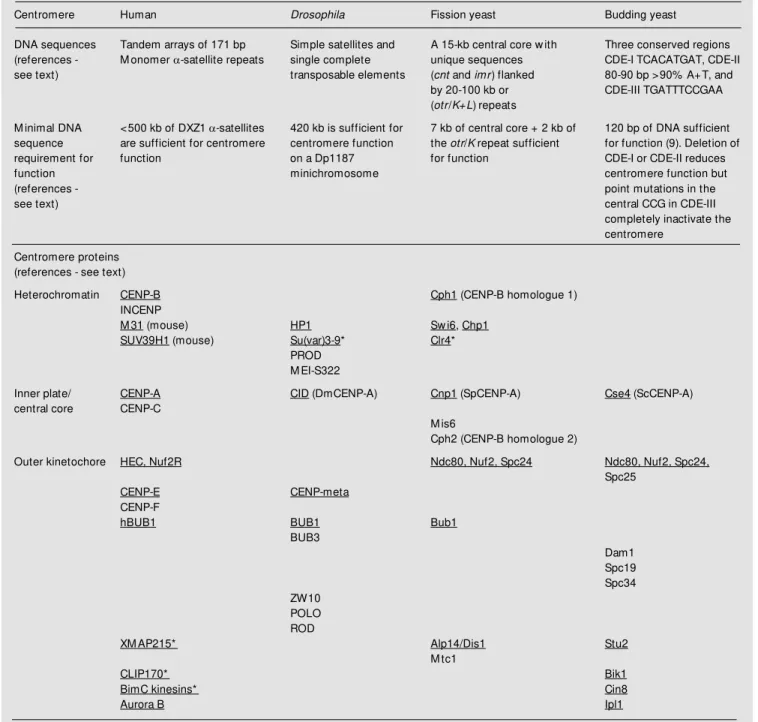
Ce ntro m e re do m ain structure
Recent studies have revealed that the
positions of some centromere proteins are
conserved within the multilayered
kineto-chore structures from
S. pombe
to humans
(Figure 1). In
S. pombe
the centromere
pro-teins Mis6 and Cnp1 (SpCENP-A) associate
exclusively with central core DNA, while
the Swi6 protein binds to the surrounding
repeats, suggesting that distinct protein
in-teraction domains exist within the
S. pombe
centromeres (35). Electron microscopy and
immunofluorescence light microscopy of the
precisely positioned centromeres in
inter-phase cells have revealed that the central
core and flanking regions indeed occupy
cytologically distinct positions within a
het-erochromatic domain. In addition, an
an-chor structure containing the Ndc80
puta-tive kinetochore protein was found between
this heterochromatic domain and the spindle
pole body (4). As seen by electron
micros-copy, the human metaphase centromere is
multilayered, containing several
substruc-tures: a fibrous corona, an outer and an inner
plate, as well as the space between them
(39). Beneath the inner plates is the
hetero-chromatic region.
Each of these
substruc-tures appears to comprise a distinct protein
composition. The fibrous corona contains
CENP-E, dynein and dynactin, the outer plate
contains CENP-F, the inner plate contains
CENP-C and CENP-A, and the underlying
heterochromatin contains CENP-B, INCENP,
HP1 and SUV39H1 (40-46).
Live analysis
of cells expressing CENP-A and CENP-B
green fluorescent protein fusion proteins
in-dicates that, although the human kinetochore
unfolds and refolds during interphase, the
human pre-kinetochore structure remains
or-dered in interphase. CENP-A localization is
limited to the edge of a larger CENP-B
het-erochromatin domain even before the
typi-cal double dot structure appears in G2 (47,48).
At metaphase, CENP-A is a component of
the inner plate in the human centromere
(44). The human homologue of Ndc80, HEC,
is localized to the outer part of HeLa cell
centromeres (31). There are also human
ho-mologues of additional components of the
HET
HET
SPB
A
B
C
Figure 1. A schematic represen-tation of the centromere domain organization at metaphase in humans (A), at metaphase inDrosophila (B), and the cen-tromere cluster near the SPB at interphase in Schizosaccharo-myces pombe (C). The hetero-chromatic (HET) domains (light gray), the inner kinetochore/in-ner plate/central core structures (dark gray) and the outer plate/ outer kinetochore/anchor struc-tures (black) are indicated. The vertical lines from the outer plate/kinetochore in A and B rep-resent kinet ochore m icrot u-bules. SPB = spindle pole body.
budding yeast kinetochore, i.e., XMAP215,
CLIP170 and BimC kinesins but their
local-ization in the human centromere has not
been determined (36).
Histo ne mo dificatio ns
Several different covalent modifications
of the histone proteins have been reported to
affect centromeric chromatin. These are
acetylation, methylation and
phosphoryla-tion. Acetylation of histones is carried out by
histone acetyl transferases at several
con-served lysine residues of the N-terminal tails
of histone H3 and H4. In general, histones in
heterochromatin are relatively
underacety-lated when compared to those present in
euchromatin (49,50). The opposite reaction,
the deacetylation of histone tails, is
per-formed by histone deacetylases (51). For
S.
pombe
,
Drosophila
and human cells it has
been shown that the centromeres are
under-acetylated (50,52). Treatment of
S. pombe
and human cells with the histone
delase inhibitor trichostatin A increases
acety-lation in centromeric heterochromatin and
disrupts centromere function (50,53). The
underacetylated state of the flanking repeats
is necessary for binding of Swi6 to
S. pombe
centromeres and for binding of HP1 to
hu-man pericentric heterochromatin (50,53).
Swi6 has been shown to associate with
cen-tromeric heterochromatin in fission yeast,
and this association has been shown to
re-quire Clr4 (25,54). Recently, Clr4 belonging
to a conserved chromo- and SET-domain
family of proteins was shown to modify the
N-terminal tail of histone H3 by methylation
(55,56). Indeed H3 methylation was shown
to be required for Swi6 binding to
centro-meric
otr/K
chromatin which is methylated
at lysine 9 of histone H3 (57,58). It has been
shown that deacetylation of histone H3 by
histone deacetylases is required to allow for
the subsequent methylation of this residue
by the Clr4 histone H3 methyltransferase in
a stepwise mechanism (57). The mouse
SUV39H1 is a homologue of Clr4 with the
same enzymatic function. It has been shown
to be required for chromosome segregation
and interacts with M31 (mouse HP1
homo-logue) in pericentric heterochromatin (46).
SUV39H1 specifically accumulates in the
centromere during prometaphase but
disso-ciates from centromeric positions in anaphase
(59). Su(var)3-9 in
Drosophila
was
origi-nally defined as a gene required for position
effect variegation, i.e., silencing of the
cen-tromeric marker gene but it remains to be
tested whether the protein itself is associated
with centromeres. Thus, underacetylation of
histones H3 and H4 and methylation of lysine
9 of histone H3 are required for
heterochro-matin formation and/or proper chromosome
segregation in several different organisms.
What is then the exact function of
centro-meric heterochromatin?
being required for metaphase chromosome
alignment, kinetochore disjunction, and
chro-mosome segregation (62,63). Notably the
budding yeast homologue of Aurora kinase,
Ipl1, is present in the kinetochore of this
organism (36). The exact targets of the Ipl1
kinase at the budding yeast kinetochores are
currently not known but Ipl1 is required for
segregation of sister chromatids at anaphase
(64).
Kine to cho re asse m bly
The domain organization of the
cen-tromere described in
Drosophila
(3) is
simi-lar to the one described in
S. pombe
and
humans. These findings now allow us to
address the function of the different
cen-tromere domains in the genetic model
organ-isms
Drosophila
and
S. pombe
. To this end,
the study of Blower and Karpen indicated
that CENP-A (CID) has a very central role in
several centromere functions. Injection of
antibodies against CID and/or RNAi
inhibi-tion of CID expression resulted in a
multi-tude of centromeric defects including
prophase arrest, indicative of an assembly
defect, and lagging chromosomes which is
indicative of cohesion defect. Inhibition of
CID led to delocalization of several
cen-tromere proteins (POLO, ROD, BUB1,
CENP-meta) indicating that the
incorpora-tion of all these factors is normally
depend-ent on CID. In budding yeast Cse4 is
neces-sary for proper chromosome segregation. In
a
cse4
conditional mutant, the core
cen-tromere chromatin structure is disrupted at
the restrictive temperature (38,65). Similarly,
in fission yeast a mutation in the
mis6
+(
mis
-segregation) gene causes a failure of the
Cnp1 (SpCENP-A) protein to bind the
cen-tral core chromatin leading to massive
aneu-ploidy (29,30). Although it remains to be
investigated whether other kinetochore
com-ponents are lost from centromeres in
mis6-1
cells, the strong defects indicate that
CENP-A may be required for kinetochore assembly
also in
S. pombe
. What targets CENP-A to
centromeres? Replication timing might be
important to form a functional centromere
and has been suggested as an important
de-terminant for CENP-A targeting (66). In
Dro-sophila
it has been shown that the
cen-tromeres replicate in the early S-phase while
surrounding heterochromatin replicates later
(67). This replication pattern might be
cru-cial since a human neocentromere 10q25.3
causes the surrounding DNA to replicate
later in the S-phase as compared to a
wild-type chromosome 10 (68). However, other
investigations show that replication timing
does not matter for the incorporation of
CENP-A and formation of a functional
cen-tromere (69). In fission yeast
otr/K
repeats in
the centromeres replicate early (70) and
CENP-A is expressed early in the S-phase
(30). However, there is no evidence for an
early replication requirement for CENP-A
incorporation into
S. pombe
centromeres.
Co nclusio ns and pe rspe ctive s
Centromere DNA sequences were
re-cently defined molecularly in humans and
can be compared with those of genetic
mo-del organisms, i.e., two evolutionarily
dis-tant yeasts (
S. pombe
and
S. cerevisiae
) and
heterochromatic part of the chromosome that
contains a special chromatin structure with
underacetylated histones. It is conceivable
that this structural similarity is conserved to
accommodate the common subfunctions of
a centromere: kinetochore assembly, sister
chromatid cohesion, binding of kinetochore
microtubules, orientation of sister
kineto-chores to opposite spindle poles, and their
movement towards the spindle poles.
Re fe re nce s
1. Pluta AF, M ackay AM , Ainsztein AM , Goldberg IG & Earnshaw WC (1995). The centromere: hub of chromosomal activi-ties. Science, 270: 1591-1594.
2. Pidoux AL & Allshire RC (2000). Cen-tromeres: getting a grip of chromosomes.
Current Opinion in Cell Biology, 12: 308-319.
3. Blow er M D & Karpen GH (2001). The role of Drosophila CID in kinetochore forma-tion, cell-cycle progression and hetero-chromatin interactions. Nature Cell Biol-ogy, 3: 730-739.
4. Kniola B, O’ Toole E, M cInt osh JR, M ellone B, Allshire R, M engarelli S, Hultenby K & Ekw all K (2001). The do-main structure of centromeres is con-served from fission yeast to humans. M o-lecular Biology of the Cell, 12: 2767-2775. 5. Schueler M G, Higgins AW, Rudd M K, Gustashaw K & Willard HF (2001). Ge-nomic and genetic definition of a func-tional human centromere. Science, 294: 109-115.
6. Cancilla M R, Taint on KM , Barry AE, Larionov V, Kouprina N, Resnick M A, Sart DD & Choo KH (1998). Direct cloning of human 10q25 neocentromere DNA using transformation-associated recombination (TAR) in yeast. Genomics, 47: 399-404. 7. Depinet TW, Zackow ski JL, Earnshaw
WC, Kaffe S, Sekhon GS, Stallard R, Sullivan BA, Vance GH, Van Dyke DL, Willard HF, Zinn AB & Schw artz S (1997). Characterization of neo-centromeres in marker chromosomes lacking detectable alpha-satellite DNA. Human M olecular Genetics, 6: 1195-1204.
8. Sun X, Wahlstrom J & Karpen G (1997). M olecular structure of a functional Droso-phila centromere. Cell, 91: 1007-1019. 9. Williams BC, M urphy TD, Goldberg M L &
Karpen GH (1998). Neocentromere activ-ity of structurally acentric mini-chromo-somes in Drosophila. Nature Genetics, 18: 30-37.
10. Clarke L, Amstutz H, Fishel B & Carbon J (1986). Analysis of centromeric DNA in the fission yeast Schizosaccharomyces pombe. Proceedings of the National
Acad-emy of Sciences, USA, 83: 8253-8257. 11. Clarke L & Baum M P (1990). Functional
analysis of a centromere from fission yeast: a role for centromespecific re-peated DNA sequences. M olecular and Cellular Biology, 10: 1863-1872. 12. Chikashige Y, Kinoshita N, Nakaseko Y,
M atsumoto T, M urakami S, Niw a O & Yanagida M (1989). Composite motifs and repeat symmetry in S. pombe centro-meres: direct analysis by integration of NotI restriction sites. Cell, 57: 739-751. 13. Cottarel G, Shero JH, Hieter P &
Hege-mann JH (1989). A 125-base-pair CEN6 DNA fragment is sufficient for complete meiotic and mitotic centromere functions in Saccharomyces cerevisiae. M olecular and Cellular Biology, 9: 3342-3349. 14. Earnshaw WC & M igeon BR (1985). Three
related centromere proteins are absent from the inactive centromere of a stable isodicentric chromosome. Chromosoma, 92: 290-296.
15. Earnshaw WC & Rothfield N (1985). Iden-tification of a family of human centromere proteins using autoimmune sera from pa-tients w ith scleroderma. Chromosoma, 91: 313-321.
16. Kerrebrock AW, M oore DP, Wu JS & Orr-Weaver TL (1995). M ei-S332, a Droso-phila protein required for sister-chromatid cohesion, can localize to meiotic cen-tromere regions. Cell, 83: 247-256. 17. Logarinho E & Sunkel CE (1998). The
Dro-sophila POLO kinase localises to multiple compartments of the mitotic apparatus and is required for the phosphorylation of M PM 2 reactive epitopes. Journal of Cell Science, 111: 2897-2909.
18. Torok T, Harvie PD, Buratovich M & Bryant PJ (1997). The product of proliferation dis-rupter is concentrated at centromeres and required for mitotic chromosome conden-sation and cell proliferation in Drosophila.
Genes and Development, 11: 213-225. 19. Scaerou F, Aguilera I, Saunders R, Kane
N, Blottiere L & Karess R (1999). The rough deal protein is a new kinetochore component required for accurate chromo-some segregation in Drosophila. Journal
of Cell Science, 112: 3757-3768. 20. Williams BC, Gatti M & Goldberg M L
(1996). Bipolar spindle attachments affect redistributions of ZW10, a Drosophila cen-tromere/kinetochore component required for accurate chromosome segregation.
Journal of Cell Biology, 134: 1127-1140. 21. Basu J, Logarinho E, Herrmann S,
Bous-baa H, Li Z, Chan GK, Yen TJ, Sunkel CE & Goldberg M L (1998). Localization of the
Drosophila checkpoint control protein Bub3 to the kinetochore requires Bub1 but not Zw 10 or Rod. Chromosoma, 107: 376-385.
22. Yucel JK, M arszalek JD, M cIntosh JR, Goldstein LS, Cleveland DW & Philp AV (2000). CENP-meta, an essential kineto-chore kinesin required for the mainte-nance of metaphase chromosome align-ment in Drosophila. Journal of Cell Biol-ogy, 150: 1-11.
23. Saunders WS, Chue C, Goebl M , Craig C, Clark RF, Pow ers JA, Eissenberg JC, Elgin SC, Rothfield NF & Earnshaw WC (1993). M olecular cloning of a human homologue of Drosophila heterochromatin protein HP1 using anti-centromere autoantibod-ies w ith anti-chromo specificity. Journal of Cell Science, 104: 573-582.
24. Henikoff S, Ahmad K, Platero JS & van Steensel B (2000). Heterochromatic depo-sition of centromeric histone H3-like pro-teins. Proceedings of the National Acade-my of Sciences, USA, 97: 716-721. 25. Ekw all K, Javerzat JP, Lorentz A, Schmidt
H, Cranston G & Allshire R (1995). The chromodomain protein Sw i6: a key com-ponent at fission yeast centromeres. Sci-ence, 269: 1429-1431.
26. Doe CL, Wang G, Chow C, Fricker M D, Singh PB & M ellor EJ (1998). The fission yeast chrom o dom ain encoding gene chp1(+) is required for chromosome seg-regation and show s a genetic interaction w ith alpha-tubulin. Nucleic Acids Re-search, 26: 4222-4229.
segregation. Genes and Development, 13: 1664-1677.
28. Bernard P, Hardw ick K & Javerzat JP (1998). Fission yeast bub1 is a mitotic cent rom ere prot ein essent ial f or t he spindle checkpoint and the preservation of correct ploidy through mitosis. Journal of Cell Biology, 143: 1775-1787. 29. Saitoh S, Takahashi K & Yanagida M
(1997). M is6, a fission yeast inner cen-tromere protein, acts during G1/S and forms specialized chromatin required for equal segregation. Cell, 90: 131-143. 30. Takahashi K, Chen ES & Yanagida M
(2000). Requirement of M is6 centromere connector for localizing a CENP-A-like pro-tein in fission yeast. Science, 288: 2215-2219.
31. Wigge PA & Kilmartin JV (2001). The Ndc80 complex from Saccharomyces ce-revisiae contains conserved centromere components and has a function in chro-mosome segregation. Journal of Cell Biol-ogy, 152: 349-360.
32. Nabetani A, Koujin T, Tsutsumi C, Haragu-chi T & Hiraoka Y (2001). A conserved protein, Nuf2, is implicated in connecting the centromere to the spindle during chro-mosome segregation: a link betw een the kinetochore function and the spindle checkpoint. Chromosoma, 110: 322-334. 33. Garcia M A, Vardy L, Koonrugsa N & Toda
T (2001). Fission yeast ch-TOG/XM AP215 hom ologue Alp14 connect s m it ot ic spindles w ith the kinetochore and is a com ponent of t he M ad2-dependent spindle checkpoint. EM BO Journal, 20: 3389-3401.
34. Nakaseko Y, Goshima G, M orishita J & Yanagida M (2001). M phase-specific ki-netochore proteins in fission yeast: mi-crotubule-associating Dis1 and M tc1 dis-play rapid separation and segregation dur-ing anaphase. Current Biology, 11: 537-549.
35. Partridge JF, Borgstrom B & Allshire RC (2000). Distinct protein interaction do-mains and protein spreading in a complex centromere. Genes and Development, 14: 783-791.
36. He X, Rines DR, Espelin CW & Sorger PK (2001). M olecular analysis of kinetochore-microtubule attachment in budding yeast.
Cell, 106: 195-206.
37. Lechner J & Carbon J (1991). A 240 kd multisubunit protein complex, CBF3, is a major component of the budding yeast centromere. Cell, 64: 717-725.
38. M eluh PB, Yang P, Glow czew ski L, Koshland D & Smith M M (1998). Cse4p is a component of the core centromere of
Saccharomyces cerevisiae. Cell, 94: 607-613.
39. Pluta AF, Cooke CA & Earnshaw WC (1990). Structure of the hum an cen-tromere at metaphase. Trends in Bio-chemical Sciences, 15: 181-185. 40. Cooke CA, Schaar B, Yen TJ & Earnshaw
WC (1997). Localization of CENP E in the fibrous corona and outer plate of mamma-lian kinetochores from prom etaphase through anaphase. Chromosoma, 106: 446-455.
41. Yao X, Anderson KL & Cleveland DW (1997). The microtubule-dependent mo-t or cenmo-t rom ere-associamo-t ed promo-t ein E (CENP-E) is an integral component of ki-netochore corona fibers that link cen-tromeres to spindle microtubules. Journal of Cell Biology, 139: 435-447.
42. Saitoh H, Tomkiel J, Cooke CA, Ratrie 3rd H, M aurer M , Rothfield NF & Earnshaw WC (1992). CENP-C, an autoantigen in scleroderma, is a component of the hu-man inner kinetochore plate. Cell, 70: 115-125.
43. Warburton PE, Cooke CA, Bourassa S, Vafa O, Sullivan BA, Stetten G, Gimelli G, Warburton D, Tyler-Smith C, Sullivan KF, Poirier G & Earnshaw WC (1997). Immu-nolocalization of CENP-A suggests a dis-tinct nucleosome structure at the inner kinetochore plate of active centromeres.
Current Biology, 7: 901-904.
44. Vafa O & Sullivan KF (1997). Chromatin containing CENP A and alpha satellite DNA is a major component of the inner kinetochore plate. Current Biology, 7: 897-900.
45. Cooke CA, Bernat RL & Earnshaw WC (1990). CENP-B: a major human cen-tromere protein located beneath the kine-tochore. Journal of Cell Biology, 110: 1475-1488.
46. Aagaard L, Laible G, Selenko P, Schmid M , Dorn R, Schotta G, Kuhfittig S, Wolf A, Lebersorger A, Singh PB, Reuter G & Jenuw ein T (1999). Functional mamma-lian homologues of the Drosophila PEV-modifier Su(var)3-9 encode centromere-associated proteins w hich complex w ith the heterochromatin component M 31.
EM BO Journal, 18: 1923-1938.
47. Pudenko AS, Kudryavtsev IS, Zatsepina OV & Chentsov Yu S (1997). Spatial asso-ciation of prekinetochores and chromo-centres in the interphase nuclei of mouse cultured fibroblasts. M embrane and Cell Biology, 11: 449-461.
48. Sugim oto K, Fukuda R & Him eno M (2000). Centromere/kinetochore localiza-tion of hum an centrom ere protein A
(CENP-A) exogenously expressed as a fu-sion to green fluorescent protein. Cell Structure and Function, 25: 253-261. 49. Jeppesen P & Turner BM (1993). The
in-active X chromosome in female mammals is distinguished by a lack of histone H4 acetylation, a cytogenetic marker for gene expression. Cell, 74: 281-289.
50. Ekw all K, Olsson T, Turner BM , Cranston G & Allshire RC (1997). Transient inhibi-tion of histone deacetylainhibi-tion alters the structural and functional imprint at fission yeast centromeres. Cell, 91: 1021-1032. 51. Taunton J, Hassig CA & Schreiber SL
(1996). A mammalian histone deacetylase related to the yeast transcriptional regula-tor Rpd3p. Science, 272: 408-411. 52. Turner BM , Birley AJ & Lavender J (1992).
Histone H4 isoforms acetylated at specif-ic lysine residues define individual chro-mosomes and chromatin domains in Dro-sophila polytene nuclei. Cell, 69: 375-384. 53. Taddei A, M aison C, Roche D & Almouzni G (2001). Reversible disrupt ion of pericent ric het erochrom at in and cen-tromere function by inhibiting deacetyl-ases. Nature Cell Biology, 3: 114-120. 54. Ekw all K, Nimmo ER, Javerzat JP,
Borg-strom B, Egel R, Cranston G & Allshire R (1996). M utations in the fission yeast si-lencing factors clr4+ and rik1+ disrupt the localisation of the chromo domain protein Sw i6p and impair centromere function.
Journal of Cell Science, 109: 2637-2648. 55. Ivanova AV, Bonaduce M J, Ivanov SV &
Klar AJ (1998). The chromo and SET do-mains of the Clr4 protein are essential for silencing in fission yeast. Nature Genet-ics, 19: 192-195.
56. Rea S, Eisenhaber F, O’Carroll D, Strahl BD, Sun ZW , Schm id M , Opravil S, M echt ler K, Pont ing CP, Allis CD & Jenuw ein T (2000). Regulation of chroma-tin structure by site-specific histone H3 methyltransferases. Nature, 406: 593-599.
57. Nakayama J, Rice JC, Strahl BD, Allis CD & Grew al SI (2001). Role of histone H3 lysine 9 methylation in epigenetic control of heterochromatin assembly. Science, 292: 110-113.
58. Bannister AJ, Zegerman P, Partridge JF, M iska EA, Thomas JO, Allshire RC & Kouzarides T (2001). Selective recognition of methylated lysine 9 on histone H3 by the HP1 chromo domain. Nature, 410: 120-124.
tran-sient accumulation at mammalian cen-tromeres. Journal of Cell Science, 113: 817-829.
60. Bernard P, M aure JF, Partridge JF, Genier S, Javerzat JP & Allshire RC (2001). Re-quirement of heterochromatin for cohe-sion at centromeres. Science, 294: 2539-2542.
61. Zeitlin SG, Barber CM , Allis CD & Sullivan K (2001). Differential regulation of CENP-A and histone H3 phosphorylation in G2/ M . Journal of Cell Science, 114: 653-661. 62. Adam s RR, Eckley DM , Vagnarelli P, Wheatley SP, Gerloff DL, M ackay AM , Svingen PA, Kaufmann SH & Earnshaw WC (2001). Human INCENP colocalizes w ith the Aurora-B/AIRK2 kinase on chro-mosomes and is overexpressed in tumour cells. Chromosoma, 110: 65-74. 63. Adams RR, M aiato H, Earnshaw WC &
Carmena M (2001). Essential roles of
Dro-sophila inner cent rom ere prot ein (INCENP) and aurora B in histone H3 phos-phorylat ion, m et aphase chrom osom e alignment, kinetochore disjunction, and chromosome segregation. Journal of Cell Biology, 153: 865-880.
64. Biggins S, Bhalla N, Chang A, Smith DL & M urray AW (2001). Genes involved in sis-ter chromatid separation and segregation in the budding yeast Saccharomyces ce-revisiae. Genetics, 159: 453-470. 65. St oler S, Keit h KC, Curnick KE &
Fitzgerald-Hayes M (1995). A mutation in CSE4, an essential gene encoding a novel chromatin-associated protein in yeast, causes chromosome nondisjunction and cell cycle arrest at mitosis. Genes and Development, 9: 573-586.
66. Shelby RD, Vafa O & Sullivan KF (1997). Assembly of CENP-A into centromeric chromatin requires a cooperative array of
nucleosomal DNA contact sites. Journal of Cell Biology, 136: 501-513.
67. Ahmad K & Henikoff S (2001). Centro-meres are specialized replication domains in heterochromatin. Journal of Cell Biol-ogy, 153: 101-110.
68. Lo AW, Craig JM , Saffery R, Kalitsis P, Irvine DV, Earle E, M agliano DJ & Choo KH (2001). A 330 kb CENP-A binding do-main and altered replication timing at a human neocentromere. EM BO Journal, 20: 2087-2096.
69. Sullivan B & Karpen G (2001). Centromere identity in Drosophila is not determined in vivo by replication timing. Journal of Cell Biology, 154: 683-690.
70. Kim SM & Huberman JA (2001). Regula-tion of replicaRegula-tion timing in fission yeast.