TOWARDS A STANDARDISED METHOD TO ACQUIRE
AND STORE LIVER SAMPLES AND GUIDELINES TO
IMPROVE QUALITY CONTROL AND EXCHANGE OF
RELATIVE EXPRESSION DATA
Frank M. Riemers, Jan Rothuizen, Louis C. Penning
Department of Clinical Sciences of Companion Animals, Faculty of Veterinary Medicine, Utrecht, the Netherlands.
Disclosure:
No potential conlict of interest
Citation:
EMJ Hepatol. 2013;1:78-84.
ABSTRACT
The current ‘state-of-the-art’ molecular techniques are extremely sensitive and consequently prone to
false results. Even more so than in the past, today’s hepatology research depends on high quality samples,
especially for the molecular analyses. In all steps, starting with specimen sampling, ixation, storage,
molecular processing and inally data calculation, variations in procedures between research laboratories
may have a profound efect on the inal conclusions. At the end of the day, this is an enormous drawback
once data from diferent research institutes need to be reproduced, compared and/or combined. To improve
standardisation, the so-called MIQE guidelines (Minimum Information for Publication of Quantitative
Real-Time PCR Experiments) were presented for quantitative PCR (qPCR) studies.
1,2Furthermore, around the
same time, recommendations were presented regarding human biospecimen collection, storage and
processing, the so-called BRISQ-guidelines (Biospecimen Reporting for Improved Study Quality).
3Finally,
the editors of
The Journal of Pathology
as well as
Histopathology
required in the December 2012 issue of
The Journal of Pathology
that researchers needed to follow the BRISQ guidelines in their papers in order to
improve the sample quality in biomedical research.
4These initiatives hold great promise to improve the comparison and independent reproduction of data
acquired in diferent research centres. Pancreas, gall bladder and liver research will especially beneit from
the standardisation protocols since these organ systems are highly vulnerable to post-biopsy autolytic
degradation. This comment illustrates that standardisation in molecular liver research is not yet at the point
where experiments can be easily replicated, and data can be compared and combined.
Keywords: Quantitative PCR, MIQE-precise, normalisation, reference genes.
INTRODUCTION
Molecular expression studies on biospecimen can gain
insight into the etiology of a disease, and may lead to
information on therapeutic efects and potentially facilitate
biomarker studies. These samples need to be acquired,
stored and processed in such a way that
laboratory-to-laboratory comparison is possible and independent
reproduction can be achieved. Standardisation of
protocols in all three steps mentioned above is a way to
come to meaningful comparisons. Cost-efective scientiic
progress can be achieved by diferent means, for instance,
by combining data and data-comparisons of diferent
research groups. High quality data is crucial in this respect.
Space limitations often hamper detailed description of
materials and methods, and consequently comparisons
between laboratories, not to mention meta-analyses, are
often lawed. For biopsies the BRISQ guidelines exist
and there are guidelines to standardise quantitative PCR
(qPCR) expression studies (MIQE-precise guidelines).
1-3to acceptance and implementation in the scientiic
community. This chapter is an initiative to raise awareness
of the cost-efective progress molecular liver research can
make once data are calculated and presented in such a
way that experiments can be easily repeated and data can
be combined and compared.
Scientists prefer their biopsies, taken at surgery not
under time pressure or other forms of stress, to be ixed
speciically for their individual research questions which
can be either at tissue, cellular or molecular level. However,
these separate research questions require diferent
ixation and storage methods. Such a complexity of tissue
handling is clearly prone to the introduction of mistakes,
leading to biospecimen of potentially lesser quality for
a speciic analysis. Although RNA is far less stable then
DNA some studies indicate that the RNA integrity is not
largely inluenced even up to 48 hours on ice.
5,6The last
study included tonsil and liver samples. In contrast, two
studies exempliied the efects of variations in liver tissue
sampling on subsequent mRNA expression studies.
7,8One
study described the inluence of the biopsy needle size
in rat liver biopsies on the RNA quality in a subsequent
micro-array expression study.
7The second study assessed
diferent sampling techniques, ixation methods, and
storage procedures for canine liver tissue to optimise the
use of a single liver biopsy for histological and molecular
(qPCR) measurements.
8Not only can total RNA be subject to degradation (usually
measured as a RNA Integrity Number (RIN) based on 18S
and 28S) during the sampling, storage and processing,
but mRNA (only 2-5% of total RNA, but most often the
compound of interest) can also be degraded. One way to
correct for mRNA degradation, and other steps in mRNA
expression studies is the inclusion of so-called reference
genes (previously erroneously called housekeeping genes)
to normalise for mRNA input and PCR eiciency. The
assumption here is that the expression of reference genes
is always constant, irrespective of variations in samples,
experimental conditions etc. In fact this assumption has
been debated for about one decade now.
9Obviously data
comparison in molecular liver research faces an enormous
hurdle if reference gene stability is either not evaluated
nor are other parts of the sample and data processing
are not described in detail. Whether this molecular deicit
indeed exists in molecular liver research was not reported
previously, and is investigated in this chapter. As it turned
out, based on a PubMed search (http://www.ncbi.nlm.
nih.gov/), the crucial step in expression studies, viz,
evaluation in reference gene stability, was often omitted
in molecular liver studies. This book chapter therefore is a
clear advocacy to implement MIQE-precise guidelines as
soon as possible.
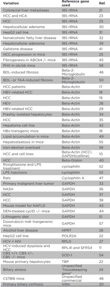
Variation Reference gene used Ref.
Colorectal liver metastases 18S rRNA 21
HCC and HCA 18S rRNA 23
HCC 18S rRNA 25
Hepatocellular adenoma 18S rRNA 30
HepG2 cell line 18S rRNA 31
Nonalcoholic fatty liver disease 18S rRNA 32 Hepatocellular adenoma 18S rRNA 39 Gallstone disease 18S rRNA 41 HCC progression in mice 18S rRNA 25 Fibrogenesis in ABCb4 /- mice 18S rRNA 45 PHX in ob/ob mice 18S rRNA 58
BDL-induced ibrosis Beta-2-Microglobulin 46
BDL- or TAA-induced ibrosis
Beta-2-Microglobulin 50
HCC patients Beta-Actin 17
HBV-related HCC Beta-Actin 18
HCC Beta-Actin 19
HEV Beta-Actin 28
HBV-related HCC Beta-Actin 29 Freshly isolated hepatocytes Beta-Actin 34
HCC Beta-Actin 35
Hepatoma cell line Beta-Actin 42 HBx-transgenic mice Beta-Actin 18 Lipid accumulation in mice Beta-Actin 49 Hepatosteatosis in mice Beta-Actin 55 Iron-dextran overload Beta-Actin 56
HCC and cell lines Beta-Actin (HCC) GAPDH(cellline) 10
HCC Beta-Globin 40
Rosiglitazone and LPS
treatment cyclophilin 52
LPS injections cyclophilin 53
Rats Cyclophilin A 47
Primary malignant liver tumor GAPDH 33
NASH GAPDH 36
HCC GAPDH 37
HCC GAPDH 38
Mouse model for NAFLD GAPDH 43 DEN-treated cyclD -/- mice GAPDH 44
Lithogenic diet GAPDH 51
Doxorubicin mdr trangenenic
mice GAPDH 57
Alcohol liver disease HPRT 26
HepG2 cell line POLR2A 22
HCV + HIV RPL0 27
HCV-induced dysplasia and
HCC RPL41 and SFRS4 11
CBS +/+, CBS +/-,
CBS -/- mice SOD-1 54
Mouse primary hepatocytes TBP 22
Biliary atresia
Unspeciied “housekeeping gene”
24
C57Bl6 mice Unspeciied
commercial 48 Primary biliary cirrhosis Villin 20
MATERIAL AND METHODS
A PubMed search was performed via http://www.ncbi.
nlm.nih.gov/ on Tuesday March 19
th11am CET on the
terms ‘human AND quantitative PCR AND expression
AND hepatology’. The search was limited to
Hepatology
and the
Journal of Hepatology
only, the two highest
top-ranked journals in the ISI-ield of ‘Gasteroenterology
and Hepatology’ speciically for hepatology. Moreover
both are oicial journals of the American Association for
the Study of Liver Diseases (AASLD) and the European
Association for the Study of the Liver (EASL) respectively.
A similar search was performed on ‘(murine OR mouse)
AND quantitative PCR AND expression AND hepatology’.
Finally, a PubMed search on papers evaluating reference
expression stability in liver samples from human and
other mammalian species was performed to reveal which
reference genes were evaluated under what kind of
research samples, and which freeware was used to indicate
expression stability and consequently which were most
reliable reference gene under that speciic condition.
RESULTS
Approximately the irst 50 hits on the combined terms
‘human AND quantitative PCR AND expression AND
hepatology’ and ‘(mouse OR murine) AND quantitative
PCR AND expression AND hepatology’ were screened to
establish which presumed stable reference gene was used
(Table 1)
. The preference for the classical reference genes,
viz
, beta-Actin, GAPDH or 18S rRNA, was obvious. Thirteen
times was normalised against beta-actin, eleven times
with 18S rRNA, and nine times with GAPDH. In one paper
for the clinical samples normalisation was performed
with beta-actin, whereas in cell lines GAPDH was used.
10None of these papers provided information on whether
or not the indicated reference gene was expressed at a
stable level. Most surprising was the observation that in all
papers analysed, except for one, only one reference gene
was used for normalisation. The exception included two
independent reference genes: SFRS4 and RPL41.
11Even
worse, in view of data comparison, was the number of other
reference genes used, including beta-2-microglobulin,
beta-globin, cyclophilin A, villin, POLR2A, RPLP0, SOD-1,
cyclophilin, TBP, or HPRT. There were no calculations on
the expression stability of the reference genes included in
any of the papers summarised in
Table 1
.
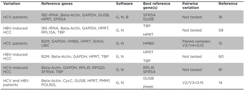
Six papers described the evaluation of reference gene
expression stability in human samples as depicted in
Table
2
. GAPDH, beta-actin and HPRT, were included in ive out
of six studies, TBP was used three times, SFRS4, GUSB,
18S rRNA and B2M were include twice. RPL13A, HMBS,
SDHA, RPL41, CYCC, RPS0, UBC, PMM1 and POLR2L were
evaluated once. GeNorm analysis
12and Norminder
13were
used to evaluate expression levels and depending on the
paper, either GUSB (twice), HPRT (twice) or TBP (twice)
performed the best, exhibiting the highest stability of
expression. SFRS4, HMBS, RPL41 and PMM1 turned out to
be the best only once. The three most frequently used
reference genes (beta-Actin, 18S rRNA or GAPDH) never
ranked as most stably expressed reference genes
(Table
2)
. The GeNorm algorithm allows us to calculate the set
of reference genes minimally required to normalise the
expression of genes of interest. This analysis (‘pairwise
variation’) has been included in as little as two of the six
papers described above. Romanowski et al.
14concluded
that two reference genes,
viz
GUSB and PMM1, were
suicient to obtain a pairwise variation below 0.15,
the recommended threshold to calculate the number
Variation Reference genes Software Best reference gene(s)
Pairwise variation
Reference
HCV patients 18S rRNA, Beta-Actin, GAPDH, GUSB,
HPRT, SFRS4 G, N, B
SFRS4
GUSB Not tested 16
HBV-induced HCC
18S rRNA, Beta-Actin, GAPDH, HPRT,
RPL13A, TBP G, N
TBP
HPRT
Not tested 59
HCC patients B2M, GAPDH, HMBS, HPRT, SDHA,
UBC G, N HMBS
Paired samples: V3/V4<0.15 15
HBV-induced
HCC B2M, Beta-Actin, GAPDH, HPRT, TBP G, N
HPRT
TBP
Not tested 60
HCV-induced HCC
Beta-Actin, GAPDH, RPL41, RPS20,
SFRS4, TBP G, N
RPL41
SFRS4 Not tested 61
HCV and HBV patients
Beta-Actin, CycC, GUSB, HPRT, PMM1,
POLR2L G, N
GUSB
PMM1
V2/V3<0.15 14
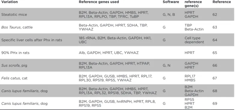
Variation Reference genes used Software Best reference gene(s)
Reference
Steatotic mice B2M, Beta-Actin, GAPDH, HMBS, HPRT, RPL13A, RPLPO, TBP, TFRC, TuBP G, N, B HPRTGAPDH 62
Bos Taurus, cattle Beta-Actin, GAPDH, HPRT, SDHA, TBP,
YWHAZ G
TBP
Beta-Actin 63
Speciic liver cells after Phx in rats 18S rRNA, B2M, Beta-Actin, GAPDH, HK1, UBC G Cell type dependent 64
90% PHx in rats Alb, GAPDH, HPRT, UBC, YWHAZ HPRT 65
Sus scrofa, pig B2M, Beta-Actin, GAPDH, HPRT, HTPAP, RPL13A G, N GAPDHHPRT 66
Felis catus, cat B2M, GAPDH, GUSB, HMBS, HPRT, RPL17, RPL30, RPS19, RPS5, YWHAZ G
RPL17
HMBS 67
Canis lupus familiaris, dog B2M, Beta-Actin, GAPDH, HMBS, HPRT,
RPL13A, RPL32, RPS18, SDHA, TBP, YWHAZ G
B2M Beta-Actin GAPDH
68
Canis lupus familiaris, dog B2M, GAPDH, GUSB, hnRNPH, HPRT, RPL8,
RPS19, RPS5 G
RPS5 HPRT B2M
69
Table 3. Papers reporting on the evaluation of expression stability of potential reference genes in
mammalian non-human liver samples.
Abbreviations in the software column: G=GeNorm, N=NormFinder.
of reference genes minimally required.
12Combining
tumourous and non-tumourous tissues revealed that
at least four reference genes were needed.
15The paper
by Congiu et al.
16clearly showed that a diferent set of
reference genes were most stably expressed if the groups
were arranged according to the levels of inlammation,
or the levels of steatosis or ibrosis. Unfortunately, it was
not indicated by pair-wise variation which number of
reference genes was optimal for each speciic condition.
The situation is similarly disturbing once the expression
stability is evaluated in liver samples from other
mammalian species like mice, rats, pigs, cats, dogs and
cattle
(Table 3)
. Again, a large list of potentially
stably-expressed reference genes evaluated for their respective
expression stability, including the favourable, but not
necessarily the most stably expressed, human reference
genes beta-actin, GAPDH and HPRT.
DISCUSSION
For relative expression levels of gene products,
normalisation is needed. The expression of reference
genes, of which the expression is to be stable amongst
diferent conditions, is then used to standardise. The
stability of their expression is tacitly presumed to be high.
Analysis of the expression stability, by the inclusion of
several independent reference genes, showed that this
assumption does not always hold true. The few calculations
on the minimal number of reference genes needed to
properly normalise relative mRNA expression levels
showed that, depending on the experimental comparison,
at least two and sometimes more reference genes are
needed. The plethora of various reference genes and the
variable outcome in the papers evaluating reference gene
expression stability, made one point clear: there are no
standardised descriptions incorporated in the papers, nor
are relevant details for data comparison, experimental
repetition or data combination provided in most
liver-related expression studies. Is this a purely academic
ine-tuning issue? This is a rhetorical question. What are the
cost-beneits for the inclusions of more reference genes?
Imagine a simple
in vivo
experiment, two groups of six
1. Bustin S A, Benes V, Garson J A, et al. The MIQE guidelines: minimum information for publication of quantitative real-time PCR experiments. Clin Chem. 2009;55:611-22. 2. Bustin S A, Beaulieu J F, Huggett J, et al. MIQE précis: practical implementation of minimum standard guidelines for luorescence-based quantitative real-time PCR experiments. BMC Mol. Biol. 2010;11:74.
3. Moore H M, Kelly A B, Jewell S D, et al. Biospecimen reporting for improved quality (BRISQ). Cancer Cytopathol. 2011;119:92-101.
4. Simeon-Dubach D, Burt AD, Hall PA. Quality really matters: the need to improve specimen quality in biomedical research. J Pathol. 2012;228:431-33. 5. Micke P, Ohshima M, Tahmasebpoor S, et al. Biobanking of fresh frozen tissue: RNA is stable in nonixed surgical specimens. Lab Invest. 2006;86:202-11. 6. Van Maldegem F, de Wit M, Morsink A, et al. Efects of processing delay, formalin ixation, and immunohistochemistry on RNA recovery from formalin-ixed parain-embedded tissue sections. Diagn Mol Pathol. 2008;17:51-8.
7. Takemura F, Inaba N, Miyoshi E, et
al. Optimization of liver biopsy RNA sampling and use of reference RNA for cDNA microarray analysis. Anal Biochem. 2005,337;224-34.
8. Hofmann G, IJzer J, Brinkhof B, et al. Comparison of diferent methods to obtain and store liver biopsies for molecular and histological research. Comp Hepatol. 2009;8:3.
9. Tricarico C, Pinzani P, Bianchi S, et al. Quantitative real-time reverse transcription polymerase chain reaction: normalization to rRNA or single housekeeping genes is inappropriate for human tissue biopsies. Anal Biochem. 2002;309:293-300.
10. Wang S, Jiang W, Chen X, et al. Alpha-fetoprotein acts as a novel signal molecule and mediates transcription of Fn14 in human hepatocellular carcinoma. J Hepatol. 2012;57:322-29.
11. Wurmbach E, Chen Y B, Khitrov G, et al. Genome-wide molecular proiles of HCV-induced dysplasia and hepatocellular carcinoma. Hepatology. 2007;45:938-47. 12. Vandesompele J De Carter K, Pattyn F, et al. Accurate normalization of real-time quantitative RT-PCR data by geometric averaging of multiple
internal control genes. Genome Biol. 2002;3:RESEARCH0034.
13. Andersen C L, Jensen J L, Orntoft T F. Normalization of real-time quantitative reverse transcription-PCD data: a model-based variance estimation approach to identify genes suited for normalization, applied to bladder and colon cancer data sets. Cancer Res. 2004;64:5245-50. 14. Romanowski T, Sikorska K, Bialawski K P. GUS and PMM1 as suitable reference genes for gene expression analysis in the liver tissue of patients with chronic hepatitis. Med Sci Monit. 2008;14:147-52. 15. Cicinnati V R, Shen Q, Solitropoulos G C, et al. Validation of putative reference genes for gene expression studies in human hepatocellular carcinoma using real-time quantitative RT-PCR. BMC Cancer. 2008;8:350.
16. Congiu M, Slavin J L, Desmond P V. Expression of common housekeeping genes is afected by disease in human hepatitis C virus-infected liver. Liv Intern. 2011;31:386-90.
17. Agostini J, Benoist S, Serman M, et al. Identiication of molecular pathways involved in oxaliplatin-associated sinusoidal dilatation. J Hepatol.
REFERENCES
data will be much more reliable, and since reference
gene expression stability was evaluated and recorded a
comparison of these expression data with other reports
becomes feasible. The investment of just $100 will save a
multitude of this amount once one can avoid a repetition
of the experiment due to a lack of proper information on
the stability of the included reference genes.
BRISQ-guided standardisation for histological research
and biobanking is obligatory in leading pathological
journals at present. Liver research can make great progress
if an improved standardisation can be accomplished for
molecular investigations. The proposed MQIE guidelines
and MQIE-precise guidelines, including proper reference
gene expression stability evaluation, ofer an easy way
to make the presented data easy to repeat, allow data
comparison, and facilitate manuscript reviewing.
1,2Alb, albumin
BDL, bile duct ligation B2M, beta-2-microglobulin CycC, cyclophillin C
GAPDH, glyceraldehyde-3 phosphate dehydrogenase
GUSB, beta-Glucoronidase HBC, hepatitis B virus
HCC, hepatocellularcarcinoma HCV, hepatitis C virus
HEV, hepatitis E virus
HIV, human immunedeicinecy virus HMBS, hydroxymethyl-bilane synthase
HPRT, hypoxanthine phosphoribosyl-transferase
HTPAP, PPAP2 domain-containing protein 1B
LPS, lipopolysaccaride
NAFLD,nonalcoholic fatty liver disease
NASH, nonalcoholic steatohepatitis PHx, partial hepatectomy
PMM1, Phosphomannomutase 1 POLR2, polymerase (RNA) II polypeptide L
RPL0, Ribosomal Protein Large0 RPL17, Ribosomal Protein Large17 RPL31A, Ribosomal Protein Large13A
RPL41, Ribosomal Protein Large41 RPS5, Ribosomal Protein Small SDHA, succinate dehydrogenase complex, subunit A
SFRS4, splicing factor serine/ arginine-rich 4
SOD1, Super Oxide Dismutase-1 TBP, TATA Box Binding Protein TFRC, transferrin receptor TuBP, tubulin alpha 4a UBC, Ubiquitin C
YWHAZ, tyrosine 3-monooxygenase/ tryptophan 5-monooxygenase activation protein, zeta polypeptide
2012;56:869-76.
18. Nault J C, Fabre M, Couchy G, et al. GNAS-activating mutations deine a rare subgroup of inlammatory liver tumors characterized by STAT3 activation. J Hepatol. 2012;56:184-91.
19. Frau M, Ladu S, Calvisi D F, et al. Mybl2 expression is under genetic control and contributes to determine a hepatocellular carcinoma susceptible phenotype. J Hepatol. 2011;55:111-19.
20. Pelletier L, Rebouissou S Paris A, et al. Loss of hepatocyte nuclear factor 1alpha function in human hepatocellular adenomas leads to aberrant activation of signaling pathways involved in tumorigenesis. Hepatology. 2010;51:557-66.
21. Lucifora J, Durantel D, Testoni B, et al. Control of hepatitis B virus replication by innate response of HepaRG cells. Hepatology. 2010;51:63-72.
22. Li H, Fang Q, Gao F, et al. Fibroblast growth factor 21 levels are increased in nonalcoholic fatty liver disease patients and are correlated with hepatic triglyceride. J Hepatol. 2010;53: 934-40. 23. Bioulac-Sage P, Rebouissou S, Thomas C, et al. Hepatocellular adenoma subtype classiication using molecular markers and immunohistochemistry. Hepatology. 2007;46:740-48.
24. Castro J, Amigo L, Miguel J F, et al. Increased activity of hepatic microsomal triglyceride transfer protein and bile acid synthesis in gallstone disease. Hepatology. 2007;45:1261-66.
25. Roderfeld M, Rath T, Voswinckel R, et al. Bone marrow transplantation medullar origin of CD34+ ibrocytes and ameliorates hepatic ibrosis in Abcb4-/- mice. Hepatology. 2010;51:267-76. 26. Redaelli CA, Semela D, Carrick F E, et al. Efect of vascular endothelial growth factor on functional recovery after hepatectomy in lean and obese mice. J Hepatol. 2004;40:305-12.
27. Kao Y H, Chen C L, Jawan B, et al. Upregulation of hepatoma-derived growth factor is involved in murine hepatic ibrogenesis. J Hepatol. 2010;52:96-105. 28. Popov Y, Patsenker E, Stickel F, et al. Integrin aplhavbeta6 is a marker of the progression of biliary and portal liver ibrosis and a novel target for antiibrotic therapies. J. Hepatol. 2008;48:453-64. 29. Huang Y, Tai A W, Tong S, et al. HBV core promoter mutations promote cellular proliferation through E2F1-mediated upregulation of S-phase kinase-associated protein 2 transcription. J Hepatol. 2013;58:1068-73.
30. Huang J F, Guo Y J, Zhao C X, et al. HBx-related lncRNA Dreh inhibits hepatocellular carcinoma metastasis by targeting the intermediate ilament
protein vimentin. Hepatology. 2013;57:1882-92.
31. Liu L, Chen X, Xie S, et al. Variant 1 of KIAA0101, overexpressed in hepatocellular carcinoma, prevents doxorubicin-induced apoptosis by inhibiting p53 activation. Hepatology. 2012;56:1760-9.
32. Bose P D, Das B C, Kumar A, et al. High viral load and deregulation of the progesterone receptor signaling pathway associated with hepatitis E-related poor pregnancy outcome. J Hepatol. 2011;54:1107-13.
33. Xiang W Q, Feng W F, Ke W, et al. Hepatitis B virus X protein stimulates IL-6 expression in hepatocytes via a MyD88-depedent pathway. J Hepatol. 2011;54:26-33.
34. Jouan L, Melancon P, Rodrigue-Gervais I G, et al. Distinct antiviral signaling pathways in primary human hepatocytes and thier diferential disruption by HCV NS3 protease. J Hepatol. 2010;52:167-75. 35. Chew V, Tow C, Teo M, et al. Inlammatory tumour microenvironment is associated with superior survival in hepatocellular carcinoma patients. J Hepatol. 2010;52:370-9.
36. Koga H, Harada M, Ohtsubo M, et al. Troglitazone indues p27Kip1-associated cell-cycle arrest through down-regulating Skip2 in hepatoma cell. Hepatology. 2003;37:1086-96.
37. Ma K L, Ruan X Z, Powis S H, et al. Inlammatory stress exacerbates lipid accumulation in hepatic cells and fatty livers of apolipoprotein E knockout mice. Hepatology. 2008;48:770-81.
38. Kremer M, Hines I N, Milton R J, et al. Favored T helper 1 response in a mouse model of hepatosteatosis is associated with enhanced T cell-mediated hepatitis. Hepatology. 2006;44:650-7.
39. Troadec M B, Courselaud B, Detivaud L, et al. Iron overload promotes Cyclin D1 expression and alters cell cycle in mouse hepatocytes. J Hepatol. 2006;44:391-9. 40. Pang E Y, Bai A H, To K F, et al. Identiication of PFTAIRE protein kinase 1, a novel cell division cycle-2 related gene, in the motile phenotype of hepatocellular carcinoma cells. Hepatology. 2007;46:436-45.
41. Ghose R, Mulder J, von Furstenberg R J, et al. Rosiglitazone attenuates suppression of RXRalpha-dependent gene expression in inlamed liver. J Hepatol. 2007;46:115-23.
42. Wegenka U M, Dikopoulos N, Reimann J, et al. The murine liver is a potential target organ for 19, 20 and IL-24:type I interferons and LPS regulate the expression of IL-20R2. J Hepatol. 2007;46:257-65.
43. Yang R Z, Park S, Reagan W J, et al. Alanine aminotransferase isoenzymes:
molecular cloning and quantitative analysis of tissue expression in rats and serm elevation in liver toxicity. Hepatology. 2009;49:598-607.
44. Heringlake S, Hofdmann M, Fiebeler A, et al. Identiication and expression analysis of the aldo-ketoreductasae 1-B10 gene in primary malignant liver tumours. J Hepatol. 2010;52:220-7.
45. Cheung O, Puri P, Eicken C, et al. Nonalcoholic steatohepatitis is associated with altered hepatic MicroRNA expression. Hepatology. 2008;48:1810-20.
46. Zeng W, Gouw A S, van den Heuvel M C, et al. The angiogenic makeup of human hepatocellular carcinoma does not favor vascular endothelial growth factor/angiopoietin-driven sprouting neovascularization. Hepatology. 2008;48:1517-27.
47. Fransvea E, Angelotti U, Antonaci S et al. Blocking transforming growth factor-beta up-regulates E-cadherin and reduces migration and invasion of hepatocellular carcinoma cells. Hepatology. 2008;47:1557-66.
48. Pathil A, Mueller J, Warth A, et al. Ursodeoxycholyl lysophosphatidylethanolamide improves steatosis and inlammation in murine models of nonalcoholic fatty liver disease. Hepatology. 2012;55:1369-78.
49. Urbanik T, Boger R J, Longerich T, et al. Liver speciic deletion of CYCLDexon7/8 induces severe biliary damage, ibrosis and increases hepatocarcinogenesis in mice. J Hepatol. 2012;57:995-1003.
50. Kovacs P, Kress R, Rocha J, et al. Variation of the gene encoding the nuclear bile salt receptor FXR and gallstone susceptibility in mice and humans. J Hepatol. 2008;48:116-24.
51. Barraud L, Merle P, Soma E, et al. Increase of doxorubicin sensitivity by doxorubicin-loading into nanoparticles for hepatocellular carcinoma cells in vitro and in vivo. J Hepatol. 2005;42:736-43. 52. Trepo E, Gustot T, Degre D, et al. Common polymorphism in the PNPLA3/ adiponutrin gene confers higher risk of cirrhosis and liver damage in alcoholic liver disease. J Hepatol. 2011;55:906-12. 53. Goldstein I, Ezra O, Rivlin N, et al. P53: a novel regulator of lipid metabolism pathways. J Hepatol. 2012;56:656-62. 54. Berzsenyi M D, Woollard D J, McLean C A, et al. Down-regulation of intra-hepatic T-cell signaling associated with Gb virus C in a HCV/HIV co-infected group with reduced liver disease. J Hepatol. 2011;55:536-44.
Hepatol. 2007;46:151-9.
56. Omenetti A, Bass L M, Anders R A, et al. Hedgehog activity, epithelial-mesenchymal transitions, and biliary dysmorphogenesis in biliary atresia. Hepatology. 2011;53:1246-58.
57. Wu J, Meng Z, Jiang M, et al. Hepatitis B virus suppresses tool-like receptor-mediated innate immune responses in murine parenchymal and nonparaenchymal liver cells. Hepatology. 2009;49:1132-40.
58. Dilger K, Hohenster S, Winkler-Budenhofer U, et al. Efect of ursodeoxycholoic acid on bile acid proiles and intestinal detoxiication machinery in primary biliary cirrhosis and health. J Hepatol. 2012;57:133-40.
59. Fu L Y, Jia H L, Dong Q Z, et al. Suitable reference genes for real-time PCR in human HBV-related hepatocellular carcinoma with diferent clinical prognoses. BMC Cancer. 2009;6:49.
60. Gao Q, Wang X Y, Fan J, et al. Selection of reference genes for real-time PCR in
human hepatocelllaur carcinoma tissues. J Cancer Res Clin Oncol. 2008;134:979-86.
61. Waxman S, Wurmbach E. De-regulation of common housekeeping genes in hepatocellular carcinoma. BMC Genomics. 2007;8:243.
62. Xu L, Ma X, Cui B, et al. Selection of reference genes for qRT-PCR in high fat diet-induced hepatic steatosis mice model. Mol Biotechnol. 2011;48:255-62. 63. Lisowski P, Pierzchala M, Goscik J, et al. Evaluation of reference genes for studies of gene expression in the bovine liver, kidney, pituitary, and thyroid. J Appl Genet. 2008;49:367-72.
64. Wang G P, Xu C S. Reference gene selection for real-time RT-PCR in eight kinds of rat regenerating hepatic cells. Mol Biotechnol. 2010;46:49-57.
65. Xing W, Deng M, Zhang J, et al. Quantitative evaluation and selection of reference genes in a rat model of extended liver resection. J Biomol Tech. 2009;20:109-15.
66. Skovgaard K, Mortensen S, Poulsen K T, et al. Validation of putative reference genes for qRT-PCR normalization in tissues and blood from pigs infected with Actinobacillus pleuropneumoniae. Vet Immunol Immunopathol. 2007;118:140-6. 67. Penning L C, Vrieling H E, Brinkhof B, et al. A validation of 10 feline reference genes for gene expression measurement in snap-frozen tissues. Vet Immunol Immunopathol. 2009;132:160-6.
Robust protection against recurrent
episodes of hepatic encephalopathy
1
INTERNATIONAL ABBREVIATED PRESCRIBING INFORMATION: XIFAXAN®/
TARGAXAN® 550 mg (rifaximin)
Presentation: Blister pack containing 14 fi lm-coated, pink tablets of 550 mg rifaximin for oral administration. Indication: Reduction in recurrence of episodes of overt hepatic encephalopathy in patients ≥ 18 years of age.
Dosage and administration: Recommended dose: 550 mg twice a day orally with a glass of water, with or without food. No specifi c dosing adjustment is necessary for patients with hepatic insuffi ciency or for the elderly. Contraindications: Hypersensitivity to rifaximin, any rifamycin antimicrobial agents or any of the excipients. Warnings and precautions:
The safety and effectiveness of XIFAXAN® for the prevention of recurrence
of hepatic encephalopathy have not been established in patients under 18 years of age. Clostridium diffi cile-associated diarrhoea (CDAD) has been reported with use of nearly all antibacterial agents, including rifaximin. The potential association of rifaximin treatment with CDAD and pseudomembranous colitis (PMC) cannot be ruled out. Caution is advised in patients with impaired renal function. Concomitant administration of rifaximin with other rifamycins is not recommended. Caution should be exercised when administering XIFAXAN® to patients with severe
hepatic impairment (Child-Pugh C) and in patients with MELD (Model for End-Stage Liver Disease) score >25. Interactions: Due to the negligible gastrointestinal absorption of orally administered rifaximin, the systemic drug interaction potential is low. In vitro studies have shown that rifaximin did not inhibit cytochrome P450 isozymes 1A2, 2A6, 2B6, 2C9, 2C19, 2D6, 2E1 and CYP3A4 at concentrations up to 200 ng/mL (at least 10 times the clinical Cmax). Rifaximin is not expected to inhibit
these enzymes in clinical use. The effectiveness of oral oestrogenic contraceptives could decrease after rifaximin administration. Additional
content is less than 50 μg. Pregnancy and lactation: Nonclinical studies of placental transfer of rifaximin/metabolites have not been conducted. There was no evidence of teratogenicity in pregnant rats or rabbits treated with rifaximin during the period of organogenesis. It is unknown whether rifaximin/metabolites are excreted in human milk. A risk to the child cannot be excluded. A decision must be made whether to discontinue breast-feeding or to discontinue/abstain from rifaximin therapy. Use of rifaximin during pregnancy is not recommended. Undesirable effects:
The adverse effects identifi ed from the pivotal clinical trial most likely to be associated with rifaximin treatment (incidence ≥10%) are: nausea, dizziness, ascites, oedema peripheral. The following adverse reactions have been identifi ed during post approval use of rifaximin. Common (≥1/100 to <1/10): Depression, dizziness, headache, dyspnoea, abdominal pain upper, abdominal distension, diarrhoea, nausea, vomiting, ascites, rashes, pruritus, muscle spasms, arthralgia. Prescribers should consult country approved prescribing information for further information in relation to undesirable effects. Overdose: No case of overdose has been reported. In patients with normal bacterial fl ora, rifaximin in dosages of up to 2,400 mg/day for 7 days did not result in any relevant clinical symptoms related to the high dosage. In case of accidental overdosage, symptomatic treatments and supportive care are suggested. Price and pack sizes: PVC-PE-PVDC/Aluminium foil blisters in cartons of 28 or 56 tablets. Contact local distributor for price. Legal category:
POM. Prescribing information: Medicinal product subject to medical prescription. Marketing authorisation holder: Norgine Pharmaceuticals Ltd. Norgine House, Widewater Place, Moorhall Road, Harefi eld, Middlesex UB9 6NS, UK. Product licence number: PL20011/0020. ATC code:
A07AA11. Date International Prescribing Information prepared:
XIFAXAN® has varying availability and licensing internationally.
Before prescribing, consult your country approved prescribing information, available from your local distributor or Norgine Ltd.
References: 1. Bass, N.M., et al. N Engl J Med, 2010; 362(12): 1071-81. 2. Norgine data on fi le. 3. Sanyal, A., et al. Aliment Pharmacol Ther, 2011; 34(8): 853-61. 4. XIFAXAN® 550 Summary of Product Characteristics,
Dec 2012.
XIFAXAN® 550 is indicated for the reduction in recurrence of episodes of
overt hepatic encephalopathy in patients ≥ 18 years of age.4
Rifaximin-α is licensed under the Trade Names of XIFAXAN®, TARGAXAN®,
and others. Please note that Trade Names and licensed indications may vary throughout Europe and between countries.
Product under licence from Alfa Wassermann S.p.A. XIFAXAN and TARGAXAN are registered trademarks of the Alfa Wassermann group of companies. XIFAXAN® and TARGAXAN® are licensed for HE to the
Norgine group of companies.
INT/XIF/0313/0182 † p<0.001 ‡ p=0.01
* >90% were receiving concurrent lactulose in both treatment arms
Adverse events should be reported to your regulatory agency. Adverse events should also be reported to your local distributor or Norgine Limited, Norgine House, Moorhall Road, Harefi eld, Uxbridge, Middlesex UB9 6NS, United Kingdom. Email: [email protected]