v. 47 – no.2 – abr./jun. 2010 Arq Gastroenterol
184
GASTROENTEROLOGIA EXPERIMENT
AL/ EXPERIMENT
AL GASTROENTEROLOGY
ARQGA/1486
COMPARATIVE RESULTS OF
GASTRIC SUBMUCOSAL INJECTION WITH
HYDROXYPROPYL METHYLCELLULOSE,
CARBOXYMETHYLCELLULOSE AND
NORMAL SALINE SOLUTION IN A
PORCINE MODEL
Luciano
LENZ
, Veruska
Di SENA
, Frank S.
NAKAO
, Gustavo Paulo de
ANDRADE
,
Maria Rachel da Silveira
ROHR
and Angelo Paulo
FERRARI Jr.
ABSTRACT – Context - Endoscopic mucosal resection is an established modality for excision of sessile lesions in the gastrointestinal tract. Submucosal luid injection creates a cushion and may prevent thermal injury and perforation. Objectives- This blind study investigated the performance of three different solutions to create submucosal luid cushions in porcine stomach. Methods - Three solutions were injected in the stomach of nine pigs BR1: normal saline solution, carboxymethylcellulose 0.5% and hydroxypropyl methylcellulose 0.25%. In each pig, submucosal injections with 6 mL per test-solution were performed. One drop of methylene blue was added to all injections for better visualization. The time for the bleb to disappear was recorded. Results - The overall median time of visible submucosal cushion was 37 minutes (range 12–60 min) for hydroxypropyl methylcellulose, 31 minutes for carboxymethylcellulose (range 10–43 min) and 19 minutes for normal saline solution (range 8–37 min). There was no statistically signiicant difference neither between normal saline solution and carboxymethylcellulose (P = 0.146) nor carboxymethylcellulose and hydroxypropyl methylcellulose (P = 0.119) but the median duration of hydroxypropyl methylcellulose was signiicantly longer than normal saline solution (P = 0.039). Conclusions - The length of hydroxypropyl methylcellulose submucosal luid cushion is longer in comparison with normal saline solution. The median time for carboxymethylcellulose was not longer than normal saline solution. Hydroxypropyl methylcellulose, in the concentration of 0.25%, may be a durable alternative for submucosal injection.
HEADINGS – Endoscopy, digestive system. Gastrointestinal neoplasms. Saline solution, hypertonic. Carboxymethylcellulose. Methylcellulose. Swine.
INTRODUCTION
Since 1984, endoscopic mucosal resection (EMR) has been used in the treatment of early cancers and precancerous lesions of the gastrointestinal tract(1, 11,
12, 14, 21, 28). The objectives of performing EMR are to
remove supericial neoplastic lesions and to obtain specimens for accurate pathologic staging(20).
One of the major complications of EMR is perforation(15, 16). The most effective method of preventing
perforation is to create an adequate submucosal luid cushion (SFC) between the lesion and the muscle layer by submucosal injection(4). A SFC lifts the mucosa
around the target lesion facilitating resection techniques and protecting from deeper tissue injury.
Normal saline solution (NSS) is the most popular solution for use in EMR, it is considered safe and has low cost(9, 22). Even though, it is dificult to maintain a
suitable level of tissue elevation after injection of NSS(18).
A long-lasting SFC is necessary in lengthy procedures or in piecemeal resection of large sessile polyps. Many solutions have been tested for submucosal injection during EMR(2, 3, 7, 13, 19, 21, 23, 24, 25, 26). Recently, solutions
with high viscosity, such as sodium hyaluronate (SH), hydroxypropyl methylcellulose (HPMC) and ibrinogen have been used. Although the duration of mucosal elevation has improved, the ideal solution has not been founded yet(8). SH is reported to create longer lasting
SFCs(4, 5, 6, 25). However, it is expensive, needs speciic
storage requirements and has to be manipulated before
There are no identifiable conflicts of interested (financial or otherwise) related to products mentioned in this article. Universidade Federal de São Paulo – UNIFESP, São Paulo, SP, Brazil.
Lenz L, Di Sena V, Nakao FS, Andrade GP, Rohr MRS, Ferrari Jr AP. Comparative results of gastric submucosal injection with hydroxypropyl methylcellulose, carboxymethylcellulose and normal saline solution in a porcine model
Arq Gastroenterol 185
v. 47 – no.2 – abr./jun. 2010
use. Hence, it is necessary to ind a less expensive and long-lasting solution as an alternative to SH.
This blind study compared the durability of three cheap solutions with SH, regarding the length of SFCs in porcine stomach. There are no previous reports on the use of HPMC and carboxy methylcellulose (CMC) in this concentration to evaluate its duration. Both solutions, which have high viscosity, have been used in ophthalmology.
METHODS
The gastric wall of nine female “BR1” pigs, about 35 kg, was injected with each of the three tested solutions. Endoscopy was performed with diagnostic endoscopes (GIF-v2- Olympus, Melville, NY, United States). The animals were kept in a liquid diet for 12 hours and submitted to tracheal intubation and general anesthesia with acepromazin, ketamine, thiopental and halothane. In each pig, three injections (6 mL per test-solution) were administered into submucosa at separate sites (minimal distance of 2 cm) in the animal stomach.
The stomach was chosen as the test site because it is technically easy and it is possible to study more than one SFC simultaneously. A standard 23-gauge, working length of 200 cm with outer diameter of 2.3 mm (Boston Scientiic®,
Natick, MA, United States) catheter injection needle was used. Three solutions were studied: NSS, CMC and HPMC, in concentration 0.9%, 0.5% and 0.25%, respectively. All solutions were at room temperature. One drop of methylene blue was added to each solution for better visualization of submucosal diffusion. If the mucosa did not elevate after injection of 2 mL, the needle was reinserted at a different place until a successful SFC was created.
A SFC was deined as the prompt appearance of a spreading and enlarging bleb of solution with a semitransparent appearance (Figure 1). Timing began after the speciic test solution (6 mL) was injected into the submucosa. Timing was stopped when the bleb had completely lattened. SFC time was reported rounded off to the nearest minute.
The same endoscopist evaluated all bleb duration and he did not have knowledge which solution was being injected. This study was approved by Ethic Committee of our institution. All animals were painless killed at the end of study in accordance with the Internationals Norms of Protection to the Animals.
Statistical analysis
Continuous variables were expressed as mean (95% conidence interval) and median. Submucosal cushions time were plotted using the Kaplan-Meier method and compared with log-rank test(10). The statistical analysis was made with
Minitab 12.2 (Minitab, State College, PA). P values less than 0.05 were considered statistically signiicant.
RESULTS
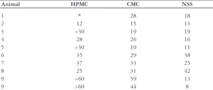
Normal saline cushions had median duration of 19 min (range 8-37 min). The median length for CMC injection was
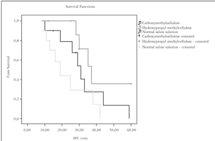
31 min (range 10-43 min). The time of HPMC injection was 37 min (median) with 12 to 60 minutes. There was no difference between SFC length between NSS and CMC (P = 0.146) or between CMC and HPMC (P = 0.119). However, the median length of HPMC was signiicantly longer than NSS (P = 0.039). Table 1 shows individual results and Figure 2 shows the comparative analysis using survival curve according to the Kaplan-Meier method.
FIGURE 1. Gastric submucosal injection
TABLE 1. Durability of submucosal luid cushions (SFC) in minutes
Animal HPMC CMC NSS
1 * 28 18
2 12 15 13
3 >30 19 19
4 28 26 16
5 >30 10 11
6 35 29 38
7 37 33 25
8 25 31 42
9 >60 59 13
9 >60 44 8
HPMC = hydroxypropyl methylcellulose; CMC = carboxymethylcellulose; NSS = normal saline solution
DISCUSSION
Medical technology development and improvement in medical knowledge enable us to choose a minimally invasive treatment for early cancer patients(5). EMR was developed
Lenz L, Di Sena V, Nakao FS, Andrade GP, Rohr MRS, Ferrari Jr AP. Comparative results of gastric submucosal injection with hydroxypropyl methylcellulose, carboxymethylcellulose and normal saline solution in a porcine model
Arq Gastroenterol
186 v. 47 – no.2 – abr./jun. 2010
Lenz L, Di Sena V, Nakao FS, Andrade GP, Rohr MRS, Ferrari Jr AP. Resultados comparativos de injeção submucosa gástrica com hidroximetil celulose, carboximetilcelulose e soro isiológico em modelo suíno. Arq Gastroenterol. 2010;47(2):184-7.
RESUMO – Contexto - A ressecção endoscópica mucosa é uma modalidade estabelecida para a excisão de lesões sésseis no trato gastrointestinal. A injeção de luídos na submucosa cria uma coxim que pode prevenir lesão térmica e perfuração. Objetivo - Este estudo cego investiga o desempenho de três diferentes soluções para criar um coxim luído submucoso no estômago suíno. Métodos - Três soluções foram injetadas no estômago de nove porcos BR1: soro isiológico, carboximetilcelulose 0.5% e hidroxipropil metilcelulose 0.25%. Em cada porco, injeções submucosas com 6 mL por solução-teste foram realizadas. Uma gota de azul de metileno foi adicionada a cada injeção para melhor visualização. O tempo de desaparecimento de cada coxim foi registrado. Resultados - O tempo mediano total do coxim submucoso visível foi de 37 minutos (faixa 12–60 min) para hidroxipropil metilcelulose, 31 minutos para carboximetilcelulose (faixa 10–43 min) e 19 minutos para soro isiológico (faixa 8–37 min). Não houve signiicância estatística entre soro isiológico e carboximetilcelulose (P = 0.146), assim como entre carboximetilcelulose e hidroxipropil metilcelulose (P = 0.119), mas a duração mediana de hidroxipropil metilcelulose foi signiicativamente maior que a do soro isiológico (P = 0.039). Conclusão - A duração do coxim submucoso com hidroxipropil metilcelulose é maior em comparação com o do soro isiológico. O tempo mediano da carboximetilcelulose não foi maior que do soro isiológico. A hidroxipropil metilcelulose, na concentração de 0.25%, pode ser uma alternativa durável para injeção submucosa.
DESCRITORES – Endoscopia do sistema digestório. Neoplasias gastrointestinais. Solução salina hipertônica. Carboximetilcelulose. Metilcelulose. Suinos.
introduced. Improvements in endoscopic resection techniques determined good results regarding local cure and long-term outcome after resection(25).
A predictable SFC is essential for safe EMR. Repeated injection is frequently necessary during EMR to isolate the mucosal tissues and to prevent deep tissue injury. Then, a more durable SFC may result in safer procedures. Normal saline is the usual solution used for EMR. However, it diffuses quickly with consequent disappearance of the bleb(5). To overcome
this problem, several other solutions were tested, but the ideal injection solution has yet to be identiied. According to Gostout(6), an ideal solution should be: cheap, nontoxic,
readily obtainable in bulk, storable at room temperature, not requiring any mixing other than dilution, highly luid outside of the body, easily injected through any standard needle catheter and almost impenetrable when in place.
Several studies have compared various solutions regarding their ability to maintain mucosal elevation during EMR, but
most of them were not blinded or was not in vivo. These two aspects can be a potential source of bias.
Although SH should be used as a irst-line solution, its high cost limits its use in clinical practice. Besides that, SH is a synthetic product that could potentially cause antigenic reactions(4). HPMC has been identiied as an economical
alternative to SH, being similarly effective at a dramatically lower cost, with no storage requirement, no need for reconstitution before use and minimal tissue reaction(3).
HPMC is cellulose derivate with viscoelastic characteristics. We used HPMC in lower concentration (0.25%) than were used in previous studies(3, 8).
CMC has high viscosity and is used as eye drops and in ophthalmologic practice. Recently, Yamasaki et al.(27) showed
that CMC was technically eficient method for dissection of gastric lesions and did not cause damage to muscular layer and surrounding. However, this study had used high concentration (2.5%), which was dificult to inject through a standard needle. Thus, we preferred a lower concentration. In spite of the SFC median time for CMC was not signiicantly longer than NSS, CMC is inexpensive, nontoxic, storable at room temperature, not requiring any mixing other than dilution, easily injected through any standard needle catheter and easy to buy in Brazil.
In conclusion, in this present study, the length of HPMC submucosal luid cushion is longer in comparison with NSS. HPMC in the concentration of 0.25% may be a durable and available alternative for submucosal injection. This is the irst blinded and further comparative studies will be necessary to ind the best solution for submucosal injection.
ACKNOWLEDGMENT
This study was supported in part by a grant from FAPESP (05/53739-7).
FIGURE 2. SFC Survival curve according to the Kaplan-Meier method
Carboxymethyloellulose Hydroxypropyl methylcellulose Normal saline solution Carboxymethyloellulose censored Hydroxypropyl methylcellulose - censored Normal saline solution - censored Survival Functions
Cum Survival
1,0
0,8
0,6
0,4
0,2
0,0
Lenz L, Di Sena V, Nakao FS, Andrade GP, Rohr MRS, Ferrari Jr AP. Comparative results of gastric submucosal injection with hydroxypropyl methylcellulose, carboxymethylcellulose and normal saline solution in a porcine model
Arq Gastroenterol 187
v. 47 – no.2 – abr./jun. 2010
REFERENCES
1. Chen YJ, Lee MD, Chen PH. Diagnosis and treatment of supericial oesophageal carcinoma. J Gastroenterol Hepatol. 1997;12:778-84.
2. Conio M, Rajan E, Sorbi D, Norton I, Herman L, Filiberti R, Gostout CJ. Comparative performance in the porcine esophagus of different solutions used for submucosal injection. Gastrointest Endosc. 2002;56:513-6.
3. Feitoza AB, Gostout CJ, Burgart LJ, Burkert A, Herman LJ, Rajan E. Hydroxypropyl methylcellulose: a better submucosal luid cushion for endoscopic mucosal resection. Gastrointest Endosc. 2003;57:41-7.
4. Fujishiro M, Yahagi N, Kashimura K, Mizushima Y, Oka M, Matsuura T, Enomoto S, Kakushima N, Imagawa A, Kobayashi K, Hashimoto T, Iguchi M, Shimizu Y, Ichinose M, Omata M. Different mixtures of sodium hyaluronate and their ability to create submucosal luid cushions for endoscopic mucosal resection. Endoscopy. 2004;36:584-9.
5. Fujishiro M, Yahagi N, Nakamura M, Kakushima N, Kodashima S, Ono S, Kobayashi K, Hashimoto T, Yamamichi N, Tateishi A, Shimizu Y, Oka M, Ogura K, Kawabe T, Ichinose M, Omata M. Successful outcomes of a novel endoscopic treatment for GI tumors: endoscopic submucosal dissection with a mixture of high-molecular-weight hyaluronic acid, glycerin, and sugar. Gastrointest Endosc. 2006;63:243-9.
6. Gostout CJ. Ode to the submucosal luid cushion. Endoscopy. 2004;36:638-9. 7. Hirao M, Masuda K, Asanuma T, Naka H, Noda K, Matsuura K, Yamaguchi
O, Ueda N. Endoscopic resection of early gastric cancer and other tumors with local injection of hypertonic saline- epinephrine. Gastrointest Endosc. 1988;34:264-9.
8. Hyun JJ, Chun HR, Chun HJ, Jeen YT, Baeck CW, Yu SK, Kim YS, Lee HS, Um SH, Lee SW, Choi JH, Ryu HS, Hyun JH. Comparison of the characteristics of submucosal injection solutions used in endoscopic mucosal resection. Scand J Gastroenterol. 2006;41:488-92.
9. Inoue H, Takeshita K, Hori H, Muraoka Y, Yoneshima H, Endo M. Endoscopic mucosal resection with a cap-itted panendoscope for esophagus, stomach, and colon mucosal lesions. Gastrointest Endosc. 1993;39:58-62.
10. Kaplan E, Meier P. Nonparametric estimation from incomplete observations. J Am Stat Assoc. 1958;53:157-81.
11. Karita M, Tada M, Okita K. The successive strip biopsy partial resection technique for large early gastric and colon cancers. Gastrointest Endosc. 1992;38:174-8. 12. Lee DK, Lee SW, Kwon SO, Jang WI, Shim YH, Cho MY. Endoscopic mucosectomy
using an esophageal variceal ligation device for minute gastric cancer. Endoscopy. 1996;28:386-9.
13. Lee SH, Cho WY, Kim HJ, Kim YH, Chung IK, Kim HS, Park SH, Kim SJ. A new method of EMR: submucosal injection of a ibrinogen mixture. Gastrointest Endosc. 2004;59:220-4.
14. Noda M, Kodama T, Atsumi M, Nakajima M, Sawai N, Kashima K, Pignatelli M. Possibilities and limitations of endoscopic resection for early gastric cancer.
Endoscopy. 1997;29:361-5.
15. Ohkuwa M, Hosokawa K, Boku N, Ohtu A, Tajiri H, Yoshida S. New endoscopic treatment for intramucosal gastric tumors using an insulated-tip diathermic knife. Endoscopy. 2001;33:221-6.
16. Ono H, Kondo H, Gotoda T, Shirao K, Yamaguchi H, Saito D, Hosokawa K, Shimoda T, Yoshida S. Endoscopic mucosal resection for treatment of early gastric cancer. Gut. 2001;48:225-9.
17. Roukos D. Current advances and changes in treatment stragery may improve survival and quality of life in patients with potentially curable gastric cancer. Ann Surg Oncol. 1999;6:46-56.
18. Sakai P, Maluf-Filho F, Iryia K, Moura EG, Tomishigue T, Scabbia A, Ishioka S. An endoscopic technique for resection of small gastrointestinal carcinomas. Gastrointest Endosc. 1996;44:65-8.
19. Schneider AR, Kriener S, Hoepffner N. Submucosal injection of autologous bood before endoscopic resection- an option to Nacl and Hyaluronate?[abstract] Gastrointest Endosc. 2006;63:ab80.
20. Soetikno RM, Gotoda T, Nakanishi Y, Soehendra N. Endoscopic mucosal resection. Gastrointest Endosc. 2003;57:567-79.
21. Tada M, Murata M, Murakami F, Shimada M, Mizumachi S, Arima K, et al. Development of the strip-off biospy [in Japanese with English abstract]. Gastroenterol Endosc. 1984;26:833-9.
22. Takekoshi T, Baba Y, Ota H, Kato Y, Yanagisawa A, Takagi K, Nogushi Y. Endoscopic resection of early gastric carcinoma: results of a retrospective analysis of 308 cases. Endoscopy. 1994;26:352-8.
23. Torii A, Sakai M, Kajiyama T, Kishimoto H, Kin G, Inoue K, Koizumi T, Ueda S, Okuma M. Endoscopic aspiration mucosectomy as curative endoscopic surgery; analysis of 24 cases of early gastric cancer. Gastrointest Endosc. 1995;42:475-9. 24. Uraoka T, Fujii T, Saito Y, Sumiyoshi T, Emura F, Bhandari P, Matsuda T, Fu KI, Saito D. Effectiveness of glycerol as a submucosal injection for EMR. Gastrointest Endosc. 2005;61:736-40.
25. Yamamoto H, Yube T, Isoda N, Sato Y, Sekine Y, Higashizawa T, Ido K, Kimura K, Kanai N. A novel method of endoscopic mucosal resection using sodium hyaluronate. Gastrointest Endosc. 1999;50:251-6.
26. Yamamoto H, Sekine Y, Higashizawa T, Kihira K, Kaneko Y, Hosoya Y, Ido K, Saito K, Sugano K. Successful en bloc resection of a large supericial gastric cancer by using sodium hyaluronate and electrocautery incision forceps. Gastrointest Endosc. 2001;54:629-32.
27. Yamasaki M, Kume K, Yoshikawa I, Otsuki M. A novel method of endoscopic submucosal dissection with blunt abrasion by submucosal injection of sodium carboxymethylcellulose: an animal preliminary study. Gastrointest Endosc. 2006;64:958-65.
28. Yasuda K. Endoscopic ultrasonic probes and mucosectomy for early gastric carcinoma. Gastrointest Endosc. 1996;43:s29-31.