Fibrotic Tissue and Middle Turbinate Exhibit
Similar Mechanical Properties. Is Fibrosis a
Solution in Nasal Polyposis?
Luciano Gregório
1Rogério Pezato
1Rafael Souza Felici
1Eduardo Macoto Kosugi
11ENT Research Laboratory, Department of ENT, Universidade Federal
de São Paulo, São Paulo, SP, Brazil
Int Arch Otorhinolaryngol 2017;21:122–125.
Address for correspondenceRogerio Pezato, MD, Department of ENT, Universidade Federal de Sao Paulo, Rua Pedro de Toledo, Sao Paulo, Sao Paulo 04023-900, Brazil (e-mail: [email protected]).
Introduction
Nasal polyposis (NP) is a chronic inflammatory condition of the upper airway characterized by overgrowth of nasal mucosa caused by the influx of inflammatory cells. Patients with NP experience a severe imbalance in immunomodulation1and an abnormal remodeling process, characterized by low production of transforming growth factor-β(TGF-β) and a lack of collagen, when compared with healthy subjects.2,3Similar results have been found for the proteoglycans biglycan, lumican,4tenascin,
andfibronectin.5The altered composition of extracellular matrix in NP creates a mechanical dysfunction in the nasal mucosa, which impairs the interstitial hydrostatic response during inter-stitial extravasation of volume in polypoid tissue.6
During the inflammatory process the balance between oncotic and hydrostatic pressure [(capillary osmotic pres-sure–interstitial osmotic pressure)–(hydrostatic
intersti-tial pressure–hydrostatic capillary pressure)] is critical to
limit edema formation. Given that the nasal polypoid tissue does not increase the interstitial hydrostatic pressure
Keywords
►
nasal polyps
►
mucosa
►
sinusitis
Abstract
Introduction
Nasal polyposis (NP) is a chronic in
fl
ammatory condition of the upper
airway characterized by overgrowth of nasal mucosa. Recent studies have shown a
mechanical dysfunction in the nasal polyp tissue.
Objective
This study aims to evaluate the mechanical properties of nasal
fi
brotic
tissue.
Method
This study was an institutional review board approved translational study in
20 participants (8 patients with NP, 7 patients with nasal synechiae, and 5 subjects
without sinus disease (control group). We used Controlled Disc Stimulation equipment
to compare the curve Pressure/Volume created during the saline solution infusion.
Results
The increase of pressure in response to solution injection was lower in the
nasal polyp group when compared with control middle turbinate group and
fi
brotic
group. No signi
fi
cant difference was found in the pressure response during solution
injection between
fi
brotic group and control middle turbinate group.
Inferior turbinate group showed signi
fi
cant difference when compared with control
middle turbinate group.
Conclusion
The mechanical dysfunction found in the nasal mucosa of patients with NP
provides new insight into this condition. These data allow the belief that the
fi
brosis has
a potential role in increasing interstitial hydrostatic pressure and, consequently,
mitigating edema formation in NP.
received July 26, 2016 accepted
September 6, 2016 published online October 26, 2016
DOI http://dx.doi.org/ 10.1055/s-0036-1593728. ISSN 1809-9777.
Copyright © 2017 by Thieme-Revinter Publicações Ltda, Rio de Janeiro, Brazil Original Research
properly, as found in different inflamed tissues, a greater volume is necessary to achieve the balance between the pressures (the polyp requires three times more infusion of saline solution to reach the same interstitial hydrostatic pressure found in healthy middle meatus mucosa).6
The interstitial hydrostatic pressure response depends on the tissue mechanical properties which in turn are related to the extracellular matrix.
Fibrotic tissue is characterized by high concentration of collagen. In this context, it should be expected that variations in the extracellular matrix could have an impact on tissue mechanical properties and, consequently, on interstitial hydrostatic pressure response.
The objective of this manuscript aims to evaluate the mechanical properties of nasalfibrotic tissue and compare it to nasal polypoid tissue and healthy middle meatus mucosa.
Material and Methods
Study Population
We selected participants among the patients of the Rhinology Section between 2014 and 2015. The study population was formed by 8 patients with NP, 7 patients with nasal synechiae, and 5 controls without nasal synechiae or sinus disease. All participants underwent an otorhinolaryngological assess-ment including nasal endoscopy.
Inclusion Criteria
European Position Paper on Rhinosinusitis and Nasal Polyps (EPOS) 2012 NP definition for research (presence of two or more symptoms, visualization of polyps in both middle meatus at nasal endoscopy, and confirmation by CT-scans) was used in this study as presence of NP.7
We considered nasal synechiae when endoscopically diagnosed and confirmed by palpation with a Cottle elevator, regardless of symptoms.
Control group was formed by patients submitted to sep-toplasty/turbinectomy, without sinus alterations at CT-scans or nasal synechiae under endoscopic visualization.
Exclusion Criteria
We excluded patients with age below 18 years old from the study.
Study Design
The local ethics committee approved this study under proto-col number 01510612.8.0000.5505.
After observing inclusion and exclusion criteria, this study included 8 patients with NP, 7 patients with nasal synechiae, and 5 controls subjects without nasal synechiae or sinus disease. The main measurement of this study was the submu-cosal local pressure. Thereby, we formed four groups: Nasal Polyps, Synechiae, Middle Turbinate, and Inferior Turbinate.
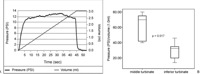
We measured the pressures within the submucosa during infusion of 0.2 to 1 ml of saline solution, presenting data as mean pressure /volume plots (►Fig. 1- illustration of a single
measurement). A 22-gauge needle coupled to a Controlled Disc Stimulation device (CDS, Smith & Nephew, Memphis, TN, USA) was used to inject the controlled volume within the submucosa to measure local pressure.6 All patients were under general anesthesia, and received no intranasal drugs before the saline injection.
Statistical Analysis
We analyzed study data in PASW Statistics for Windows, Version 18 (SPSS Inc., Chicago, IL, USA). We used the Mann-Whitney U test to evaluate between-group differences. P values of<0.05 were deemed significant.
Results
There was no difference in age between the synechiae and NP groups (p¼0.9). We found a significant difference in age
between the control group and the NP group (p¼0.04).
Fig. 1 (A) Graphic illustration of increase in interstitial pressure during volume infusion in a single patient. (B) Comparison of pressure increase in
healthy middle turbinate versus healthy inferior turbinate after saline infusion. The pressure increase in the middle turbinate is greater than in the inferior turbinate.
Increase in Pressure versus Volume (PSI/ml)
Thefirst part of the pressure-volume (P/V) plot demonstrated linear variation (►Fig. 1A); we therefore used the mean P/V to
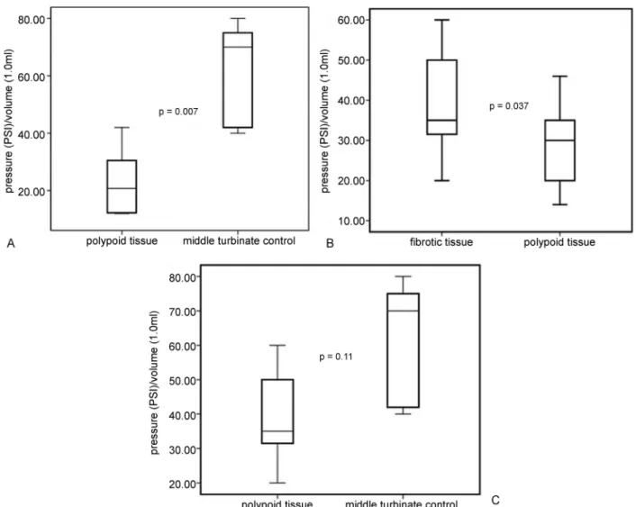
compare the hydrostatic pressure created during infusion. We observed that the mean P/V was lower in the submu-cosa of polypoid tissue than in the submusubmu-cosa of middle turbinates (►Fig. 2A) andfibrotic tissue (►Fig. 2B). We found
no significant difference in mean P/V betweenfibrotic tissue and healthy middle turbinate mucosa (►Fig. 2C).
When we compared turbinates in the control group, we found a lower mean P/V in inferior turbinates as compared with middle turbinates (►Fig. 1B).
Discussion
The mechanical dysfunction found in the nasal mucosa of patients with NP provides new insight into this condition. In this study, the concept of NP is more focused on the conse-quences caused by the effects of a chronic inflammation in
patients prone to developing NP than on understanding the different types of inflammatory process involved (orchestrated by Th1 or Th2, predominance of eosinophils or neutrophils).
Consequently, new forms of treatment can be suggested. The interstitial hydrostatic pressure (IHP) during volume extravasation is lower in patients with NP than in healthy nasal mucosa, creating an imbalance in the pressures (osmot-ic and hydrostat(osmot-ic) responsible for water return into the capillaries.
In theory, mechanisms that increase the IHP could partially prevent edema formation in NP, and one plausible hypothesis would be to shift the remodeling process in a morefibrotic direction. To evaluate this hypothesis, we investigated the IHP response during volume infusion into nasal tissues.
Our finding of higher pressure in the middle turbinate from healthy nasal mucosa than in nasal polypoid tissue corroborates a previous study.6
The higher IHP found infibrotic tissue (synechiae) after saline infusion when compared with polypoid nasal mucosa
Fig. 2 (A) Comparison of pressure increase in nasal polypoid tissue versus healthy nasal mucosa (middle turbinate), the pressure increase is
greater in healthy middle turbinate than in polypoid tissue. (B) Comparison of pressure increase infibrotic tissue (synechia) versus nasal polypoid tissue, the pressure increase is greater infibrotic tissue than in polypoid tissue. (C) Comparison of pressure increase infibrotic tissue (synechia) versus healthy nasal mucosa (middle turbinate). There is no significant difference in pressure.
International Archives of Otorhinolaryngology Vol. 21 No. 2/2017
supports, from a mechanical standpoint, the typical histolog-ical findings (high concentration of extracellular matrix, especially collagen, infibrotic tissue versus soft tissue with a lack of extracellular matrix in NP).2,4,5,8,9
The mechanical properties found infibrotic tissue seem to be more similar to healthy nasal mucosa than to nasal polypoid tissue (►Fig. 2), makingfibrosis a potential option
to interfere with the abnormal remodeling process found in NP.
Although we found a significant difference in age between the control group and the NP group, our focus in this experi-ment was to assess thefibrotic tissue. The Synechiae group (fibrotic tissue) did not have a significant difference in age when compared with Nasal Polyps group and control groups (Middle Turbinate and Inferior Turbinate group).
Finally, our data provide further support for the notion that the inferior turbinate is a different structure from the middle turbinate (►Fig. 1B).10Other authors have already
demonstrated this fact in terms of histology,11 airflow physiology,12 and nasal nitric oxide levels.13 In view of thesefindings, data generated through the use of inferior turbinate tissue as control in NP research should be ana-lyzed very carefully.
We believe thatfibrosis has a potential role in increasing IHP and, consequently, mitigating edema formation in NP.
Conclusion
Fibrotic tissue exhibits a greater increase in IHP in response to acute volume infusion than nasal polypoid tissue. This increase of IHP infibrotic tissue closely resembles the response seen in healthy nasal mucosa, suggesting that a tissue remodeling process prone to a more fibrotic direction could slow down the NP progression.
Conflict of Interest
The authors declare no conflict of interest.
References
1 Pezato R, de Almeida DC, Bezerra TF, et al. Immunoregulatory effects of bone marrow-derived mesenchymal stem cells in the nasal polyp microenvironment. Mediators Inflamm 2014;2014:583409 2 Van Bruaene N, Derycke L, Perez-Novo CA, et al. TGF-beta signaling
and collagen deposition in chronic rhinosinusitis. J Allergy Clin Immunol 2009;124(2):253–259, 259.e1–259.e2
3 Balsalobre L, Pezato R, Perez-Novo C, et al. Epithelium and stroma from nasal polyp mucosa exhibits inverse expression of TGF-β1 as compared with healthy nasal mucosa. J Otolaryngol Head Neck Surg 2013;42:29
4 Lee SH, Park JH, Oh BH, et al. Analysis of proteoglycan gene messages in human nasal mucosa and nasal polyp using dot blot hybridization. Acta Otolaryngol 2001;121(3):398–402
5 Liu Z, Gao Q, Zhang S, You X, Cui Y. [Expression of tenascin and fibronectin in nasal polyps]. Zhonghua Er Bi Yan Hou Ke Za Zhi 2002;37(3):173–176
6 Pezato R, Voegels RL, Pinto Bezerra TF, Perez-Novo C, Stamm AC, Gregorio LC. Mechanical disfunction in the mucosal oedema forma-tion of patients with nasal polyps. Rhinology 2014;52(2):162–166 7 Fokkens WJ, Lund VJ, Mullol J, et al. EPOS 2012: European position
paper on rhinosinusitis and nasal polyps 2012. A summary for otorhinolaryngologists. Rhinology 2012;50(1):1–12
8 Yang YC, Zhang N, Van Crombruggen K, Hu GH, Hong SL, Bachert C. Transforming growth factor-beta1 in inflammatory airway dis-ease: a key for understanding inflammation and remodeling. Allergy 2012;67(10):1193–1202
9 Pezato R, Voegels RL. Why do we notfind polyps in the lungs? Bronchial mucosa as a model in the treatment of polyposis. Med Hypotheses 2012;78(4):468–470
10 Pezato R, Voegels RL, Stamm AC, Gregório LC. Why we should avoid using inferior turbinate tissue as control to Nasal Polyposis studies. Acta Otolaryngol 2016;136(9):973–975
11 Berger G, Finkelstein Y, Ophir D, Landsberg R. Old and new aspects of middle turbinate histopathology. Otolaryngol Head Neck Surg 2009;140(1):48–54
12 Wang Y, Lee HP, Gordon BR. Impacts offluid dynamics simulation in study of nasal airflow physiology and pathophysiology in realistic human three-dimensional nose models. Clin Exp Otorhi-nolaryngol 2012;5(4):181–187
13 Takeno S, Yoshimura H, Kubota K, Taruya T, Ishino T, Hirakawa K. Comparison of nasal nitric oxide levels between the inferior turbinate surface and the middle meatus in patients with symp-tomatic allergic rhinitis. Allergol Int 2014;63(3):475–483