doi: 10.7213/academica.7699 ISSN 0103-989X Lic
enciado sob uma Lic
ença C
reative C
ommons
[A]
Marcelo Piagentini[a], Nereu Carlos Prestes[b], Carla Fredrichsen Moya-Araujo[c], Gustavo Henrique Marques Araujo[d], Regina Kiomi Takahira[e], Eunice Oba[f]
Biochemical and hormonal proile of the
amniotic luid from Nelore calves conceived by
different reproduction biotechnologies
[I]
Peril bioquímico e hormonal do líquido amniótico de bezerros
Nelore concebidos por diferentes biotecnologias
[a] Veterinarian, Phd in Animal Reproduction, Department of Animal Reproduction and Veterinary Radiology, School of Veterinary Medicine and Animal Science (FMVZ) (Unesp), Botucatu, SP - Brazil, e-mail: [email protected] [b] Veterinarian, Phd in Animal Reproduction, Department of Animal Reproduction and Veterinary Radiology, professor at
the School of Veterinary Medicine and Animal Science (FMVZ) (Unesp), Botucatu, SP - Brazil, e-mail: [email protected]
[c] Veterinarian, Phd in Animal Reproduction, professor at the Faculdades Integradas of Ourinhos, Ourinhos, SP - Brazil, e-mail: [email protected]
[d] Veterinarian, Phd in Animal Reproduction, professor at the Unicastelo, Descalvado, SP - Brazil, e-mail:[email protected]
[e] Veterinarian, Phd in Clinical Medicine, professor at the School of Veterinary Medicine and Animal Science (FMVZ) (Unesp), Botucatu, SP - Brazil, e-mail: [email protected]
[f ] Veterinarian, Phd in Animal Reproduction, Department of Animal Reproduction and Veterinary Radiology, professor at the School of Veterinary Medicine and Animal Science (FMVZ) (Unesp), Botucatu, SP - Brazil,
e-mail:[email protected]
[R
Abstract
This study aimed to quantify biochemical constituents and hormonal concentrations in the amniotic fluid
of Nelore calves conceived by different reproduction biotechnologies. Sixty animals were used and
divi-ded into: 1 – Twenty Nelore cows pregnant of calves obtained by artificial insemination; 2 – Twenty cows pregnant with Nelore calves obtained by embryo transference; 3 – Twenty cows pregnant with Nelore
calves obtained by in vitro production. During the expulsion phase, the amnion was punctured and 15 mL
of fluid were collected. The biochemical evaluation was performed with commercial kits in accordance to
the manufacturer’s recommendations. It was used the radioimmunoassay to assess the progesterone and
testosterone levels. The statistical analysis used was the variance analysis and Tukey (5% significance level). Mean urea and creatinine when comparing the three groups showed that group 3 had higher values
of these two metabolites, suggesting that the kidney maturity is suitable. For gamma-glutamyltransferase,
group 3 had a lower value compared to group 1. This enzyme is related to metabolism, may be inferred
that the metabolic activity is high in animal production in vitro. The glucose values did not differ between the groups, suggests that the maternal-fetal exchanges were suitable for the power supply to the fetus.
Lower chloride concentration was showed in group 3. Sodium and potassium were found in greater quan
and progesterone. The results obtained in biochemical analysis can conclude that the renal and liver acti-vity and maternal-fetal exchange are normal in the different groups.
Keywords: Amniotic fluid. Laboratorial analyses. Nelore.
]
Resumo
O presente trabalho teve por objetivo quantificar a concentração dos constituintes bioquímicos e hormo
-nal no líquido amniótico de fetos Nelore, oriundos de diferentes biotécnicas reprodutivas. Utilizaram-se 60 vacas gestantes divididas em: 1 – 20 vacas prenhes de bezerros oriundos de inseminação artificial con
-vencional (IA); 2 – 20 vacas prenhes de bezerros oriundos de transferência de embriões (TE) e 3 – 20 vacas prenhes de bezerros oriundos de produção in vitro (PIV). Durante a fase de expulsão, o âmnion foi puncionado e colheram-se 15 mL de líquido amniótico. As determinações bioquímicas foram con
-duzidas por kits comerciais. A progesterona e a testosterona foram determinadas por meio de radioi
-munoensaio (RIA) em fase sólida. Empregou-se a análise de variância e teste de Tukey com 5% de signi
-ficância. O teor de ureia e creatinina no G3 foi maior que nos outros grupos e significativo (p < 0,05) em relação ao G1. Para gamaglutamiltransferase (GGT), o G3 apresentou menor valor em relação ao G1 (p < 0,05). Os valores de glicose não diferiram entre os grupos. O cloreto apresentou maior concentração em G1 e G2 que no G3 (p < 0,05). O sódio e o potássio foram encontrados em maior quantidade no G 3 em relação aos demais grupos. Na análise hormonal não foi detectada diferença significativa para testosterona e progestero
-na. Pelos teores de ureia e creatinina, concluiu-se que a presença destes, sugere maturidade renal adequada, que a GGT (relacionada ao metabolismo animal) está elevada nos bezerros de produção in vitro e que os gru
-pos tiveram a concentração de glicose adequada ao fornecimento de energia ao feto. Os resultados obtidos na análise bioquímica indicam que a atividade renal e hepática, além das trocas materno-fetal, está normal nos diferentes grupos. [P]
Palavras-chave: Líquido amniótico. Análise laboratorial. Nelore.
Introduction
The amniotic fluid is an important indicator of fe -tal health and its contents are factors that can help to
clarify gestational problems. This fluid can be submit -ted to a variety of tests including biochemistry, cytology, biophysics and immunology assays. These evaluations can determine the maturity of fetal pulmonary, renal, and epidermal systems. In addition, they can detect
ge-netic abnormalities and other disorders (KJELDSBERG; KNIGHT, 1993).
The fluid allows the fetus to adopt an adequate po -sition prior to parturition, providing protection against traumas, dehydration and temperature variations. It also enables the fetus to grow and move without
caus-ing any harm to the uterus. Not only that, but the fluid
promotes cervix, vagina and vulva dilatation during par-turition, lubricating the vagina after the chorioallantois disruption helping the calf to pass through the vaginal canal. Apart from having bactericidal proprieties, these
fluids also avoid skin adhesions (PRESTES; LANDIM-ALVARENGA, 2006 ; TONIOLLO; VICENTE, 1995).
The fetal fluids components and their relation to
both the concept viability and maturity have been stud-ied in different species, but in the bovine species there are only a few recent reports of the biochemical and
hormonal profile of these fluids. Hence, the purpose of
the present study was to quantify biochemical constit-uents, progesterone and testosterone concentration at
amniotic fluid at the delivery time of Nelore calves that
were conceived by either in vitro production (IVP),
con-ventional embryo transfer (ET) or artificial insemina -tion (AI), and compare them.
Materials and methods
of Clinical Veterinary Medicines at the School of Veterinary Medicine and Animal Science (FMVZ),
Botucatu, São Paulo, Brazil (22o
51’S/48o26’W).
Sixty animals were kindly offered by private farms that were located in the rural area of Avaré,
São Paulo, Brazil (23o03’S/48o55’W) in order to per
-form the sample collection of amniotic fluid. Herd
health assessment and vaccination program were performed according to the farms’ management schedule. All animals were kept on pasture and
sup-plemented with silage, industrial feed and water ad
libitum. The cows were divided into three groups: 1-Twenty Nelore pregnant cows with calves obtained
by AI; 2-Twenty pregnant cows of ET with Nelore
calves obtained by superovulation of embryo donors
and 3-Twenty pregnant cows with Nelore calves ob -tained by IVP after follicular aspiration.
Close to the expected calving date the cows were transferred to a calving paddock to facilitate labor observation. During the expulsion phase of the labor,
the amnion was punctured and 15 mL of fluid were collected using a 40 × 12G needle and a 20 mL sy
-ringe. The material was deposited in plastic 50 mL tubes and frozen in regular freezer (-20 °C) for lat
-er analysis. Biochemical analyses of the amniotic flu -id samples were performed at the Clinic Laboratory of the Department of Clinical Veterinary Medicine – FMVZ, Botucatu, São Paulo, Brazil. The hormonal assays were performed at the Endocrinology Laboratory of the Animal Reproduction and Veterinary Radiology of the same School.
It was used commercial kits according to the manufacturer recommendation in order to eva-luate the biochemical parameters as follows: Glucose – (mg/dL) colorimetric method
(gluco-se SL; Celm®; Barueri, SP) spectrophotometer Celm SB-190; Urea – (mg/dL) colorimetric method (urea ES; Celm®; Barueri, SP) spectrophotometer Celm SB-190; Creatinine–(mg/dL) kinetic method (ki
-netic creatinine; Celm®; Barueri, N= SP) – spectro -photometer Celm SB-190;
Gamma-glutamyltrans-ferase (GGT) – (UI/L) kinetic method (gamma
GT; Celm®; Barueri-SP) spectrophotometer Celm E-210S; Sodium – (mmol/L) flame photometry (so
-dium; Celm®; Barueri, SP) flame photometer Celm FC-280; Potassium – (mmol/L) flame photometry (potassium; Celm®; Barueri, SP) flame photome -ter Celm FC-280; Chloride – (mmol/L) colorime
-tric method (chlorides; Labtest®; Lagoa Santa, MG)
spectrophotometer Celm SB–190; Total Protein – (g/dL) colorimetric method (total proteins;
Labtest®; Lagoa Santa, MG) – spectrophotometer
Celm SB-190. Progesterone and testosterone con
-centrations in the amniotic fluid were determined
by radioimmunoassay (RIA) using commercial kits in solid phase (Diagnostic Products Corporation –
DPC; MedLab®; São Paulo, SP). Results were analyzed with Gama Count Cobra II and it was used
the variance analysis and Tukey test (5% of signifi -cance level) for the statistical study.
Results
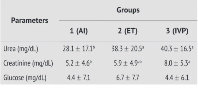
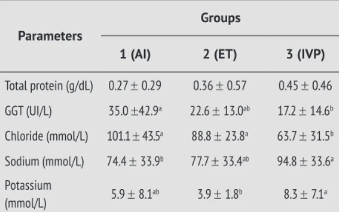
Mean values and standard deviation for the
bio-chemical parameters of the amniotic fluid, of the
fetuses conceived by AI, ET or IVP are described in the Table 1.
It was observed that group 1 presented lower urea
mean value when compared to groups 2 and 3. As for both creatinine and sodium, group 3 had higher lev
-els than group 1 (p < 0.05), and group 2 presented in -termediated levels of such substances, and similar
val-ues when compared to other groups’ valval-ues (p > 0.05).
Glucose and total protein levels did not differ among groups. For group 1, GGT levels were statically
high-er than for group 3, and group 2 showed inthigh-ermediat -ed levels, which were similar to the other both groups.
Chloride levels were higher for group 3 (p < 0.05) and potassium levels were higher for group 2 (p < 0.05).
Table 2 shows mean and standard error of the pro -gesterone and testosterone for all groups. The hor-mone concentration analysis disregarded the fetal gender. The results did not show any relevant
statisti-cal difference among groups (p > 0.05).
Table 1 - The mean values and standard deviation for
the biochemical parameters obtained from the amniotic luid at calving in the different groups
Parameters
Groups
1 (AI) 2 (ET) 3 (IVP)
Urea (mg/dL) 28.1 ± 17.1b 38.3 ± 20.5a 40.3 ± 16.5a
Creatinine (mg/dL) 5.2 ± 4.6b 5.9 ± 4.9ab 8.0 ± 5.3a
Glucose (mg/dL) 4.4 ± 7.1 6.7 ± 7.7 4.4 ± 6.1
Parameters
Groups
1 (AI) 2 (ET) 3 (IVP)
Total protein (g/dL) 0.27 ± 0.29 0.36 ± 0.57 0.45 ± 0.46
GGT (UI/L) 35.0 ±42.9a 22.6 ± 13.0ab 17.2 ± 14.6b
Chloride (mmol/L) 101.1 ± 43.5a 88.8 ± 23.8a 63.7 ± 31.5b
Sodium (mmol/L) 74.4 ± 33.9b 77.7 ± 33.4ab 94.8 ± 33.6a
Potassium
(mmol/L) 5.9 ± 8.1
ab 3.9 ± 1.8b 8.3 ± 7.1a
Source: Research data.
Note: a,b =Values within rows with different superscripts are significantly
different (p < 0.05).
Table 2 - The mean values and standard deviation for the hormonal concentration obtained from the am -niotic luid at calving in the different groups.
Parameters
Group
01 (AI) 02 (ET) 03 (IVP)
Progesterone (ng/mL) 7.9 ± 6.1 8.1 ± 6.3 6.1 ± 5.4
Testosterone (ng/dL) 7.8 ± 6.6 8.7 ± 11.4 5.1 ± 4.6
Source: Research data. Note: (p > 0.05).
Discussion
Creatinine is one of the most studied biochemi-cal compound and its level can be used to deter-mine the fetal kidney maturation during gestation.
It can be detected in the fluid due to the gradient
difference among the umbilical cord, digestory mucosa and bronchial mucosa, by simple diffusion. Creatinine is a non protein nitrogen substance
ex-creted via glomerullar filtration, as the kidney
gets more mature it excretes higher concentra-tions of creatinine, increasing its levels in the
am-niotic fluid. The urea concentration level is used
to evaluate the kidney maturation at veterinary and human medicine, in parallel to the creatinine
level (HERVEY; SLATER, 1967; VOTTA, 1975).
Creatinine mean concentration observed in groups
1 and 2 was lower than the one found in the bo
-vine species (BAETZ et al., 1976), but for group 3
mean concentration values were similar. Urea con-centration values for group 1 were similar to the
ones found for the bovine (BAETZ et al., 1976) and ovine (PRESTES et al., 2001) species. These creati -nine and urea concentration values suggested sat-isfactory kidney maturation.
Glucose is the main sugar at the fetuses’ blood;
therefore it is the main energy source. Large amount of the produced glucose is obtained via fa-cilitated transport across the maternal and the fe-tal placenta. The mean concentration values of glu-cose in the three groups were higher than the ones
found in the ovine species (PRESTES et al., 2001). The finding of no statistical difference in the re -sults indicates that the maternal-fetal glucose transport were at normal levels.
The GGT is an enzyme found in high concentra-tion in the kidney, liver and pancreas, acting at met-abolic level mediating a large number of physiologi-cal functions. In relation to the mean concentration of GGT, this experiment had shown that the three groups had a higher level of this enzyme than the one
reported in ovine species (PRESTES et al., 2001).
Potassium and sodium urine excretion are regulated by aldosterone that is produced on the fetal adrenal gland, and the main re-sults are the increase in the potassium and de-crease in the sodium concentrations, and this is sufficient to indicate the fetal kidney ma-turity. The amniotic fluid’s sodium and chlo-ride concentrations depend on the oronasal secretions during gestation (EVRARD et al.,
1997). The mean sodium concentration in the
amniotic fluid in the three groups was lower than other results found in the reviewed
litera-ture (BRACE et al., 2004; GAGNON et al., 2002; PRESTES et al., 2001) for the ovine species. The
potassium concentration in group 1 was similar to the one described in the ovine species by Brace et al.
(2004), but its value was lower in group 2 and its value was higher in group 3. However, other
authors described a higher potassium level than
three groups (PRESTES et al., 2001). Baetz et al. (1976), in the bovine species, described potas -sium concentration was similar in group 1, but
(CONCLUSION) Table 1 - The mean values and standard deviation for
EVRARD, V. et al. Intra-uterine tracheal obstruction
de-creases amniotic fluid sodium and chloride concentra -tion in the fetal lamb. American Journal of Obstetrics & Gynecology, v. 176, n. 1, p. 171, 1997.
GAGNON, R.; HARDING, R.; BRACE, R.A. Amniotic fluid
and fetal urinary responses to severe placental
insu-fficiency in sheep. American Journal of Obstetrics & Gynecology, v. 186, n. 5, p. 1076-1084, 2002.
HERVEY, E. J.; SLATER, J. S. The source of sheep fetal
fluids in the later stages of gestation. The Journal of Physiology, v. 22, n. 1, p. 40-41, 1967.
KJELDSBERG, C.; KNIGHT, J. Body fluid: laborato -ry examination of amniotic, cerebroespin, serus and synovial fluids. In: American Society for Clinical Pathology. Chicago: Theid, 1993.
MAIA, M. et al. Concentração dos hormônios esteróides no plasma sanguíneo fetal e nos líquidos amniótico e alantoideano no terço inicial, médio e final da gestação de vacas. ARS Veterinária, v. 20, n. 3, p. 353-360, 2004. PRESTES, N. C.; CHALHOUB, M. C. L.; LOPES, M. D.
Amniocentesis and biochemical evaluation of
amniot-ic fluid in ewes at 70, 100 and 145 days of pregnancy. Small Ruminant Research, v. 39, n. 3, p. 277-281, 2001. PRESTES, N. C; LANDIM-ALVARENGA, F. C. Medicina Veterinária – Obstetrícia Veterinária. Rio de Janeiro:
Guanabara-Koogan, 2006.
TONIOLLO, G. H.; VICENTE, W. R. R. Manual de ob-stetrícia veterinária. São Paulo: Livraria Varela,
1995. 86p.
VOTTA, R. A. Aplicaciones clínicas del estudo de la creatinina del líquido amniótico. In: VOTTA, R. A.
Investigaciones clínicas al conocimiento del estado fetal. México: Panamericana, 1975 p. 59-63.
Received: 07/19/2012
Recebido: 19/07/2012
Approved: 10/23/2012
Aprovado: 23/10/2012
its value was lower in group 2 and its value was higher in group 3.
Brace et al. (2004) described an ovine chlo -ride level similar to the one for animals in group 1
and higher than the results for group 2 and 3.
However, other authors in the ovine species also
(GAGNON et al., 2002) had shown a lower chloride concentration when compared to the findings from
group 1 and a higher concentration when
com-pared to groups 2 and 3, in contradiction to the results from Evrard et al. (1997), which showed a
higher level of chloride.
The testosterone concentration level found for the three groups during this experiment was lower than the one described in another paper
(MAIA et al., 2004). On the other hand the pro -gesterone concentration level showed in this experiment was higher than the concentration
reported by Maia et al. (2004). These results dif -fered probably due to hormonal kit used.
Conclusion
The results obtained in biochemical analysis can conclude that the renal and liver activity and mater-nal-fetal exchange are normal in the different groups.
The amniotic fluid is an important source for fetal
evaluation through biochemical and hormonal analy-sis that allow the determining process of hepatic and renal maturity in the fetus.
Acknowledgments:
Research received financial support from Fapesp.
References
BAETZ, A. L.; HUBERT, W. T.; GRAHAN, C. K. Changes of bio
-chemical constituents in bovine fetal fluids with gestational
age. American Journal of Veterinary Research, v. 37, n. 9, p. 1047-1052, 1976.
BRACE, R. A.; VERMIN, L. M.; HUIJSSOON, E. Regulation
of amniotic fluid volume: intramembranous solu-te and volume fluxes in lasolu-te gestation fetal sheep.
American Journal of Obstetrics & Gynecology,