SEROLOGICAL SURVEY FOR RABIES IN SERUM SAMPLES FROM VAMPIRE BATS (Desmodus rotundus) IN BOTUCATU REGION, SP, BRAZIL
LANGONI H (1, 2), SOUZA LC (1), ZETUN CB (1), SILVA TCC (3), HOFFMANN JL (1, 2), SILVA RC (1)
(1) Zoonosis Research Center, NUPEZO, Veterinary Medicine and Animal Husbandry School, São Paulo State University, UNESP, Botucatu, São Paulo State, Brazil; (2) Department of Tropical Diseases, Botucatu Medical School, São Paulo State University, UNESP, Botucatu, São Paulo State, Brazil; (3) Department of Animal Health and Production, School of Dentistry, São Paulo State University, UNESP, Araçatuba, São Paulo State, Brazil.
ABSTRACT: The chiropterans constitute 25% of the world’s mammal fauna. Due to the destruction of their natural ecosystem, the vampire bats have moved from nature to artificial roosts closer to man and domestic animals. This phenomenon has happened particularly in rural areas. Rabies is a viral anthropozoonosis, 100% lethal, and vampire bats (Desmodus rotundus) represent an important role in its epidemiology. D. rotundus were captured at night with mesh nets in partnership with the Botucatu Defense Office and sent to the Zoonosis Diagnostic Service, at the School of Veterinary Medicine and Animal Husbandry, UNESP. Serum samples from 204 bats were analyzed by enzyme-linked immunosorbent assay (ELISA) and fluorescent antibody viral neutralization test (FAVN) for rabies antibody detection. The results showed 7.4% of sera with titers higher or equal to 0.5 U for rabies antibodies, which demonstrated viral flow circulation among the studied region. Data suggest a need for constant monitoring accomplished by epidemiological and sanitary measures.
KEY WORDS: Desmodus rotundus, vampire bats, rabies virus, LPC-ELISA, RFFIT.
CONFLICTS OF INTEREST: There is no conflict.
FINANCIAL SOURCE: FAPESP 2005/02682-5.
CORRESPONDENCE TO:
INTRODUCTION
The chiropterans are one of the most distinguished mammals as they present special
features that make them capable of true flight (19). These animals play an important
role in nature, specifically, in several tropical and subtropical areas, since they are
responsible for spreading seeds, flower pollination and insect population control.
They constitute somewhat wide range of mammal fauna, in the number of species
and population density, representing approximately a quarter of mammal fauna
worldwide (23).
In spite of their ecological function, bats can be transmitters of many diseases like
arbovirus, rickettsiosis, criptococosis (17), histoplasmosis, Chagas’s disease,
brucellosis, salmonelosis, candidiasis (8) and rabies to humans and other animals.
The latter is the most severe disease that can be transmitted by bats. D. rotundus,
the most common vampire bat of the Americas, can be found in colonies of 20 to 100
animals (16). They live in caves, tree holes, abandoned mines, home basements,
drainpipes or roosts (4).
Vampire bats present some adaptations to feed on blood: their saliva enzymes inhibit
blood coagulation and two ducts on each side of their tongues enable them to suck
blood. Each bat ingests 15 to 25 mL of blood per night and usually feeds on the
same prey and the same wound for many nights in a row; additionally, a parasitized
animal can be exploited by more than one bat during the same night (18).
As they feed only on blood, they are potential transmitters of rabies, which may
cause damage to equine and cattle flocks due to numerous animal deaths (14).
Humans, in turn, indiscriminately kill chiropterans, vampires or not, threatening the
ecological balance (18).
Attacks on humans and rabies transmission by D. rotundus have increased in recent
decades, especially in the Amazon area, in Peru and Brazil. Some of these
outbreaks have been preceded by local changes, including rapid domestic animal
removal, mineral extraction or deforestation, activities that can change the natural
environment where the D. rotundus lives (20).
An important fact regarding the epidemiological vigilance of this disease in Botucatu,
and its surrounding cities, is the Castelo Branco highway that presents many
embankments and, consequently, numerous areas of water drainage through large
and livestock. Although there are reforestation and agricultural areas in the bordering
municipalities, which usually show some human activity, there are also piping,
abandoned rural buildings and caves where bats can take shelter (22).
Wild animals constitute the natural reservoirs and transmit the virus to domestic
animals. The most common transmission route of rabies is through infected animal
saliva, especially by biting. The infection may also occur if the saliva reaches any
mucous membrane or open skin wound or scratch (1). The actions necessary to
hinder the disease are well known and there have been many improvements, for
example, in feline and canine control in urban areas (13). On the other hand, the
chiropteran population has grown in non-rural areas due to lack of metropolitan
planning with respect to architectural and landscape projects. Together with the
chiropteran population growth, acts of epidemiological vigilance were intensified. As
a result, the number of chiropteran rabies cases increased and the virus was found
even in areas where the disease had been thought to be under control (12).
Due to the disease expansion, D. rotundus has interfered in the aerial cycle, causing
countless rabies cases in non-vampire bats, as well as in the urban cycle, being
responsible by eight out of fourteen cases of the disease recorded among dogs and
cats (7).
Fifteen rabies cases in humans transmitted by bats were observed in the Americas
from 1995 to 2000. Bats rank second in transmission cases in Latin America. In the
USA and Canada, they were the only transmitters of autochthonous human rabies
(2). An outbreak has been recently detected in Pará State, Brazil, with 15 cases of
rabies in humans caused by bats within a month. It has been the most serious
outbreak in humans in Brazil, the largest ever registered in such short a period (3).
The subclinical infection may occur in bats, when viruses multiply in their fatty tissue
without invading the central nervous system (CNS). This makes them efficient
reservoirs that may cause infections for months or years by secretion and saliva
elimination (5). The main clinical rabies signs in D. rotundus are: daytime eating
habit, hyperexcitability, aggressiveness, incoordination, muscle spasms, paralysis
and death. The fatal infection does not seem common among chiropterans. The
infection cycles originate from bites (15).
The viral transmission among bats occurs through bites, breathing, scratches or
bat migration, which leads to the viral spreading (21).The intraspecies dissemination
may cause acute disease in some individuals, while in others the abortive infection
results in seroconversion. Serological tests permit the evaluation of the
epidemiological situation, since the seropositivity in a colony may simply demonstrate
an exposure to the virus, whereas the high prevalence associated with high antibody
titers can indicate recent outbreaks (15).
Due to the importance of vampire bats in South America, this study aimed to
evaluate the presence of serum antibodies in healthy free-living bats from the
Botucatu region of São Paulo State.
MATERIALS AND METHODS
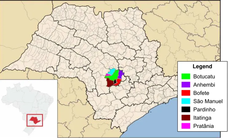
Two-hundred and four vampire bats were captured in the following municipalities:
Bofete (36), Pardinho (10), São Manuel (45), Pratânia (24), Botucatu (16), Itatinga
(28) and Anhembi (45) (Figure 1). The captures were performed during the evening,
between 6 and 11 p.m., using mesh nets set up in front of roosts, caves, tree holes
as well as inside piping (under highways or railways). Captured animals were sent to
the Zoonosis Diagnostic Laboratory in the Veterinary Medicine and Animal
Husbandry School, UNESP, Brazil, where they were properly anesthetized for blood
collection via intracardiac puncture. The blood samples were centrifuged at 1600 x g for 10 minutes and the sera were frozen at –20°C. The animals were killed and, then, their brain tissues were examined for Negri bodies by direct immunofluorescence
(DIF) according to Dean et al. (10).
Liquid-phase competitive enzyme-linked immunosorbent assays (LPC-ELISA) and
rapid fluorescent focus inhibition test (RFFIT) were performed according to Cardoso
et al. (6) with some modifications. CVS23 (virus standard) propagated in the chicken
embryo related (CER) cell line was inactivated and used for antigen preparation for
ELISA (11) and for RFFIT (6). Both methods are recognized by the World
Organization for Animal Health (OIE) as standard tests, with specificity and sensitivity
higher than 80% and presenting no cross-reaction with other antigens except for
rabies virus particles. A positive control serum of dog origin titrated to 132 IU/mL, obtained by the US Centers for Disease Control (CDC), stored at –20°C and diluted
to 5 IU/mL was used for all assays. As negative control, another dog serum with
In order to calculate ELISA titers, the two-graph receiver operating characteristic
(TG-ROC) technique was performed. Serum ranging between 0.10 and 0.20 was
considered weak positive; 0.20 to 0.30, positive; and greater than 0.30, high positive.
Each high positive serum was tested by RFFIT and the titers were calculated through
the analysis of 20 microscopic fields in only one pool, for each serum dilution
assessed in the presence of infected cells using a fluorescence microscope (Carl
Zeiss Inc., USA) (160x magnification). Titers were calculated using the method of
Reed and Muench in which the respective titers ≥ 0.5 IU/mL were considered
positive.
Botucatu
São Manuel
Itatinga Pratânia Bofete Anhembi Legend
Pardinho
Figure 1. Cities comprising the Botucatu region – in São Paulo State, Brazil –where
the vampire bats were captured in 2008.
Approximate scale: 1:600,000. Adapted from Wikimedia Commons, available from:
RESULTS AND DISCUSSION
All animals were negative for Negri bodies by DIF.
In the LPC-ELISA, from 204 serum samples analyzed, 92 (45.10%) reacted weakly
(titer from 0.10 to 0.20); 19 (9.31%) presented average reactions (titer between 0.20
and 0.3); 22 (10.78%) reacted strongly (titer greater than 0.30); and 71 samples did
not react. Every serum with titer higher than 0.3 was examined by RFFIT; among this
group, 15 samples (7.35%) were positive, presenting titers greater than 0.5 IU/mL
(Table 1).
In another study performed in Rio Preto, Minas Gerais State, Brazil, by Piccinini et al.
(19), 59 vampire bats – 15 (60%) out of 25 males and 17 (50%) out of 34 females –
had presented the virus in brain and/or in gland tissues, which revealed rabies
infection. These results show a significant difference when compared to the 7.35%
positivity of the current report, which presumably indicates a higher viral circulation in
both distinct regions.
Souza et al. (22) found, in the same Botucatu region, only 0.1% positivity among 895
studied bats by employing the immunofluorescence technique for brain tissue. On the
other hand, Cortês et al. (9), in another study in the same area, found 0.9% positivity.
The results of the present study may indicate subclinical disease, without Negri
bodies detected by the immunofluorescence technique. The animals presenting
subclinical infections constitute efficient reservoirs because the virus can multiply in
their fatty tissues without affecting the central nervous system. In reservoir animals,
the rabies virus can endure and is eliminated through both respiratory secretion and
saliva, so that they are capable of infecting other bats, humans as well as domestic
and wild animals.
The epidemiological characteristics of the disease are still poorly understood.
Consequently, more research and implementation of an epidemiologic vigilance
system are necessary (14). D. rotundus plays an important role in rabies circulation
Table 1. Numbers and percentages of D. rotundus bats infected by rabies (RFFIT
serology), in Botucatu region, 2008
Origin Number of captured
animals
Number of positive
animals Positivity (%)
Bofete 36 2 5.56
Pardinho 10 1 10.00
São Manuel 45 3 6.67
Pratânia 24 0 0
Botucatu 16 0 0
Itatinga 28 4 14.29
Anhembi 45 2 4.44
Total 204 15 7.35
CONCLUSIONS
The studied region presents viral flow circulation through bats, thus a constant
monitoring accomplished by epidemiological and sanitary measures is necessary to
maintain the control of rabies.
REFERENCES
1 ACHA PN., SZYFRES B. Zoonosis y enfermedades transmisibles comunes al
hombre y a los animales: clamidiosis, rickettsiosis y virosis. 3.ed. Washington:
Organización Panamericana de la Salud, 2003. 480p.
2 BELOTTO AJ. Raiva transmitida por morcegos nas Américas: impacto na saúde
pública e na produção. In: SEMINÁRIO INTERNACIONAL MORCEGOS COMO
TRANSMISSORES DA RAIVA, 2001, São Paulo, SP. Programas e resumos. São
Paulo: Secretaria da Saúde, 2001. 24-5p.
3 BRASIL. Ministério da Saúde. Secretaria de Vigilância em Saúde. Surto de raiva
humana transmitida por morcegos no município de Portel, Pará, março/abril de 2004.
Boletim Epidemiológico, ano 4. Available in: http://www.saude.gov.br/svs. Access in:
4 BREDT A., ARAÚJO FAA., CAETANO-JÚNIOR J., RODRIGUES MGR.,
YOSHIZAWA M., SILVA MMS., HARMANI NMS., MASSUNAGA PNT., BÜRER, SP.,
PORTO VAR., UIEDA W. Morcegos em áreas urbanas e rurais: manual de manejo e
controle. Brasília: Fundação Nacional de Saúde, 1996. 117p.
5 CAMPBELL JB., CHARLTON KM. Eds. Rabies. London: Kluwer Academic, 1988.
431p.
6 CARDOSO TC., QUEIROZ DA SILVA LH., DA SILVA SEL., ALBAS A., PARDO
PE., TANAKA AH., COSSY LB., PERRI SHV. Chicken embryo related (CER) cell line
for quantification of rabies neutralizing antibody by fluorescent focus inhibition test.
Biologicals, 2006, 34, 29-32.
7 CARRIERI ML., FAVORETTO SRL., CARNIELI P., QUEIROZ, LH., SOUZA
MCAM., PANACHÃO MRI., TAKAOKA NY., HARMANI NMS., KOTAIT I. Desmodus
rotundus como transmissor da raiva canina e felina, no Estado de São Paulo,
1998-2000. In: SEMINÁRIO INTERNACIONAL DE RAIVA, São Paulo, 1998-2000. Resumos...
São Paulo: Instituto Pasteur de São Paulo, 2000. 42-43p.
8 CONSTANTINE DG. Transmission of pathogenic microorganisms by vampire bats.
In: GREENHALL, AM., SCHMIDT, U. Ed. Natural history of vampire bats. Boca
Raton: CRC Press, 1988. 167-89p.
9 CORTÊS VA., SOUZA LC., UIEDA W., FIQUEIREDO AC. Abrigos diurnos e
infecção rábica em morcegos de Botucatu, São Paulo, Brasil. Vet. Zootec., 1994, 6,
179-86.
10 DEAN, DJ., ABELSETH, MK., ATANASIU, P. Routine laboratory procedures: The
fluorescent antibody test. In: MESLIN FX., KAPLAN MM., KOPROWSKY H. Ed.
Laboratory techniques in rabies. 4.ed. Genebra: World Health Organization, 1996.
88-95p.
11 FACHIN N., CARVALHO BA., CARDOSO TC. A comparison of serological
methods for detecting the immune response after rabies vaccination in dogs and
cows from rabies-endemic areas in Brazil. Intern. J. Appl. Res. Vet. Med., 2005, 3,
199-203.
12 KOTAIT I., AGUIAR EAC., CARRIERI ML., HARMANI NMS. Manejo de
13 LANGONI H., LIMA K., MENOZZI BD., SILVA RC. Rabies in the big fruit-eating
bat Artibeus lituratus from Botucatu, southeastern Brazil. J. Venom. Anim. Toxins
incl. Trop. Dis., 2005, 11, 84-7.
14 MAYEN F. Haematophagous bats in Brazil, their role in rabies transmission,
impact on public health, livestock industry and alternatives to an indiscriminate
reduction of bat population. J. Vet. Med., 2003, 50, 469-72.
15 MEGID J. Raiva. In: CUBAS, ZS.; SILVA, JCR.; CATÃO DIAS, JL. Eds. Tratado
de animais selvagens – medicina veterinária. São Paulo: Roca, 2007. 785-98p.
16 NOWAK RM. Walker’s bats of the world. Maryland: John’s Hopkins University
Press Ltd., 1994. 287p.
17 PAMSITT JR., VALDIVIESO D. Los murciélagos y la salud pública: estudio con
especial referencia a Puerto Rico. Bol. Oficina Sanit. Panam., 1970, 69, 122-40.
18 PERACCHI AL., LIMA IP., REIS NR., NOGUEIRA MR., ORTÊNCIO FILHO H.
Ordem Chiroptera. In: REIS, NR., PERACCHI, AL., PEDRO, WA., LIMA, IP. Eds.
Mamíferos do Brasil. Londrina: N. R. dos Reis, 2006. 153-230p.
19 PICCININI RS., GITTI CB., SILVA SB., BASTOS P. Presença do vírus rábico em
uma colônia de morcegos hematófagos Desmodus rotundus no município de Rio
Preto, Minas Gerais, Brasil. Rev. Bras. Med. Vet., 1996, 18, 106-9.
20 SCHNEIDER MC., ARON J., SANTOS-BURGOA C., UIEDA W., RUIZ VELAZCO
S. Common vampire bat attacks on humans in a village of the Amazon region of
Brazil. Cad. Saúde Pública, 2001, 17, 1531-6.
21 SERRA-COBO J., AMENGUAL B., ABELLÁN C., BOURHY H. European bat
lyssavirus infection in Spanish bat populations. Emerg. Infect. Dis., 2002, 8, 413-20.
22 SOUZA LC., LANGONI H., SILVA RC., LUCHEIS SB. Vigilância epidemiológica
da raiva na região de Botucatu-SP: importância dos quirópteros na manutenção do
vírus na natureza. Ars Veterinária, 2005, 21, 62-8.
23 TADDEI VA. Morcegos: Algumas considerações sistemáticas e biológicas.