Cellular/Molecular
Proliferative Defects and Formation of a Double Cortex in
Mice Lacking
Mltt4
and
Cdh2
in the Dorsal Telencephalon
Cristina Gil-Sanz,
1Bruna Landeira,
2Cynthia Ramos,
1Marcos R. Costa,
2and Ulrich Mu¨ller
11Department of Molecular and Cellular Neuroscience, Dorris Neuroscience Center, The Scripps Research Institute, La Jolla, California 92037, and2Brain
Institute, Federal University of Rio Grande do Norte, Natal, Rio Grande do Norte 59056-450, Brazil
Radial glial cells (RGCs) in the ventricular neuroepithelium of the dorsal telencephalon are the progenitor cells for neocortical projection neurons and astrocytes. Here we show that the adherens junction proteins afadin and CDH2 are critical for the control of cell proliferation in the dorsal telencephalon and for the formation of its normal laminar structure. Inactivation of afadin or CDH2 in the dorsal telenceph-alon leads to a phenotype resembling subcortical band heterotopia, also known as “double cortex,” a brain malformation in which heterotopic gray matter is interposed between zones of white matter. Adherens junctions between RGCs are disrupted in the mutants, progenitor cells are widely dispersed throughout the developing neocortex, and their proliferation is dramatically increased. Major subtypes of neocortical projection neurons are generated, but their integration into cell layers is disrupted. Our findings suggest that defects in adherens junctions components in mice massively affects progenitor cell proliferation and leads to a double cortex-like phenotype.
Key words: afadin; CDH2; double cortex; neocortex; progenitor; radial glia
Introduction
Radial glial cells (RGCs) of the dorsal telencephalon are the pro-genitor cells for neocortical projection neurons and astrocytes. Newly born neurons and astrocytes migrate from their place of birth near the ventricle toward the meninges to form the neocor-tical cell layers (Kriegstein and Noctor, 2004;Ayala et al., 2007; Franco and Mu¨ller, 2013;Greig et al., 2013). Defects in cell pro-liferation migration and differentiation lead to brain malforma-tions, such as periventricular heterotopia, lissencephaly, and subcortical band heterotopia (SBH;des Portes et al., 1998;Sheen et al., 2004;Ferland et al., 2009). In SBH, also known as “double cortex,” heterotopic gray matter is interposed between zones of white matter (Barkovich et al., 1994;Dobyns et al., 1999). The mechanisms that cause SBH have been proposed to involve de-fects in both the migration of neurons into the developing neo-cortical cell layers and the differentiation and proliferation of RGCs (Ross and Walsh, 2001;Bielas et al., 2004;Cappello et al., 2012).
RGCs project apical processes toward the ventricle and basal processes toward the meninges (Fishell and Kriegstein, 2003;
Ra-kic, 2003). The end feet of the apical processes are connected by adherens junctions that require for their formation CDH2, -catenin, and ␣E-catenin (Brault et al., 2001;Machon et al., 2003;Junghans et al., 2005;Lien et al., 2006;Woodhead et al., 2006;Kadowaki et al., 2007;Tang et al., 2009). Inactivation of the small GTPase RhoA in the dorsal telencephalon of mice results in the disruption of adherens junctions and the formation of a dou-ble cortex (Cappello et al., 2012), suggesting a mechanistic link between perturbation in the adhesive interactions between RGCs and cortical lamination defects.
In epithelial cells, the localization and activity of cadherins is promoted by nectins, which are members of the Ig superfamily (Ikeda et al., 1999;Takahashi et al., 1999;Miyahara et al., 2000; Sato et al., 2006). The adaptor protein afadin links nectins and cadherins by binding to the cytoplasmic domains of nectins and associating with p120-catenin and␣-catenin (Takahashi et al., 1999;Tachibana et al., 2000;Pokutta et al., 2002;Hoshino et al., 2005), which in turn interact with the cytoplasmic domains of cadherins (Ozawa et al., 1989,1990;Herrenknecht et al., 1991; Nagafuchi et al., 1991;Hirano et al., 1992;Knudsen and Whee-lock, 1992;Reynolds et al., 1992). Without afadin, cadherin clus-tering is perturbed, leading to defects in the formation of adherens junctions (Ikeda et al., 1999;Sato et al., 2006). Although cadherins/catenins are required for adherens junction formation between RGCs, the role of afadin in this process is less clear. Afadin inactivation in RGCs at approximately E11.5 using
nestin–Cre leads to progressive loss of adherens junctions (Yamamoto et al., 2013). The phenotype is restricted to some brain regions, and the severe hydrocephalus of the mutants has complicated mechanistic studies.
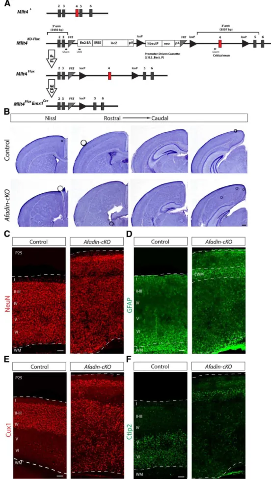
To further define the function of afadin in the neocortex, we generated a mouse line carrying a floxed allele of its gene (named
Received May 2, 2014; revised June 10, 2014; accepted June 13, 2014.
Author contributions: C.G.-S., B.L., M.R.C., and U.M. designed research; C.G.-S., B.L., and C.R. performed research; C.G.-S., B.L., and U.M. analyzed data; C.G.-S., B.L., M.R.C., and U.M. wrote the paper.
This work was supported by National Institutes of Health Grants NS046456 and HD070494 (U.M.), the Dorris Neuroscience Center (U.M.), the Skaggs Institute for Chemical Biology (U.M.), Ministry of Education Grant EX2009-0416 (C.G.-S.), Generalitat Valenciana Grant APOSTD/2010/064 (C.G.-S.), California Institute of Regenerative Med-icine (C.G.-S.), and CAPES (Coordenação de Aperfeiçoamento de Pessoal de Nível Superior) Foundation/Ministry of Education Grant PDSE 7640-13-7 (B.L.).
Correspondence should be addressed to Ulrich Mu¨ller, Dorris Neuroscience Center, The Scripps Research Institute, 10550 North Torrey Pines Road, La Jolla, CA 92037. E-mail: [email protected].
DOI:10.1523/JNEUROSCI.1793-14.2014
Copyright © 2014 the authors 0270-6474/14/3410475-13$15.00/0
Mtll4). We then inactivatedMtll4 expression usingEmx1–Cre
mice, which induces recombination in the dorsal telencephalon starting 1 day earlier than achieved withnestin–Cre(Gorski et al., 2002;Li et al., 2003;Dubois et al., 2006). The mutant offspring showed widespread disruption of adherens junctions between RGCs and a dramatic increase in progenitor proliferation, a phe-notype that was even more pronounced in mice lackingCdh2
in the dorsal telencephalon. Adult mice with a dorsal telencephalon-specific inactivation of eitherMttl4orCdh2 ex-hibited a double cortex-like phenotype. These findings suggest that adherens junctions are critical for the control of progenitor cell proliferation during neocortical development and that mu-tations that affect adherens junction proteins can lead to a double cortex-like phenotype in mice.
Materials and Methods
Mice.Experiments using mice were performed under the oversight of an institutional review board. Mice carrying a floxedMllt4gene were generated using ES cell clones from European Conditional Mouse Mutagenesis Program (EUCOMM; HEPD0591_8_E01) with knock-out first mutation (HEPD0591_8_E01;Ryder et al., 2013; http://www. mousephenotype.org/martsearch_ikmc_project/martsearch/ikmc_project/ 79371). ES clones were injected into C57BL/6J blastocysts, and the resulting chimeras were mated to C57BL/6J females to obtain germ-line transmission. Offspring were genotyped by PCR using the following primers: G5arm, CATGTTTATT CTTGGTTTCAGCTGGG; G3arm, AACGACTTCACACCTTGACTAAGG; and LAR3, CAACGGGTTCTTCTG TTAGTCC. The sizes of the PCR products are 549 bp (WT band) and 411 bp (mutant band). Heterozygous F1 mice (Mllt4KO-flox/⫹) were mated with
B6.Cg-Tg(ACTFLPe)mice (stock #005703; The Jackson Laboratory) to remove the promoter-driven selection cassette of the knock-out first mutation, and the resulting offspring (Mllt4flox-FLP/⫹) were subsequently
mated to C57BL/6J mice to segregate theFLPetransgene. Heterozygous offspring (Mllt4flox/⫹) were crossed to generateMllt4flox/floxmice, and genotyping was performed by PCR (G5arm, CATGTTTATTCTTG-GTTTCAGCTGGG; and G3arm, AACGACTTCACACCTTGACTAA GG). The sizes of the PCR products are 549 bp (WT band) and 650 bp (flox band).Mllt4flox/floxmice were mated toEMX1Cretransgenic mice (Gorski et al., 2002) to generateMllt4flox/⫹EMX1Cre/⫹mice, which were
crossed withMllt4flox/floxmice to obtain the animals used in the experi-ments. The presence of CRE was analyzed by PCR using previously pub-lished methods (Gorski et al., 2002).Cdh2flox/floxmice (stock #007611; The Jackson Laboratory) were crossed withEmx1Cremice to generate
Cdh2flox/⫹Emx1Cre/⫹, which were crossed withCdh2flox/floxto generate the animals used in the experiments.
Histology and immunostaining.Nissl staining and immunohistochem-istry was performed as described previously (Franco et al., 2011,2012; Gil-Sanz et al., 2013). Embryonic brains were dissected and fixed in 4% paraformaldehyde (PFA) overnight at 4°C. Adult brains were transcar-dially perfused with 4% PFA, and brains were dissected and postfixed in 4% PFA overnight at 4°C. Brains were sectioned coronally at 50 or 100 m with a vibrating microtome (VT1200S; Leica) or at 12m with a cryostat (CM 3050S; Leica). Sections used for 5-bromo-2⬘-deoxyuridine (BrdU) immunostaining were first treated with 2N HCl for 20 min and washed twice with borate buffer, pH 8.0, to equilibrate. Antibodies used for immunostaining are as follows: anti-␣E-catenin rabbit polyclonal (Cell Signaling Technology), afadin rabbit polyclonal (Sigma), anti--catenin mouse monoclonal (BD Biosciences), anti-BrdU mouse monoclonal (BD Biosciences), anti-BrdU rat monoclonal (AbD Sero-tec), anti-CDH2 mouse monoclonal (Sigma), anti-Ctip2 rat monoclonal (Abcam), Cux1 rabbit polyclonal (Santa Cruz Biotechnology), anti-GFAP rabbit polyclonal (Dako), anti-Ki67 rabbit polyclonal (Abcam), anti-Ki67 rat monoclonal (AbD Serotec), anti-L1 rat polyclonal (Milli-pore), anti-NeuN mouse monoclonal (Milli(Milli-pore), anti-Pax6 rabbit poly-clonal (Covance), anti-phospho-Histone 3 (pH3) rabbit polypoly-clonal (Cell Signaling Technology), anti-TAG1 mouse monoclonal (Developmental Studies Hybridoma Bank, National Institute of Child Health and Human
Development, and University of Iowa Department of Biology), anti-Tbr2 chicken (Millipore), anti-Tbr2 rabbit polyclonal (Abcam), and anti-Tuj1 mouse monoclonal (Covance). Nuclei were stained with DAPI (Thermo Fisher Scientific) and sections were mounted on slides with Prolong Gold mounting medium (Thermo Fisher Scientific). Images were captured using a Nikon-C2 or a Nikon-A1 laser-scanning confocal microscope and an Olympus AX70 microscope for bright-field images.
Quantifications and cell cycle analysis.At least three histological sec-tions from three different animals at three distinct rostrocaudal levels for each genotype were analyzed for each immunostaining. Confocal optical sections were used for quantification. Cells were counted in columns from the ventricular zone (VZ) to the marginal zone (MZ). Values are mean⫾SEM. To measure the density of Pax6, Tbr2, or Ki67 cells, the number of positive cells was determined in an area including a column of the cortical primordium (from MZ to VZ) and normalized to 104m2. For quantifications inCdh2conditional knock-out (Cdh2-cKO) attrib-utable to the severe hyperplasia that generates a dramatic increase of the thickness of the cortex, we evaluated the differences in the number of Pax6, Tbr2, or Ki67 cells in radial columns of the same thickness as in controls from the VZ to the MZ. To follow proliferating cells, we injected pregnant females intraperitoneally with BrdU (Sigma) at 150g/g. Em-bryos were collected 30 min after the injection (to label cells during S-phase) and 24 h after the injection (to label cells in S-phase and some postmitotic neurons). pH3 labels cells in M-phase. Ki67 labels the cells in any phase of the cell cycle. For cell cycle quantifications, three parameters were analyzed: (1) quitting fraction, the proportion of BrdU-positive/ Ki67-negative (BrdU⫹/Ki67⫺) cells among all BrdU⫹cells, 24 h after
the injection; (2) BrdU labeling index, the proportion of BrdU⫹cells
among all dividing Ki67⫹cells, 30 min after the injection; and (3) mitotic
index, proportion of the pH3⫹cells among all dividing Ki67⫹cells. Cells
were counted using the count tool of Adobe Photoshop 6. Statistical analysis was performed using GraphPad Prism software 5.01.
Results
Double cortex-like phenotype in adultAfadin-cKOmice Mice with a null mutation in theMttl4gene die at approximately E10 (Ikeda et al., 1999), preventing an analysis of afadin function in neocortical development with a simple knock-out mouse line. To evaluate genetically the role of afadin during the development of the neocortex, we generated a mouse line carrying a floxed allele of theMttl4gene (Fig. 1A). To inactiveMttl4expression, we crossedMttl4-floxmice withEmx1-Cremice that induced recom-bination as early as E10.5 in progenitor cells of the dorsal telen-cephalon (Gorski et al., 2002;Iwasato et al., 2004). For simplicity, we will refer to the mutant offspring (Mttl4flox/fox;EmxCre/⫹) as Afadin-cKOmice. For all experiments, we compared the pheno-type ofAfadin-cKOmice with the phenotype of littermates that did not express Cre or contained only one floxedMttl4allele.
Afadin-cKOmice were born in the expected Mendelian fre-quency and survived into adulthood. In whole mounts, the dis-sected brain of the mutant mice at P25 appeared larger than in controls (data not shown). In Nissl stainings of cortical sections at several histological levels along the rostrocaudal axis of the brain, the most obvious difference was the increased size and severe disorganization of the neocortex in theAfadin-cKOmice (Fig. 1B). In control animals, cells in the neocortex were orga-nized into cell layers above the cell-sparse white matter that con-tains axonal projections (Fig. 1B). In contrast, the cortex of
Afadin-cKOmice contained a thin layer of cells near the meninges and a disorganized mass of cells heterotopically localized deeper within the cortex (Fig. 1B). This mass of heterotopic cells was flanked above and below by relatively cell sparse regions (Fig. 1B). Hippocampal structure was overall pre-served but less well organized.
rons (Fig. 1C). Immunostaining with the astrocyte marker GFAP that labels fibrous astrocytes within the white matter revealed the accumulation of GFAP⫹cells in the relatively cell-sparse regions
flanking the heterotopia (Fig. 1D). The histological features in
Afadin-cKOmice resemble the phenotype described for SBH/ double cortex, a defect originally associated with defects in neu-ronal migration (Ross and Walsh, 2001;Bielas et al., 2004) but more recently also linked to alterations in the function of RGCs (Cappello et al., 2012).
Next we used immunohistochemistry to analyze the distribu-tion of Cux1⫹upper layer neurons and Ctip2⫹lower layer
neu-rons inAfadin-cKOmice. Cux1⫹cells were abundant in both the
thin layer of cells close to the meninges and the heterotopia that were situated deeper in the neocortex (Fig. 1E). Ctip2⫹cells were
prominently present in the thin band of cells close to the menin-ges and scattered throughout the ectopia (Fig. 1F). These find-ings suggest that at least some subtypes of cortical projection neurons are generated inAfadin-cKOmice but that their assem-bly into neocortical cell layers is perturbed. The histological fea-tures of the neocortex inAfadin-cKOmice are similar to those observed in SBH/double cortex caused by mutations in other genes, such asDCXin humans (des Portes et al., 1998;Gleeson et al., 1998) and RhoA in mice (Cappello et al., 2012).
Disruption of adherens junctions inAfadin-cKOmice To define the mechanism by which afadin affects neocortical development, we analyzed its expression pattern in the develop-ing dorsal telencephalon. Usdevelop-ing immunohistochemistry on sec-tions of E13.5 mice, we confirmed that afadin is expressed throughout the developing dorsal telencephalon, with highest expression levels near the ventricle in which the end feet of RGCs are connected by adherens junctions (Fig. 2A,B). At the ventricle, afadin was colocalized with CDH2,-catenin, and␣E-catenin (Fig. 2B,C), suggesting that it cooperates with these proteins in the formation of adherens junctions. InAfadin-cKOmice, ex-pression of afadin was maintained in the VZ of the ganglionic eminence but not in the VZ of the dorsal telencephalon in which
Emx1–Creis expressed (Fig. 2D,E). However, afadin expression was maintained in blood vessels within the neocortex of the mu-tant mice (Fig. 2D, arrows).
To determine the extent to which lack of afadin in the neuro-epitelial cells disrupted adherens junctions between RGCs, we analyzed the distribution of three additional junctional proteins (CDH2,-catenin, and␣E-catenin) at E13.5. Unlike in control mice, the distribution of the three proteins was severely
per-turbed (Fig. 2E,F). Afadin expression was no longer detectable (Fig. 2E), CDH2 and-catenin were distributed more evenly around the cell surface (Fig. 2E,F), and␣E-catenin was distrib-uted diffusely throughout the cell (Fig. 2F). This redistribution of proteins is consistently observed when adherens junctions are disrupted (Junghans et al., 2005;Lien et al., 2006;Sato et al., 2006; Rasin et al., 2007; Yamamoto et al., 2013), suggesting that, in
Afadin-cKOmice, adherens junctions were not properly formed or maintained.
Dispersion of proliferating progenitor cells in Afadin-cKOmice
To determine the temporal progression of the neocortical phe-notype inAfadin-cKOmice, we analyzed histological sections of control and mutant mice at E13.5 and E16.5. At E13.5, the thick-ness of the developing neocortex was substantially increased in
Afadin-cKOmice compared with controls as revealed by staining of nuclei with DAPI (Fig. 3A). Consistent with defects in the integrity of adherens junctions, the ventricular surface was dis-rupted and irregular in the mutants (Fig. 3A). At E16.5, the neo-cortex of the Afadin-cKOmice was severely disorganized and appeared hyperplastic (Fig. 3F).
We next used molecular markers to determine the distribu-tion of progenitor cells and neurons within the developing neo-cortex ofAfadin-cKOmice. We used Pax6 as a marker for RGCs, Tbr2 as a marker for intermediate progenitors, and TuJ1 as a marker for differentiated neurons (Caccamo et al., 1989;Lee et al., 1990a,,b;Go¨tz et al., 1998;Englund et al., 2005). Staining with Ki67 at E13.5 and E16.5 revealed that proliferating cells were primarily confined to the VZ and subventricular zone (SVZ) of control mice but broadly distributed throughout the entire thick-ness of the nascent neocortex inAfadin-cKOmice (Fig. 3B,G). Consistent with this finding, in controls at E13.5, Pax6⫹RGCs
were primarily concentrated in the VZ (Fig. 3C), Tbr2⫹
interme-diate progenitors in the SVZ (Fig. 3D), and Tuj1⫹neurons in the
nascent cortical plate (Fig. 3E). In contrast, Pax6⫹RGCs and
Tbr2⫹intermediate progenitors in the mutants were dispersed
throughout the entire developing neocortex (Fig. 3C,D); Tuj1⫹
neurons were distributed irregularly with some of them occupy-ing positions within the VZ and SVZ (Fig. 3E). At E16.5, a simi-larly abnormal distribution of Pax6⫹ and Tbr2⫹progenitors
throughout the entire thickness of the developing neocortex was observed inAfadin-cKOmice (Fig. 3H,I). In addition, staining with Tuj1 revealed an abnormally thin cortical plate and accumu-lations of neurons at the surface of the ventricle and below the intermediate zone in which they formed rosette-like structures (Fig. 3J).
To further ascertain the distribution of Pax6⫹ RGCs and
Tbr2⫹ intermediate progenitors, we also performed
double-immunofluorescence microscopy (Fig. 4A). The two markers were still primarily expressed in distinct cell populations, indicat-ing that deletion of afadin did not lead to the generation of a progenitor with a mixed RGC/intermediate progenitor identity. Finally, we anticipated that the gross disruption of the laminar structure of the neocortex would affect the organization of axonal tracts in the mutant mice. Staining with antibodies to TAG1 and L1 revealed massive perturbations in axonal tracts (Fig. 4B). Al-though these perturbations are likely at least in part a secondary consequence of defects in cell numbers and cell position, we can-not exclude that afadin also plays a more direct role in axonal outgrowth or pathfinding.
4
(Figure legend continued.) floxed promoter-driven neo cassette inserted into an intron of the targeted gene. Engrailed (En2) splice acceptor disrupts gene function, resulting in a lacZ fusion for studying gene expression. Flp recombinase removes the gene trap cassette, converts the “knock-out first” allele to a conditional allele (flox). Cre recombinase deletes the floxed exon of the conditional allele resulting in a frame shift and null mutation (modified fromRyder et al., 2013). Numbered boxes represent exons. Red box represents the critical exon. PCR primers for genotyping (G5arm, G3arm, and LAR3) are indicated.B, Nissl staining showing coronal sections
of adult brains from control andAfadin-cKOmice at several histological levels along the rostro-caudal axis of the neocortex. Note the enlarged neocortex and the ectopic mass of cells sur-rounded by cell-sparse regions in the mutant mice. Scale bar, 500m.C,D, Coronal sections of
adult brains from control andAfadin-cKOmice immunostained with the neuronal marker NeuN (red;C) and with the astrocyte marker GFAP (green;D). Note the ectopic accumulation of
GFAP⫹astrocytes, presumably ectopic white matter, in the upper part of the cortex of the
mutant mice. Scale bar, 100m.E,F, Coronal sections of adult brains from control and Afadin-cKOmice immunostained with the upper layer marker Cux1 (red;E) and the lower layer marker
Defects in cell proliferation inAfadin-cKOmice
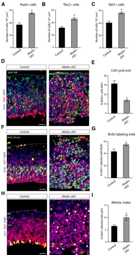
The thickness of the neocortex appeared to be increased in
Afadin-cKOmice already at embryonic ages. This could be a consequence of the generation of abnormal numbers of cells. Consistent with this hypothesis, the numbers of Ki67⫹cells
was increased at E13.5 by⬃50% in the mutant mice, with an approximately equal increase in the number of Pax6⫹RGC
and Tbr2⫹intermediate progenitors (Fig. 5A–C). Increases in
the number of proliferative cells could be caused by a
reduc-tion in the number of progenitor cells that exited the cell cycle. To test this hypothesis, we injected pregnant females at E12.5 with BrdU to label cells in S-phase and prepared histo-logical sections of the embryos 24 h later. We stained the sections for BrdU and Ki67, the latter labeling proliferating cells throughout different stages of the cell cycle. Cells that do not leave the cell cycle within the 24 h period are therefore double positive for BrdU and Ki67, whereas cells that exited the cell cycle are positive for BrdU only. The number of Figure 2. Afadin-cKOmice present disruption of the cortical adherens junctions.A, Coronal section of the brain of control mice at E13.5 immunostained for afadin protein. Note the accumulation of afadin at the ventricular surface of the pallium and subpallium. Scale bar, 100m.B, Coronal section of the E13.5 developing cortex of control mice showing the VZ immunostained for the junctional proteins CDH2 (green) and afadin (red). Nuclei stained with DAPI are shown in blue. Scale bars, 5m.C, Coronal section of the E13.5 developing cortex of control mice showing the VZ immunostained for the junctional proteins␣E-catenin (green) and-catenin (red). Nuclei stained with DAPI are shown in blue. Scale bars, 5m.D, Coronal section of the brain ofAfadin-cKOmice at E13.5 immunostained for afadin protein. Note the disappearance of afadin immunostaining in the dorsal telencephalon, whereas afadin expression is maintained in the subpallium. Arrows point toward blood vessels in the dorsal pallium, which also continue to express afadin. Scale bar, 100m.E, Coronal section of the E13.5 developing cortex ofAfadin-cKOmice showing the VZ immunostained with the junctional proteins CDH2 (green) and afadin (red). Note the absence of afadin expression and the altered localization of CDH2 at the apical–junctional complex. Nuclei stained with DAPI are shown in blue. Scale bars, 5m.F, Coronal section of the E13.5 developing cortex of Afadin-cKOmice showing the VZ immunostained with the junctional proteins␣E-catenin (green) and-catenin (red). Note the altered localization of both proteins at the apical– junctional complex. Nuclei stained with DAPI are shown in blue. Scale bars, 5m. PSB, Pallial–subpallial boundary.
BrdU⫹/Ki67⫺cells was strongly reduced (⬃43% decrease) in Afadin-cKOmice (Fig. 5D,E), suggesting an alteration of cell cycle withdrawal in the mutant mice. To measure cell cycle length, we calculated the BrdU labeling index by labeling pro-genitors for 30 min with BrdU, followed by immunostaining for BrdU and Ki67 (Fig. 5F,G). The percentage of BrdU⫹/
Ki67⫹cells was increased by⬃25%, indicative of a shortened
cell cycle in the progenitors inAfadin-cKOmice. Finally, we calculated the mitotic index, the proportion of dividing cells in M-phase. Immunostaining for Ki67 and the M-phase marker pH3 demonstrated an⬃50% increase in the number of Ki67⫹/pH3⫹cells (Fig. 5H,I). Together, these data
dem-Figure 3. Hyperplasia in the developing neocortex ofAfadin-cKOmice. Overview of the developing neocortex from control andAfadin-cKOat E13.5 and E16.5 stained with the nuclear marker DAPI (A–F), the proliferation marker Ki67 (B–G), the radial glia cell marker Pax6 (C–H), the intermediate progenitor marker Tbr2 (D–I), and the neuronal marker Tuj-1 (E–J). Note the general
onstrate that cell cycle regulation and proliferation are signif-icantly altered inAfadin-cKOmice.
Defects in adherens junctions, cell proliferation, and axonal pathfinding in CDH2 mutant mice
In epithelial cells, nectins cooperate with cadherins in the assem-bly of adherens junctions (Ikeda et al., 1999; Takahashi et al., 1999;Miyahara et al., 2000;Sato et al., 2006). The adaptor protein afadin links nectins and cadherins by binding to the cytoplasmic domains of nectins and associating with p120-catenin and ␣-catenin (Takahashi et al., 1999;Tachibana et al., 2000;Pokutta et al., 2002;Hoshino et al., 2005), which in turn interact with the
cytoplasmic domains of cadherins (Fig. 6A;Ozawa et al., 1989, 1990;Herrenknecht et al., 1991;Nagafuchi et al., 1991;Hirano et al., 1992;Knudsen and Wheelock, 1992;Reynolds et al., 1992). Without afadin, cadherin clustering is perturbed, leading to de-fects in the formation of adherens junctions (Ikeda et al., 1999; Sato et al., 2006). Therefore, we wondered whether there is a mechanistic link between afadin and cadherins in RGCs.
Adherens junctions are disrupted in mice lacking either CDH2 or␣E-catenin, but proliferative defects similar to those reported here were only reported for␣E-catenin-deficient mice (Lien et al., 2006;Kadowaki et al., 2007). This suggests that afadin and␣E-catenin might have specific functions in controlling pro-Figure 4. Progenitor distribution and axonal projections inAfadin-cKOmice.A, Coronal brain sections from control andAfadin-cKOmice at E13.5 were stained with antibodies to Pax6 (red) and Tbr2 (green). Note that the two markers are primarily expressed in non-overlapping cell populations, even in mutant animals. Scale bars: overviews, 100m; insets, 25m.B, Coronal brain sections
from control andAfadin-cKOmice at E16.5 were stained with antibodies to TAG1 (green) and L1 (red) to reveal axonal projections. Note the gross disorganization of axonal tracts in the mutants. Scale bar, 100m.
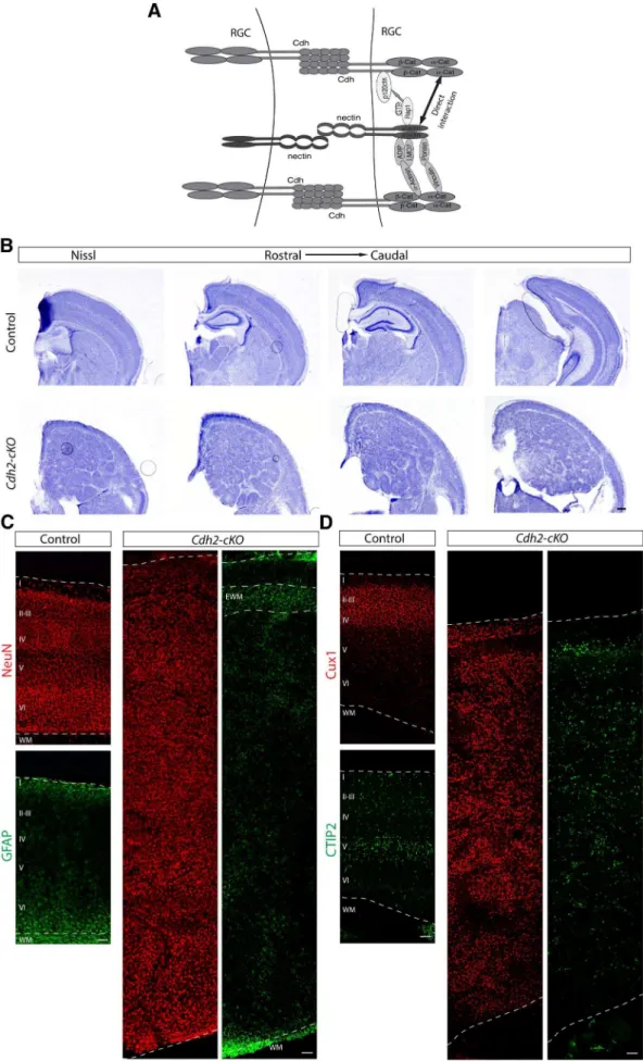
liferation. Alternatively, disruption of ad-herens junctions more generally might affect proliferation. Defects after disrup-tion of theCdh2gene might have escaped detection in previous studies (Kadowaki et al., 2007), possibly because of incom-plete gene inactivation with theCremice used in previous studies. To distinguish between these possibilities, we crossed
Cdh2flox/floxmice withEmx1–Cremice to generateCdh2-cKOmice. By P13, the cor-tex ofCdh2-cKOmice was even more hy-perplastic than inAfadin-cKOmice and vastly increased in size (Fig. 6B). The lam-inar structure was disrupted, and a mas-sive heterotopia formed below a thin band of cells near the meninges (Fig. 6B).
Neurons expressing NeuN, Cux1, and CTIP2 were observed in the thin band of cells near the meninges and in the hetero-topia (Fig. 6C,D). GFAP⫹cells
accumu-lated in the relatively cell-sparse cell regions flanking the heterotopia (Fig. 6C). Therefore, the histological features of
Cdh2-cKOmice closely resemble the phe-notype ofAfadin-cKOmice.
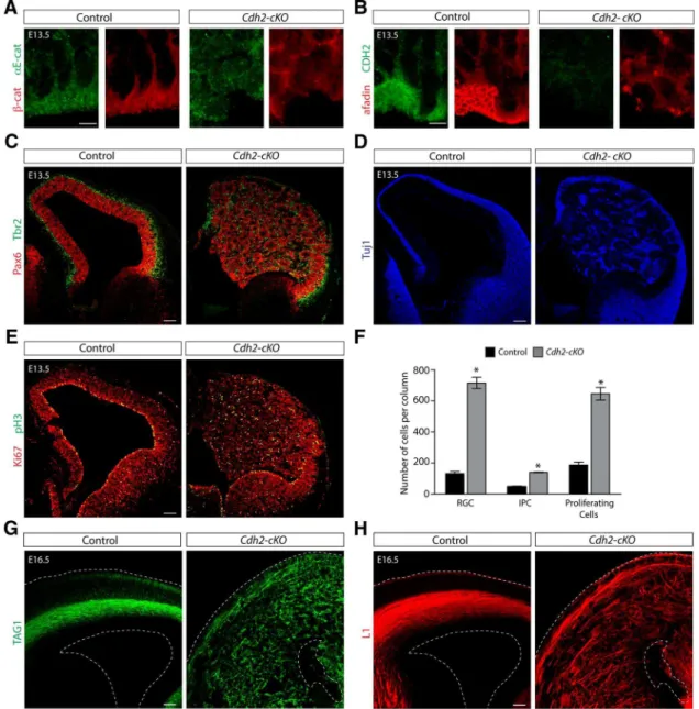
Immunohistochemical analysis at E13.5 confirmed that the localization of adherens junction proteins was perturbed in the VZ of the developing dorsal telen-cephalon ofCdh2-cKOmice (Fig. 7A,B). Afadin expression levels seemed reduced (Fig. 7B), although this could be a conse-quence of broader distribution of afadin protein throughout the cytoplasm. In ad-dition, Pax6⫹and Tbr2⫹progenitor cells
and Tuj1⫹ neurons were distributed
through the entire thickness of the abnor-mal neocortex (Fig. 7C,D). The distribu-tion of the cells was not homogenous, and numerous rosette-like structures were de-tected, which was especially obvious when sections were stained for Tuj1 (Fig. 7D).
Figure 5. Increased proliferation of cortical progenitor cells inAfadin-cKOmice.A, Quantification (mean⫾SEM) of the number
of RGCs (Pax6⫹cells) in control andAfadin-cKOmice at E13.5. *p⬍0.0001 by Student’sttest.B, Quantification (mean⫾SEM)
of the number of intermediate progenitor cells (Tbr2⫹cells) in control andAfadin-cKOmice at E13.5. *p⬍0.0005 by Student’st
test.C, Quantification (mean⫾SEM) of the number of proliferating cells (Ki67⫹cells) in control andAfadin-cKOmice at E13.5.
*p⬍0.0005 by Student’sttest.D,E, The deletion of afadin causes a decrease in cell cycle exit.D, Coronal sections of control and Afadin-cKObrains at E13.5 showing the neocortex immunostained with BrdU (green) and Ki67 (red). Nuclei stained with DAPI are shown in blue. Pregnant females were injected with BrdU 24 h before the animals were euthanized. Cells reentering the cell cycle are BrdU⫹Ki67⫹, whereas cells withdrawn from the cell cycle are BrdU⫹Ki67⫺. Scalebar,50m.E,Quantification(mean⫾SEM)
4
of the data inD. *p⬍0.0001 by Student’sttest.F,G, Cell cycle
length is decreased in progenitors fromAfadin-cKOmice at E13.5.F, Coronal sections of control andAfadin-cKObrains at E13.5 showing the neocortex immunostained with BrdU (green) and Ki67 (red). Nuclei stained with DAPI are shown in blue. The percentage of Ki67⫹progenitors labeled with BrdU
after a 30 min pulse was calculated for controls andAfadin-cKO mice. Scale bar, 50m.G, Quantification (mean⫾SEM) of
the data inF. *p⬍0.03 by Student’sttest.H,I, The number of
mitotic cells is increased inAfadin-cKOmice.H, Coronal section
of control andAfadin-cKObrains at E13.5 brains showing the neocortex immunostained with pH3 (green) and Ki67 (red). Nuclei stained with DAPI are shown in blue. The percentage of the mitotic cells, immunostained with pH3, among the total number of dividing cells, immunostained with Ki67, was de-termined in controls andAfadin-cKOmice. Scale bar, 50m.I,
Figure 6. Cdh2deletion in the cortical progenitors causes severe cortical lamination defects.A, Diagram showing the different interactions described for afadin and cadherins in adherens junction
Similarly to theafadin-cKOmice, the double immunostaining for Pax6 and Tbr2 revealed that the two markers were primarily expressed in different cell populations (Fig. 7C). Immunostain-ing usImmunostain-ing the cell cycle markers Ki67 and pH3 showed that divid-ing cells were distributed through the enlarged developdivid-ing neocortex (Fig. 7E). Quantification of Pax6, Tbr2, and Ki67 at E13.5 confirmed massive increases in progenitor cell numbers and in their proliferation (Fig. 7F). Immunostaining with the axonal markers TAG1 and L1 at E16.5 (Fig. 7G,H) revealed a massive disorganization of the fibers in theCdh2-cKOmice, sim-ilar to the alteration found for axonal tracts in Afadin-cKO. Therefore, we conclude that the phenotype ofCdh2-cKOmice closely resembles the phenotype ofAfadin-cKOmice, leading to massively enhanced proliferation of progenitor cells and defects in neocortical development that ultimately lead to the formation of a severely disorganized cerebral cortex.
Discussion
The findings presented here show that the maintenance of adhe-rens junctions between the end feet of RGCs in the VZ of the dorsal telencephalon is critical for the development of the normal laminar structure of the neocortex. When adherens junctions are genetically disrupted, by either perturbing afadin or CDH2, the structural integrity of the cortical VZ is compromised and pro-genitors disperse throughout the developing cortical wall. Pro-genitor proliferation is drastically increased, suggesting that the maintenance of adherens junctions is critical for controlled pro-genitor proliferation. We observed increases in proliferation not only in RGCs but also in intermediate progenitors, but the latter might have been affected secondarily as a result of the changes in the behavior of RGCs. Staining for Cux2 and CTIP2 revealed that projection neuron subtypes were still present but abnormally po-sitioned in the mutants, indicating that adherens junctions are not essential for the generation of several major subtypes of pro-jection neurons but for their positioning within appropriate neo-cortical cell layers.
Previous studies have demonstrated that the formation of ad-herens junctions between RGCs in the developing neocortex de-pends on CDH2,␣E-catenin, and-catenin (Machon et al., 2003; Lien et al., 2006;Kadowaki et al., 2007). We now show that afadin is also critical for this process. In its absence, key components of adherens junctions, such as CDH2,-catenin, and␣E-catenin, are no longer appropriately targeted to the apical end feet of RGCs, leading to disruptions of the neuroepithelial cell layer and dispersion of RGCs throughout the cortical wall. This suggests that afadin regulates adherens junction assembly between RGCs in similar ways as in epithelia outside the CNS in which it is critical to regulate the targeting and activity of the cadherin/ catenin complex (Fig. 6A;Takahashi et al., 1999;Hoshino et al., 2005). In epithelia, afadin connects the cadherin/catenin com-plex to nectins. Nectins are expressed in the cortical VZ (C.G.-S. and U.M., unpublished data), but their function in the formation of adherens junctions between RGCs still needs to be established.
Interestingly, the maintenance of adherens junctions between RGCs also depends on numb and numb-like, which appear to exert their function at least in part by regulating cadherin func-tion (Rasin et al., 2007). It will be interesting to test whether numb and numb-like cooperate with afadin to regulate the for-mation and maintenance of adherens junctions.
One of our most striking findings is the dramatic increase in progenitor proliferation in the absence of afadin and CDH2. Pre-vious studies have shown that cell proliferation in the cortical VZ is increased in the absence of␣E-catenin (Lien et al., 2006), but no changes have been reported in proliferation in the absence of afadin or CDH2 (Kadowaki et al., 2007). How can these differ-ences be reconciled? In previous studies, Cre mice were used that lead to recombination⬃1 day later than achieved with theEmx1– Cremouse line used here. The difference in timing is likely cru-cial. Recently, it has been demonstrated that Cre recombination mediated bynestin–Creis not efficient in RGCs at early/mid-gestation (Liang et al., 2012). In fact, it has been shown that inactivation of afadin at the later time point using nestin–Cre
leads to slow progressive and incomplete disruption of adherens junctions (Yamamoto et al., 2013), a phenotype that we have reproduced in our laboratory (C.G.-S. and U.M., unpublished data). Thus, delayed and possibly incomplete inactivation of afa-din and CDH2 (Kadowaki et al., 2007) may have masked the proliferative defects described here. Moreover, proliferation was not analyzed in these animals, and adult mice were not evaluated. Overall, the published and our new findings show that disruption of adherens junctions by targeting three of their critical compo-nents (␣E-catenin, CDH2, afadin) leads to a similar proliferative defects, strongly suggesting that the maintenance of adherens junctions is critical for the control of cell proliferation. Because adherens junctions regulate cell polarity and affect the mitotic spindle in complex ways (Lu et al., 2001;Geldmacher-Voss et al., 2003;Harris and Peifer, 2007;Inaba et al., 2010), disruption of adherens junctions could lead in an imbalance of symmetric ver-sus asymmetric cell divisions. Alternatively, disorganization of the VZ might affect how RGCs receive signals from other cells or via their cilia from the corticospinal fluid. Additional studies will be necessary to explore these possibilities.
Intriguingly, inactivation of the adherens junction compo-nent-catenin in the cortical VZ leads to decreased progenitor proliferation (Machon et al., 2003;Woodhead et al., 2006;Tang et al., 2009). Conversely, overexpression of a stabilized-catenin in neural progenitors delays their differentiation into neurons and leads to the expansion of the progenitor pool (Chenn and Walsh, 2002). These and other experiments (Chenn and Walsh, 2003) support the view that-catenin is critical for the mainte-nance of the progenitor state of RGCs. Notably,-catenin is not only a component of adherens junctions but also of the wnt sig-naling pathway (Wodarz and Nusse, 1998). Simultaneous pertur-bation of adherens junctions and wnt signaling likely affects progenitors in complex ways.-Catenin might integrate signals from adherens junctions and wnt receptors to control the ulti-mate proliferative behavior of RGCs. In the mice reported here, -catenin might be displaced from adherens junctions, leading to increased signaling to the nucleus, followed by increases in pro-liferation. Additionally, plakoblobin, which is closely related to -catenin (Fukunaga et al., 2005), might contribute to signaling, but its function in the neocortex has not yet been evaluated.
Recent studies in the cortex of primates have identified a new progenitor type with radial glial properties that is neurogenic and has a long basal process (Fietz et al., 2010;Hansen et al., 2010). This progenitor type is located in the outer SVZ (OSVZ), which is
4
(Figure legend continued.) mice. Note hyperplastic and disorganized cortex and the presence of a cell-sparse region in the upper part of the cortex on top of a hyperplastic ectopia inCdh2-cKO mice. Scale bar, 500m.C, Coronal sections of adult brains from control andCdh2-cKOmice immunostained for the neuronal marker NeuN (red) and the astrocyte marker GFAP (green). Note the presence of GFAP⫹cells, presumably ectopic white matter, in the upper part of the
expanded massively in primates and especially in humans and has been proposed to be critical for the expansion of the neocortex during primate evolution. This OSVZ progenitor appears to rely on different signals for its expansion compared with RGCs in the VZ (Hansen et al., 2010;Stenzel et al., 2014). OSVZ progenitors lack apical processes and therefore the adherens junctions that normally connect the apical end feet of RGCs progenitors. In light of the data presented here, it is conceivable that downregu-lation of adherens junction proteins might be a critical step in the formation of these OSVZ progenitors. It will be interesting to evaluate the expression and function of cadherins and their asso-ciated signaling molecules in these important OSVZ progenitors.
Cell positioning in the neocortex is massively affected in the
Afadin-cKOandCdh2-cKOmice. It seems likely that several pro-cesses contribute to this defect. The end feet of the basal propro-cesses of RGCs are attached at the meninges. When adherens junctions between the end feet of apical processes of RGCs are disrupted, the cell bodies of RGCs likely are translocated away from the ventricle toward the meninges because of an imbalance in the tensile forces within their processes. As a consequence, the radial glia fiber network is disrupted, which is expected to affect migra-tion. In addition, CDH2, nectins, and afadin are required for radial migration of cortical neurons (Kawauchi et al., 2010;Jossin and Cooper, 2011; Franco and Mu¨ller, 2013; Gil-Sanz et al., Figure 7. Disruption of adherens junctions and massively increased proliferation in the neocortex ofCdh2-cKOmice.A,B, Disruption of the apical–junctional complex inCdh2-cKOmice as visualized by the immunostaining for-catenin (red),␣E-catenin (green;A), as well as afadin (red) and CDH2 (green;B). Scale bars, 5m.C, Overview of the developing neocortex from control
andCdh2-cKOmice at E13.5 immunostained with the RGC marker Pax6 (red) and the intermediate progenitor marker Tbr2 (green). Note the cortical disorganization and severe hyperplasia in the mutant mice. Note that despite of the disorganization each marker is primarily expressed for different cell populations. Scale bar, 100m.D, Overview of the developing neocortex from control and Cdh2-cKOmice at E13.5 immunostained with the neuronal marker Tuj1. Note the presence of multiple rosette-like structures in theCdh2-cKOmice. Scale bar, 100m.E, Overview of the developing
neocortex from control andCdh2-cKOmice at E13.5 immunostained with the Ki67 (red) and pH3 (green). Note the dramatic increase in the number of proliferative cells present in the enlarged cortex of theCdh2mutant mice. Scale bar, 100m.F, Analysis of the number of Pax6⫹cells, Tbr2⫹cells, and Ki67⫹cells in controls andCdh2-cKOmice at E13.5 reveals massive increases in the number
2013), suggesting that deletion of the genes encoding CDH2 and afadin already at the progenitor stage could produce cell-autonomous defects in migrating neurons.
Our findings provide evidence that defects in adherens-junction components can lead to a phenotype resembling a dou-ble cortex. In humans, defects in neurons that affect their migration are most commonly thought to cause formation of a double cortex (Ross and Walsh, 2001;Bielas et al., 2004;Guerrini and Parrini, 2010). Surprisingly, inactivation of the murine ho-mologs of the genes mutated in human neuronal migration dis-orders, such as DCX, have failed to reproduce these malformations (Corbo et al., 2002), which could be a conse-quence of the higher complexity and larger size of the human neocortex in which cells have to migrate larger distances than in mice. However, in mice mutations in RhoA that affect RGCs lead to a double cortex-like phenotype (Cappello et al., 2012). These findings and the results presented here suggest that it will be interesting to determine whether mutations in human genes that affect adherens junctions between RGCs are mechanistically linked to the formation of a double cortex.
References
Ayala R, Shu T, Tsai LH (2007) Trekking across the brain: the journey of neuronal migration. Cell 128:29 – 43.CrossRef Medline
Barkovich AJ, Guerrini R, Battaglia G, Kalifa G, N⬘Guyen T, Parmeggiani A, Santucci M, Giovanardi-Rossi P, Granata T, D’Incerti L (1994) Band heterotopia: correlation of outcome with magnetic resonance imaging parameters. Ann Neurol 36:609 – 617.CrossRef Medline
Bielas S, Higginbotham H, Koizumi H, Tanaka T, Gleeson JG (2004) Cor-tical neuronal migration mutants suggest separate but intersecting path-ways. Annu Rev Cell Dev Biol 20:593– 618.CrossRef Medline
Brault V, Moore R, Kutsch S, Ishibashi M, Rowitch DH, McMahon AP, Sommer L, Boussadia O, Kemler R (2001) Inactivation of the beta-catenin gene by Wnt1-Cre-mediated deletion results in dramatic brain malformation and failure of craniofacial development. Development 128: 1253–1264.Medline
Caccamo D, Katsetos CD, Herman MM, Frankfurter A, Collins VP, Rubin-stein LJ (1989) Immunohistochemistry of a spontaneous murine ovar-ian teratoma with neuroepithelial differentiation. Neuron-associated beta-tubulin as a marker for primitive neuroepithelium. Lab Invest 60: 390 –398.Medline
Cappello S, Bo¨hringer CRJ, Bergami M, Conzelmann KK, Ghanem A, To-massy GS, Arlotta P, Mainardi M, Allegra M, Caleo M, van Hengel J, Brakebusch C, Go¨tz M (2012) A radial glia-specific role of RhoA in dou-ble cortex formation. Neuron 73:911–924.CrossRef Medline
Chenn A, Walsh CA (2002) Regulation of cerebral cortical size by control of cell cycle exit in neural precursors. Science 297:365–369.CrossRef Medline Chenn A, Walsh CA (2003) Increased neuronal production, enlarged
fore-brains and cytoarchitectural distortions in beta-catenin overexpressing transgenic mice. Cereb Cortex 13:599 – 606.CrossRef Medline Corbo JC, Deuel TA, Long JM, LaPorte P, Tsai E, Wynshaw-Boris A, Walsh
CA (2002) Doublecortin is required in mice for lamination of the hip-pocampus but not the neocortex. J Neurosci 22:7548 –7557.Medline des Portes V, Pinard JM, Billuart P, Vinet MC, Koulakoff A, Carrie´ A, Gelot A,
Dupuis E, Motte J, Berwald-Netter Y, Catala M, Kahn A, Beldjord C, Chelly J (1998) A novel CNS gene required for neuronal migration and involved in X-linked subcortical laminar heterotopia and lissencephaly syndrome. Cell 92:51– 61.CrossRef Medline
Dobyns WB, Truwit CL, Ross ME, Matsumoto N, Pilz DT, Ledbetter DH, Gleeson JG, Walsh CA, Barkovich AJ (1999) Differences in the gyral pattern distinguish chromosome 17-linked and X-linked lissencephaly. Neurology 53:270 –277.CrossRef Medline
Dubois NC, Hofmann D, Kaloulis K, Bishop JM, Trumpp A (2006) Nestin-Cre transgenic mouse line Nes-Nestin-Cre1 mediates highly efficient Nestin-Cre/loxP mediated recombination in the nervous system, kidney, and somite-derived tissues. Genesis 44:355–360.CrossRef Medline
Englund C, Fink A, Lau C, Pham D, Daza RAM, Bulfone A, Kowalczyk T, Hevner RF (2005) Pax6, Tbr2, and Tbr1 are expressed sequentially by radial glia, intermediate progenitor cells, and postmitotic neurons in de-veloping neocortex. J Neurosci 25:247–251.CrossRef Medline
Ferland RJ, Batiz LF, Neal J, Lian G, Bundock E, Lu J, Hsiao YC, Diamond R, Mei D, Banham AH, Brown PJ, Vanderburg CR, Joseph J, Hecht JL, Folkerth R, Guerrini R, Walsh CA, Rodriguez EM, Sheen VL (2009) Dis-ruption of neural progenitors along the ventricular and subventricular zones in periventricular heterotopia. Hum Mol Genet 18:497–516. CrossRef Medline
Fietz SA, Kelava I, Vogt J, Wilsch-Bra¨uninger M, Stenzel D, Fish JL, Corbeil D, Riehn A, Distler W, Nitsch R, Huttner WB (2010) OSVZ progenitors of human and ferret neocortex are epithelial-like and expand by integrin signaling. Nat Neurosci 13:690 – 699.CrossRef Medline
Fishell G, Kriegstein AR (2003) Neurons from radial glia: the consequences of asymmetric inheritance. Curr Opin Neurobiol 13:34 – 41.CrossRef Medline Franco SJ, Mu¨ller U (2013) Shaping our minds: stem and progenitor cell
diver-sity in the mammalian neocortex. Neuron 77:19 –34.CrossRef Medline Franco SJ, Martinez-Garay I, Gil-Sanz C, Harkins-Perry SR, Mu¨ller U (2011)
Reelin regulates cadherin function via Dab1/Rap1 to control neuronal migration and lamination in the neocortex. Neuron 69:482– 497. CrossRef Medline
Franco SJ, Gil-Sanz C, Martinez-Garay I, Espinosa A, Harkins-Perry SR, Ramos C, Mu¨ller U (2012) Fate-restricted neural progenitors in the mammalian cerebral cortex. Science 337:746 –749.CrossRef Medline Fukunaga Y, Liu H, Shimizu M, Komiya S, Kawasuji M, Nagafuchi A (2005)
Defining the roles of beta-catenin and plakoglobin in cell-cell adhesion: isolation of beta-catenin/plakoglobin-deficient F9 cells. Cell Struct Funct 30:25–34.CrossRef Medline
Geldmacher-Voss B, Reugels AM, Pauls S, Campos-Ortega JA (2003) A 90-degree rotation of the mitotic spindle changes the orientation of mitoses of zebrafish neuroepithelial cells. Development 130:3767–3780.CrossRef Medline
Gil-Sanz C, Franco SJ, Martinez-Garay I, Espinosa A, Harkins-Perry S, Mu¨ller U (2013) Cajal-Retzius cells instruct neuronal migration by coincidence signaling between secreted and contact-dependent guidance cues. Neu-ron 79:461– 477.CrossRef Medline
Gleeson JG, Allen KM, Fox JW, Lamperti ED, Berkovic S, Scheffer I, Cooper EC, Dobyns WB, Minnerath SR, Ross ME, Walsh CA (1998) Doublecor-tin, a brain-specific gene mutated in human X-linked lissencephaly and double cortex syndrome, encodes a putative signaling protein. Cell 92:63– 72.CrossRef Medline
Gorski JA, Talley T, Qiu M, Puelles L, Rubenstein JLR, Jones KR (2002) Cortical excitatory neurons and glia, but not GABAergic neurons, are produced in the Emx1-expressing lineage. J Neurosci 22:6309 – 6314.Medline
Go¨tz M, Stoykova A, Gruss P (1998) Pax6 controls radial glia differentiation in the cerebral cortex. Neuron 21:1031–1044.CrossRef Medline Greig LC, Woodworth MB, Galazo MJ, Padmanabhan H, Macklis JD (2013)
Molecular logic of neocortical projection neuron specification, develop-ment and diversity. Nat Rev Neurosci 14:755–769.CrossRef Medline Guerrini R, Parrini E (2010) Neuronal migration disorders. Neurobiol Dis
38:154 –166.CrossRef Medline
Hansen DV, Lui JH, Parker PRL, Kriegstein AR (2010) Neurogenic radial glia in the outer subventricular zone of human neocortex. Nature 464: 554 –561.CrossRef Medline
Harris TJC, Peifer M (2007) aPKC controls microtubule organization to balance adherens junction symmetry and planar polarity during develop-ment. Dev Cell 12:727–738.CrossRef Medline
Herrenknecht K, Ozawa M, Eckerskorn C, Lottspeich F, Lenter M, Kemler R (1991) The uvomorulin-anchorage protein alpha catenin is a vinculin homologue. Proc Natl Acad Sci U S A 88:9156 –9160.CrossRef Medline Hirano S, Kimoto N, Shimoyama Y, Hirohashi S, Takeichi M (1992)
Iden-tification of a neural alpha-catenin as a key regulator of cadherin function and multicellular organization. Cell 70:293–301.CrossRef Medline Hoshino T, Sakisaka T, Baba T, Yamada T, Kimura T, Takai Y (2005)
Reg-ulation of E-cadherin endocytosis by nectin through afadin, Rap1, and p120ctn. J Biol Chem 280:24095–24103.CrossRef Medline
Ikeda W, Nakanishi H, Miyoshi J, Mandai K, Ishizaki H, Tanaka M, Togawa A, Takahashi K, Nishioka H, Yoshida H, Mizoguchi A, Nishikawa S, Takai Y (1999) Afadin: a key molecule essential for structural organization of cell-cell junctions of polarized epithelia during embryogenesis. J Cell Biol 146:1117–1132.CrossRef Medline
telencephalon-specific expression of Cre recombinase in PAC transgenic mice. Genesis 38:130 –138.CrossRef Medline
Jossin Y, Cooper JA (2011) Reelin, Rap1 and N-cadherin orient the migra-tion of multipolar neurons in the developing neocortex. Nat Neurosci 14:697–703.CrossRef Medline
Junghans D, Hack I, Frotscher M, Taylor V, Kemler R (2005) Beta-catenin-mediated cell-adhesion is vital for embryonic forebrain development. Dev Dyn 233:528 –539.CrossRef Medline
Kadowaki M, Nakamura S, Machon O, Krauss S, Radice GL, Takeichi M (2007) N-cadherin mediates cortical organization in the mouse brain. Dev Biol 304:22–33.CrossRef Medline
Kawauchi T, Sekine K, Shikanai M, Chihama K, Tomita K, Kubo K, Nakajima K, Nabeshima Y, Hoshino M (2010) Rab GTPases-dependent endocytic pathways regulate neuronal migration and maturation through N-cadherin trafficking. Neuron 67:588 – 602.CrossRef Medline Knudsen KA, Wheelock MJ (1992) Plakoglobin, or an 83-kD homologue
distinct from beta-catenin, interacts with E-cadherin and N-cadherin. J Cell Biol 118:671– 679.CrossRef Medline
Kriegstein AR, Noctor SC (2004) Patterns of neuronal migration in the em-bryonic cortex. Trends Neurosci 27:392–399.CrossRef Medline Lee MK, Rebhun LI, Frankfurter A (1990a) Posttranslational modification of class
III beta-tubulin. Proc Natl Acad Sci U S A 87:7195–7199.CrossRef Medline Lee MK, Tuttle JB, Rebhun LI, Cleveland DW, Frankfurter A (1990b) The
expression and posttranslational modification of a neuron-specific beta-tubulin isotype during chick embryogenesis. Cell Motil Cytoskeleton 17: 118 –132.CrossRef Medline
Liang H, Hippenmeyer S, Ghashghaei HT (2012) A Nestin-cre transgenic mouse is insufficient for recombination in early embryonic neural pro-genitors. Biol Open 1:1200 –1203.CrossRef Medline
Lien WH, Klezovitch O, Fernandez TE, Delrow J, Vasioukhin V (2006) alphaE-catenin controls cerebral cortical size by regulating the hedgehog signaling pathway. Science 311:1609 –1612.CrossRef Medline
Li HS, Wang D, Shen Q, Schonemann MD, Gorski JA, Jones KR, Temple S, Jan LY, Jan YN (2003) Inactivation of Numb and Numblike in embry-onic dorsal forebrain impairs neurogenesis and disrupts cortical morpho-genesis. Neuron 40:1105–1118.CrossRef Medline
Lu B, Roegiers F, Jan LY, Jan YN (2001) Adherens junctions inhibit asym-metric division in the Drosophila epithelium. Nature 409:522–525. CrossRef Medline
Machon O, van den Bout CJ, Backman M, Kemler R, Krauss S (2003) Role of beta-catenin in the developing cortical and hippocampal neuroepithe-lium. Neuroscience 122:129 –143.CrossRef Medline
Miyahara M, Nakanishi H, Takahashi K, Satoh-Horikawa K, Tachibana K, Takai Y (2000) Interaction of nectin with afadin is necessary for its clus-tering at cell-cell contact sites but not for its cis dimerization or trans interaction. J Biol Chem 275:613– 618.CrossRef Medline
Nagafuchi A, Takeichi M, Tsukita S (1991) The 102 kd cadherin-associated protein: similarity to vinculin and posttranscriptional regulation of ex-pression. Cell 65:849 – 857.CrossRef Medline
Ozawa M, Baribault H, Kemler R (1989) The cytoplasmic domain of the cell adhesion molecule uvomorulin associates with three independent proteins structurally related in different species. EMBO J 8:1711–1717.Medline Ozawa M, Ringwald M, Kemler R (1990) Uvomorulin-catenin complex
for-mation is regulated by a specific domain in the cytoplasmic region of the
cell adhesion molecule. Proc Natl Acad Sci U S A 87:4246 – 4250. CrossRef Medline
Pokutta S, Drees F, Takai Y, Nelson WJ, Weis WI (2002) Biochemical and structural definition of the l-afadin- and actin-binding sites of alpha-catenin. J Biol Chem 277:18868 –18874.CrossRef Medline
Rakic P (2003) Developmental and evolutionary adaptations of cortical ra-dial glia. Cereb Cortex 13:541–549.CrossRef Medline
Rasin MR, Gazula VR, Breunig JJ, Kwan KY, Johnson MB, Liu-Chen S, Li HS, Jan LY, Jan YN, Rakic P, Sestan N (2007) Numb and Numbl are required for maintenance of cadherin-based adhesion and polarity of neural pro-genitors. Nat Neurosci 10:819 – 827.CrossRef Medline
Reynolds AB, Herbert L, Cleveland JL, Berg ST, Gaut JR (1992) p120, a novel substrate of protein tyrosine kinase receptors and of p60v-src, is related to cadherin-binding factors beta-catenin, plakoglobin and arma-dillo. Oncogene 7:2439 –2445.Medline
Ross ME, Walsh CA (2001) Human brain malformations and their lessons for neuronal migration. Annu Rev Neurosci 24:1041–1070.CrossRef Medline Ryder E, Gleeson D, Sethi D, Vyas S, Miklejewska E, Dalvi P, Habib B, Cook R,
Hardy M, Jhaveri K, Bottomley J, Wardle-Jones H, Bussell JN, Houghton R, Salisbury J, Skarnes WC, Ramirez-Solis R (2013) Molecular characteriza-tion of mutant mouse strains generated from the EUCOMM/KOMP-CSD ES cell resource. Mamm Genome 24:286 –294.CrossRef Medline Sato T, Fujita N, Yamada A, Ooshio T, Okamoto R, Irie K, Takai Y (2006)
Regulation of the assembly and adhesion activity of E-cadherin by nectin and afadin for the formation of adherens junctions in Madin-Darby ca-nine kidney cells. J Biol Chem 281:5288 –5299.CrossRef Medline Sheen VL, Ganesh VS, Topcu M, Sebire G, Bodell A, Hill RS, Grant PE, Shugart
YY, Imitola J, Khoury SJ, Guerrini R, Walsh CA (2004) Mutations in ARF-GEF2 implicate vesicle trafficking in neural progenitor proliferation and mi-gration in the human cerebral cortex. Nat Genet 36:69 –76.CrossRef Medline Stenzel D, Wilsch-Bra¨uninger M, Wong FK, Heuer H, Huttner WB (2014) Integrin␣v3 and thyroid hormones promote expansion of progenitors in embryonic neocortex. Development 141:795– 806.CrossRef Medline Tachibana K, Nakanishi H, Mandai K, Ozaki K, Ikeda W, Yamamoto Y,
Nagafuchi A, Tsukita S, Takai Y (2000) Two cell adhesion molecules, nectin and cadherin, interact through their cytoplasmic domain-associated proteins. J Cell Biol 150:1161–1176.CrossRef Medline Takahashi K, Nakanishi H, Miyahara M, Mandai K, Satoh K, Satoh A,
Nish-ioka H, Aoki J, Nomoto A, Mizoguchi A, Takai Y (1999) Nectin/PRR: an immunoglobulin-like cell adhesion molecule recruited to cadherin-based adherens junctions through interaction with Afadin, a PDZ domain-containing protein. J Cell Biol 145:539 –549.CrossRef Medline Tang M, Miyamoto Y, Huang EJ (2009) Multiple roles of beta-catenin in
controlling the neurogenic niche for midbrain dopamine neurons. Devel-opment 136:2027–2038.CrossRef Medline
Wodarz A, Nusse R (1998) Mechanisms of Wnt signaling in development. Annu Rev Cell Dev Biol 14:59 – 88.CrossRef Medline
Woodhead GJ, Mutch CA, Olson EC, Chenn A (2006) Cell-autonomous beta-catenin signaling regulates cortical precursor proliferation. J Neuro-sci 26:12620 –12630.CrossRef Medline
Yamamoto H, Maruo T, Majima T, Ishizaki H, Tanaka-Okamoto M, Mi-yoshi J, Mandai K, Takai Y (2013) Genetic deletion of afadin causes hydrocephalus by destruction of adherens junctions in radial glial and ependymal cells in the midbrain. PLoS One 8:e80356 – e80356. CrossRef Medline