145
Analysis of susceptibility profile of Pseudomonas
spp. and prevalence of bacterial samples from the
surfaces of dental consulting-rooms
Pietro, R.C.L.R.1,2*; Kashima, S.1; Almeida, A.M.F.1; Silva, C.H.P.M.3; Rocha, L.B.4; Pádua, J.M.4; Lia, R.C.C.4
1Curso de Ciências Farmacêuticas da Universidade de Ribeirão Preto,UNAERP; 2Departamento de Fármacos e Medicamentos da
Faculdade de Ciências Farmacêuticas de Araraquara, UNESP; 3
Curso de Odontologia, FAESA; 4 Curso de Odontologia da
Universidade de Ribeirão Preto, UNAERP
Recebido 15/09/05 / Aceito 21/11/05
Rev. Ciênc. Farm. Básica Apl., v. 26, n.2, p. 145-148, 2005
ISSN 1808-4532
*Corresponding author: Rosemeire C. L. R. Pietro - Curso de Ciências Far-macêuticas da Universidade de Ribeirão Preto-UNAERP. Avenida Costábile Romano, 2201, 14096-380, Ribeirão Preto, São Paulo, Brazil. E-mail: [email protected], Fone: +55-16-6036748, Fax: +55-16-6036705.
Revista de Ciências Farmacêuticas Básica e Aplicada
Journal of Basic and Applied Pharmaceutical Sciences
ABSTRACT
The aim of this research was to evaluate the susceptibility profile of Pseudomonas spp. and the prevalence of bacterial samples isolated from horizontal surfaces surrounding wash-basins used by dentists in several adjoined consulting-rooms, at points next to and at a distance from the basin, before and after surgical procedures. Our results showed a high percentage of Gram-positive cocci and Gram-negative bacilli; 34.66% were Staphylococcus spp. and 30.12% were non-fermentative Gram-negative bacilli among which
Pseudomonas spp. (40.90%) was the commonest genus.
Analysis of the susceptibility profile of Pseudomonas spp. isolates by determining the minimal inhibitory concentration (MIC) of 14 antibiotics showed a great variation among the strains and high rates of resistance to cefazolin, ceftazidime and aztreonan. Of the 14 antibiotics tested, 59.03% were found to be active against all the environmental isolates. Strains were resistant to aztreonan (62.82%), while susceptibility to third generation cephalosporins was variable.
Keywords: Pseudomonas, susceptibility, dental
consulting-room, P. aeruginosa, P. stutzeri.
INTRODUCTION
Pseudomonas aeruginosa is an opportunistic
pathogen that causes urinary tract infections, respiratory system infections, dermatitis, soft tissue infections, bacteremia and a great variety of systemic infections (Bodey et al., 1983; Agarwal et al., 2005). The typical Pseudomonas bacterium in nature might be found in a biofilm, and is one of the most vigorous, fast-swimming bacteria seen in infusions and water samples (Hardalo & Edberg, 1997; Costerton et al., 1999). This species is found all over the world, and may be present as part of the normal flora of humans, although the prevalence of colonization of healthy individuals outside the hospital is rather low (Moss, 1995). While colonization usually precedes infections by P.
aeruginosa, the exact source and mode of transmission of
the pathogen are often unclear because of its ubiquitous
presence in the environment.
The objective of this study was to analyze the susceptibility profile of P.aeruginosa, one of the most prevalent bacteria involved in cross-contamination, and the incidence of microbial contamination on surfaces in dental consulting-rooms, near to and distant from the dentists’ wash basins, with the purpose of revealing any high risks of infections that could be reduced with effective contamination control procedures.
MATERIAL AND METHODS
Sample Collection: Samples (1246 isolates) were taken from the surfaces of several adjoining dental consulting-rooms with cotton swabs. The samples were collected once before and once after clinical procedures, near to and distant from the wash basins used by the dentists.
Isolation and identification of microorganisms: By using conventional procedures to collect the samples, each sample, after bacterial enrichment in BHI broth (Brain Heart Infusion broth, Merck, Darmstadt, Germany), was streaked on 5% sheep blood agar, MacConkey agar and mannitol salt agar to allow differentiation of microorganisms. Bacterial strains were isolated and identified in accordance with the Manual of Basic Procedures in Medical Microbiology for the Control of Nosocomial Infection (Brasil, 1991), by streaking one loopful on selective and nonselective media. Microorganisms of medical importance were identified to genus, species and subspecies levels. Commercial kits (Probac do Brasil Ltda., São Paulo, SP, Brazil), Gram-negative Combo 20 panel (MicroScan System, Dade-Behring, USA) and/or traditional methods were used to identify bacterial samples (Balows et al., 1991). Testing was performed according to manufacturer ’s instructions.
Susceptibility Tests: The antimicrobial susceptibility profiles were determined by microdilution broth method. The Minimal Inhibitory Concentration (MIC) was determined using Negative MIC Plus Panel Type 3 (MicroScan system, Dade-Behring, USA) and the drugs/ MIC range (in µg/mL) tested were cefazolin/2-32 (CZ),
146
Susceptibility profile of Pseudomonas
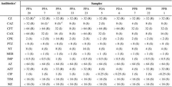
Table 2 - MIC values (µg/mL) of the 156 samples of Pseudomonas spp. isolated from the surfaces of wash basin around the sink (PP), wash basin distant from sink (PD) of dental offices before (A) and after dental surgical procedures (B) of dental offices.
aAntibiotics: cefazolin (CZ), ceftazidime (CAZ), cefotaxime (CFT), ceftriaxone (CAX), cefepime (CPE), piperacillin plus tazobactam (PTZ), netilmicin
(NT), meropenem (MER), imipenem (IMP), azlocillin (AZ), aztreonam (AZT), ciprofloxacin (CIP), ticarcillin plus clavulanic acid (TIM) and mezlocillin (MZ). b (R): resistant; c (I): intermediate; d (S): sensible.
Table 1 - Identification of Pseudomonas spp. isolated from the surfaces of wash basin around sink (PP), wash basin distant sink (PD) of dental offices before (A) and after dental surgical procedures (B).
Samples/Isolates Microorganisms PPA/20 Pseudomonas aeruginosa
PPA/7 Pseudomonas aeruginosa
PPA/32 Pseudomonas aeruginosa
PPA/33 Pseudomonas aeruginosa
PPA/23 Pseudomonas stutzeri
PPB/7 Pseudomonas stutzeri
PPB/22 Pseudomonas aeruginosa
PPB/1 Pseudomonas aeruginosa
PDA/5 Pseudomonas aeruginosa
PDA/6 Pseudomonas stutzeri
Samples Antibioticsa
PPA 20
PPA 7
PPA 32
PPA 33
PPA 23
PDA 5
PDA 6
PPB 7
PPB 22
PPB 1
CZ > 32 (R) b > 32 (R) > 32 (R) > 32 (R) > 32 (R) > 32 (R) > 32 (R) > 32 (R) > 32 (R) > 32 (R) CAZ > 32 (R) 16 (I) c 8 (S) d 8 (S) 8 (S) 2 (S) 8 (S) 4 (S) 8 (S) 8 (S)
CFT > 64 (R) > 64 (R) 32 (I) 8 (S) > 64 (R) > 64 (R) > 64 (R) 32 (I) 32 (I) 32 (I)
CAX > 64 (R) 32 (I) 16 (S) 8 (S) > 64 (R) 32 (I) 8 (S) 8 (S) 8 (S) 16 (S)
CPE 2 (S) < 2 (S) > 16 (R) 2 (S) 2 (S) < 2 (S) < 2 (S) 2 (S) < 2 (S) < 2 (S)
PTZ < 8 (S) < 8 (S) < 8 (S) < 8 (S) < 8 (S) < 8 (S) < 8 (S) < 8 (S) < 8 (S) < 8 (S)
NT 8 (S) 4 (S) 8 (S) 4 (S) 16 (I) 4 (S) 4 (S) 8 (S) 4 (S) 4 (S)
MER < 1 (S) < 1 (S) < 1 (S) < 1 (S) < 1 (S) < 1 (S) < 1 (S) < 1 (S) < 1 (S) < 1 (S)
IMP < 0.5 (S) < 0.5 (S) 1 (S) 1 (S) < 0.5 (S) < 0.5 (S) < 0.5 (S) 1 (S) < 0.5 (S) < 0.5 (S)
AZ < 64 (S) < 64 (S) < 64 (S) < 64 (S) < 64 (S) < 64 (S) < 64 (S) < 64 (S) < 64 (S) < 64 (S)
AZT > 32 (R) 4 (S) > 32 (R) 4 (S) > 32 (R) 4 (S) 4 (S) 4 (S) > 32 (R) > 32 (R)
CIP 1 (S) 1 (S) 1 (S) 1 (S) 1 (S) < 0.25 (S) < 0.25 (S) 1 (S) 1 (S) <0.25 (S)
TIM < 16 (S) < 16 (S) < 16 (S) < 16 (S) < 16 (S) < 16 (S) < 16 (S) < 16 (S) < 16 (S) < 16 (S)
MZ < 16 (S) < 16 (S) < 16 (S) < 16 (S) < 16 (S) < 16 (S) < 16 (S) < 16 (S) < 16 (S) < 16 (S) ceftriaxone/2-64 (CAX), cefepime/2-16 (CPE), piperacillin/
8-64 plus tazobactam/4 (PTZ), netilmicin/2-16 (NT), meropenem/1-8 (MER), imipenem/0.5-16 (IMP), azlocillin/ 64 (AZ), aztreonam/1-32 (AZT), ciprofloxacin/0.25-4 (CIP), ticarcillin/16-128 plus clavulanic acid/2 (TIM) and mezlocillin/16-128 (MZ). The results were classified into susceptible, intermediate or resistant, as per CLSI (Clinical and Laboratory Standard Institute) guidelines (Balows et al. 1991; NCCLS, 2000).
RESULTS
In this study, 1246 isolates were taken from the surfaces around wash-basins, close to and distant from the basins, in dental surgeries, before and after surgical procedures. The most prevalent group of bacteria found were the staphylococci (34.66%), followed by non-fermentative Gram-negative bacilli, contributing 30.12%.
The incidence of Pseudomonas spp. was 40.09% of the total Gram-negative bacilli, 145 isolates (92.94%) being collected around the basins and 7.05% (11 isolates) distant from them. (Table 1).
Given the pathogenic potential of this non-fermentative Gram-negative bacillus, some Pseudomonas strains were tested for in vitro susceptibility. The samples tested were selected on the basis of their highly variable susceptibility. Fifty percent of all antibiotics tested inhibited all Pseudomonas spp. strains. Third and fourth generation cephalosporins possessed lower activity against the isolates, which showed between 17.30 and 79.48% of resistance. The
general analysis of susceptibility profiles, carried out as recommended in M7-A5/M100-S10 (NCCLS, 2000) showed that 18.77% of strains were resistant to all tested drugs, and of 156 tests performed, 23.53% of antimicrobial agents were ineffective. To the monobactam, aztreonam, 62.82% of environmental isolates showed resistance and to cefotaxime and cefazolin the percentages were 39.10 and 100% of resistance, respectively. The drugs with high activity against all the environmental isolates of
Pseudomonas spp. were meropenem, imipenem,
147
Susceptibility profile of Pseudomonas
DISCUSSION
The transmission of infection during dental procedures can occur by direct contact with tissues and secretions or blood, by aerosols containing infectious agents and on the cutting edge of contamined instruments. The goal of our study was to demonstrate the importance of the evaluation of the presence of microorganisms in the dental surgery environment. The results showed that the isolated microorganisms were positive cocci and Gram-negative bacilli, among which the most prevalent species in each group were S. aureus and Pseudomonas spp, respectively. Pathogens such as P. aeruginosa and P. stutzeri have been isolated from some areas of dental consulting-rooms, suggesting a risk of infection associated with oral dissemination (Barbeau et al., 1998; Barbeau, 2000; Jensen et al., 1997; O’Donnell et al., 2005). In a microbiological study of selected risk areas in dental technology laboratories,
Staphylococcus spp. were most commonly isolated from
curing water baths and from air (Verran et al., 1996). In another study, bacteria were isolated from pumice slurry, the major contaminants being Pseudomonas spp.,
Staphylococcus spp. and Bacillus spp (Verran et al., 1997),
the same agents that were found in our study.
P. aeruginosa continues to be a major pathogen in
the surgical environment because it is the most prevalent species in the water used (Paviani et al., 2004) and one of the most important bacterial pathogens in cystic fibrosis-associated lung disease (Chambers et al., 2005). In the environment of the dental surgery this agent can infect dental equipment. Patients with cystic fibrosis often suffer from P. aeruginosa lung infection and although the source of the organism is not known there is a risk of contamination from dental equipment, because strains have been found both in water taken from dental equipment and clinical isolates. Additionally one case of genotypically-identified
Pseudomonas was acquired in dental sessions (Jensen et
al., 1997).
P. aeruginosa is naturally resistant to many
antibiotics (Tadeu et al., 2000) and only a few antibiotics are effective against Pseudomonas, including fluoroquinolone, gentamicin and imipenem, and even these antibiotics are not effective against all strains. In a study, Pitten et al. (2001) demonstrated resistance of P. aeruginosa to beta-lactam antibiotics including carbapenens, aztreonam, aminoglycosides and quinolones and in vitro susceptibility only to polymyxin B, in clinical and environmental samples isolated from hospital infection episode. The detection of clusters of beta-lactamases that hydrolyze broad-spectrum beta-lactams has received great attention in the combatting of cross-infection because the environmental dissemination of such agents can lead to the transference of the metallo-beta-lactamase gene among P.
aeruginosa and other Gram-negative bacilli (Panzig et al.,
1999; Tsuji et al., 2005). Our results for the environmental isolates showed similar percentages to clinical isolates in the study of Cavallo et al. (2000) supporting the hypothesis
that part of isolates of the Pseudomonas spp contamination could occur during the surgical procedures, when aerosols would be disseminated to places nearby, carrying mainly the resistant strains. Similarly, a high percentage of resistance to aztreonam was detected, and this is an optional drug alternative in therapy that involves cephalosporins (Somekh & Cordova, 2000). Furthermore, P. aeruginosa has shown resistance to other antibiotics, such as third generation cephalosporins (CAZ and CFT), as demonstrated in a study of clinical isolates (Panzig et al., 1999; Pitten et al., 2001; Blandino et al., 2004). In the present study, the results suggests that the resistance cannot be associated with areas of collection because the samples were from several dental surgeries located in different places and the same considerations apply to the surgical procedures. Interestingly, P. stutzeri was isolated near to and distant from the wash basin, with different susceptibility profile and resistance to CAX and AZT, suggesting the possibility that the strains were from surgical aerosols. The knowledge of the susceptibility profile of given bacterial groups will be important for an understanding not only of the dissemination of these agents as sources of infection but also of the ecology of this organism in the dental surgery environment. This study suggests that special attention should be paid to these areas, using, for example, hospital disinfection methods, to weaken the chain of cross-infection.
ACKNOWLEDGEMENTS
This work was supported by University of Ribeirão Preto – UNAERP.
RESUMO
Análise do perfil de susceptibilidade de Pseudomonas spp. e da prevalência de bactérias isoladas de superfícies
de consultórios odontológicos
148
Susceptibility profile of Pseudomonas
cefalosporinas de terceira geração foi variável.
Palavras-chave: Pseudomonas, susceptibilidade,
consultórios odontológicos, P. aeruginosa, P. stutzeri.
REFERENCES
Agarwal G, Kapil A, Kabra SK, Das BK, Dwivedi, N. Characterization of Pseudomonas aeruginosa isolated from chronically infected children with cystic fibrosis in India.
BMC Microbiology2005; 5:43. Avaiable at: http://
www.biomedcentral.com/1471-2180/5/43. Accessed August, 15, 2005.
Balows A, Hausler WJ, Hermann KL, Isenberg HD, Shadamytt, J. Manual of clinical microbiology. 5th ed.
Washington (DC): American Society for Microbiology, 1991.
Barbeau J, Ten Bokum L, Gauthier C, Prevost, AP. Cross contamination potential of saliva ejectors used in dentistry.
J Hosp Infect 1998; 40:303-11.
Barbeau J. Waterborne biofilms and dentistry: the changing face of infection control. J Can Dent Assoc 2000; 66:539-41.
Blandino G, Marchese A, Ardito F, Fadda G, Fontana R, Lo Cascio G, Marchetti F, Schito GC, Nicoletti G. Antimicrobial susceptibility profiles of Pseudomonas aeruginosa and
Staphylococcus aureus isolated in Italy from patients with
hospital-acquired infections. Intern J Antimicrob Agents 2004; 24:515-18.
Bodey GP, Bolivar R, Fainstein V, Jadeja L. Infections caused by Pseudomonas aeruginosa. Rev Infect Dis 1983; 5 (2):279-313.
Brasil. Ministério da Saúde, Secretaria Nacional de Assistência à Saúde. Manual de procedimentos básicos em microbiologia clínica para o controle de infecção hospitalar. Brasília: Divisão de Controle Hospitalar/SNAS; 1991. Cavallo JD, Leblanc F, Fabre R. Surveillance of
Pseudomonas aeruginosa sensitivity to antibiotics in France
and distribution of beta-lactam resistance mechanisms: 1998 GERPB study. Pathol Biol 2000; 48 (5):472-7.
Chambers D, Scott F, Bangur R, Davies R, Lim A, Walters S, Smith G, Pitt T, Stableforth D, Honeybourne D. Factors associated with infection by Pseudomonas aeruginosa in adult cystic fibrosis. Eur Respir J 2005; 26(4):651-6. Costerton JW, Stewart PS, Greenberg EP. Bacterial biofilms: a common cause of persistent infections. Science1999; 284 (5418):1318-22.
Hardalo C, Edberg SC. Pseudomonas aeruginosa: assessment of risk from drinking water. Crit
RevMicrobiol1997; 23 (1):47-75.
Jensen ET, Giwercman B, Ojeniyi B, Bangsborg JM, Hansen A, Koch C, Fiehn NE, Hoiby N. Epidemiology of
Pseudomonas aeruginosa in cystic fibrosis and the possible
role of contamination by dental equipment. J Hosp Infect 1997; 36 (2):117-22.
Moss RB. Cystic fibrosis: pathogenesis, pulmonary infection and treatment. Clin Infect Dis 1995; 21 (4):839-49. National Committee For Clinical Laboratory Standards. Methods for dilution antimicrobial susceptibility tests for bacteria that grow aerobically, 4th. ed.: approved standard,
M7-A5/M100-S10. Villanova, Pennsylvania: National Committee for Clinical Laboratory Standards; 2000. O’Donnell MJ, Tuttlebee CM, Falkiner FR, Coleman D.C. Bacterial contamination of dental chair units in a modern dental hospital caused by leakage from suction system hoses containing extensive biofilm. J Hosp Infect 2005; 59(4):348-60.
Panzig B, Schröder G, Pitten FA, Grüdling M. A large outbreak of multiresistant Pseudomonas aeruginosa strains in northeastern Germany. J Antimicrob Chemother1999; 43:415-8.
Paviani ER, Stadnik CB, Heinek I. Estudo da epidemiologia e perfil de sensibilidade da Pseudomonas aeruginosa.
Infarma 2004; 15: 66-70.
Pitten FA, Panzig B, Schröder G, Tietze K, Kramer A. Transmission of a multiresistant Pseudomonas aeruginosa strain at a German University Hospital. J Hosp Infect 2001; 47:125-30.
Somekh E, Cordova Z. Ceftazidime versus aztreonam in the treatment of pseudomonal chronic suppurative otitis media in children. Scand J Infect Dis2000; 32:197-9. Tadeu AF, Fernandes MLV, Ribeiro NF. Infecção hospitalar e suas interfaces na área de saúde. São Paulo: Editora Atheneu; v.2, 2000.
Tsuji A, Kobayashi I, Oguri T, Inoue M, Yabuuchi E, Goto S. An epidemiological study of the susceptibility and frequency of multiple-drug-resistant strains of Pseudomonas
aeruginosa isolated at medical institutes nationwide in
Japan. J Infect Chemother 2005; 11:64-70.
Verran J, Kossar, S Mccord JF. Microbiological study of selected risk areas in dental technology laboratories. J Dent 1996; 24:77-80.
Verran J, Winder C, Mccord JF, Maryan CJ. Pumice slurry as a crossinfection hazard in nonclinical (teaching) dental technology laboratories. Inst J