Bacterial translocation in rats nonfunctioning diverted distal colon
1Translocação bacteriana no coto colônico distal desfuncionalizado de ratos
Francisco Edilson Leite Pinto Júnior2, Carlos Teixeira Brandt3, Aldo da Cunha Medeiros4, Ariano José Freitas de
Oliveira5, Selma Maria Jerônimo6, Helena Marques Fonseca de Brito7
1. Research performed at Federal University of Pernambuco (UFPE), Brazil.
2. PhD, Associate Professor of Surgery, Federal University of Rio Grande do Norte (UFRN), Brazil. 3. Head Professor, Pediatric Surgery, UFPE, Brazil.
4. Head Professor, Surgery, UFRN, Brazil. 5. Fellow PhD degree, UFRN, Brazil.
6. PhD, Associate Professor of Biochemistry, UFRN, Brazil. 7. Microbiology Specialist, Rio Grande do Norte, Brazil.
ABSTRACT
Purpose:To investigate whether the alterations of the diverted colon segment mucosa, evidenced in fecal colitis, would be able to alter Bacterial Translocation (BT). Methods: Sixty-two Wistar male rats ranging from 220 to 320 grams of weight, were divided in two groups: A (Colostomy) and B (Control), with 31 animals each one. In group A, all animals underwent end colostomy, one stoma, in ascending colon; and in the 70th POD was injected in five rats, by rectal route –
diverted segment - 2ml of a 0.9% saline solution in animals (A1 subgroup); in eight it was inoculated, by rectal route, 2ml
of a solution containing Escherichia coli ATCC 25922 (American Type Culture Collection), in a concentration of 108
Colony Forming Unit for milliliters (CFU/ml) - A2 Subgroup; in ten animals the same solution of E. coli was inoculated, in a concentration of 1011 CFU/ml (A3 Subgroup); and in eight it was collected part of the mucus found in the diverted
distal colonic segment for neutral sugars and total proteins dosage (A4 subgroup). The animals from the group B underwent the same procedures of group A, but with differences in the colostomy confection. In rats from subgroups A1, A2, A3, B1, B2, and B3 2ml of blood were aspirated from the heart, and fragments from mesenteric lymphatic nodule, liver, spleen, lung and kidney taken for microbiological analysis, after their death. This analysis consisted of evidencing the presence of E. coli ATCC 25922 CFU. Mann-Whitney and ANOVA Tests were applied as analytic techniques for association of variables. Results:The occurrence of BT was evidenced only in those animals in which inoculated concentration of E.
coli ATCC 25922, reached levels of 1011CFU/ml, i.e. in Subgroups A3 and B3, although, being significantly greater
(80%) in those animals without colostomy (subgroup B3) when compared to the ones with colostomy (20%) from the subgroup A3 (P <0.05). Lung, liver and mesenteric lymphatic nodules were the tissues with larger percentile of bacterial recovery, so much in subgroup A3, as in B3. Blood culture was considered positive in 60% of the animals from subgroup B3 and in 10% of those from subgroup A3 (p <0.05). There was greater concentration of neutral sugars, in subgroup A4 - mean 27.3mg/ml -, than in subgroup B4 - mean 8.4mg/ml - (P <0.05). Conclusion: The modifications in the architecture of intestinal mucosa in colitis following fecal diversion can cause alterations in the intestinal barrier, but it does not necessarily lead to an increased frequency of BT.
Key words: Bacterial Translocation. Colitis. Rats, Wistar.
RESUMO
Objetivo: Investigar se as alterações do cólon desfuncionalizado, evidenciadas na colite de derivação fecal, seriam capazes de permitir Translocação Bacteriana (TB), ou se a mucosa intestinal atrofiada permitiria a passagem de bactérias para órgãos à distância. Métodos: Foram utilizados 62 ratos Wistar, machos, pesando entre 220 e 320 gramas, divididos em dois grupos: A (Colostomia) e B (Controle), contendo cada um 31 animais. No grupo A, os animais foram submetidos à colostomia, terminal boca única, em cólon ascendente. A partir do 70º dia de observação os seguintes procedimentos foram adotados: em cinco ratos foi injetado por via retal – no segmento desfuncionalizado - 2ml de uma solução salina
0,9% nos animais (subgrupo A1); em oito inoculou-se, por via retal, uma solução de 2ml contendo Escherichia coli
ATCC 25922 (American Type Culture Collection), na concentração de 108 Unidades Formadoras de Colônias por mililitros
(UFC/ml) - Subgrupo A2; em dez animais inoculava-se a mesma solução de E. coli, na concentração de 1011UFC/ml
(Subgrupo A3); e em oito colhia-se o muco do segmento colônico distal desfuncionalizado, para dosagens de açúcares neutros e proteínas totais (subgrupo A4). Os animais do grupo B foram submetidos aos mesmos procedimentos do grupo A, e não foram submetidos à colostomia. Nos animais dos subgrupos A1, A2, A3, B1, B2, e B3, após serem mortos,
realizou-se punção cardíaca para coleta de 2ml de sangue e retiraram-se fragmentos de tecidos de linfonodo do mesocólon, fígado, baço, pulmão e rim, para análise microbiológica. Essa análise consistia em evidenciar a presença de UFC de
Escherichia coli ATCC 25922. Os testes de Mann-Whitney e Anova foram aplicados como técnicas investigativas para associação das variáveis. Resultados: A ocorrência de TB, só foi evidenciada nos animais em que a concentração inoculada de Escherichia coli ATCC 25922, atingia níveis de 1011UFC/ml, ou seja, nos Subgrupos A3 e B3, no entanto, sendo
significantemente superior (80%) nos animais sem colostomia (subgrupo B3) quando comparados com os ratos com colostomia (20%) do subgrupo A3 (P<0,05). Pulmão, fígado e linfonodo foram os tecidos com maior percentual de recuperação bacteriana, tanto no subgrupo A3, quanto no B3. A hemocultura foi positiva em 60 % dos animais do subgrupo B3 e em 10% do subgrupo A3 (p<0,05). Houve maior concentração de açúcares neutros, no subgrupo A4 -média 27,3mg/ml -, do que no subgrupo B4 - -média 8,4mg/ml – (P<0,05). Conclusão: As modificações na arquitetura da mucosa intestinal na colite de derivação fecal podem promover alterações na barreira intestinal, mas não necessariamente conduzem a maior freqüência de TB.
Descritores: Translocação Bacteriana. Colite. Ratos Wistar.
Introduction
Bacterial translocation (BT) is the passage of viable intestinal bacteria and endotoxins through the intestine wall for the mesenteric lymphatic nodules, to other organs and systemic circulation1-4. This phenomenon is
associated with bacteremia and septicemia, renal failure and pulmonary dysfunction, which can lead to multiple organ failure, and occasionally death. In the necropsy of some cases the main focus of infection seems to have origin in the bowell2,3. It is possible to prove BT in
approximately 16% of the patients who had undergone laparotomy as septicemia is found in 41% of them.
Escherichia coli is the most prevalent bacteria in those
cases1,4. Alterations on the intestine mucosa barrier, as
well as changes in the micro biota, changes associated with: shock; total parenteral nutrition (TPN); prolonged antibiotic therapy, bowel obstruction; severe burns; use of immune suppressors and cytoxic drugs; and intestinal manipulation during surgical procedure, are considered predisposing factors for development of BT5. In addition
of digesting and absorbing nutrients, the intestinal mucosa works as an important barrier mechanism, which not allow the passage of bacteria and endotoxins, as well as other elements from the bowel tube for places outside the intestine5.Patients who undergo TPN present many
more septic complications as compared with patients who are receiving enteric nutrition. The lack of nutrients in the bowel tube can cause intestine cell malfunction, as well as mucosa atrophy, which can facilitate BT5,6. Colitis
following fecal diversion is a clinical condition found in almost 100% of patients who undergo colostomy and nonfunctioning colon; this surgical procedure leads to atrophy of colon mucosa, which can produce BT7,8. In
some cases when colostomy results in diverted dysfunctional colon colitis following fecal diversion can occur. There are few studies on this condition and none on the possible association with BT. Thus, the purpose of this study was to investigate whether the histology changes in the dysfunctional colon and the colitis following fecal diversion would facilitate the development of BT.
Methods
Sixty-two Wistar male rats were enrolled in the study. Weights ranged from 220 and 320 grams. The rats were kept in the animal house of the Experimental Operating Room “Prof. Travassos Sarinho” -. Department of Surgery - Health Sciences Center – Federal University of the Rio Grande do Norte State (UFRN), Brazil. During the study period the animals were kept in adequate environment and in individual cages with day night cycle of 12 hours. They were fed with standard diet formulation and water ad libido. Strict ethical principles aiming at protecting animals were observed and the experimental protocol was approved by the Ethical Committee – Health Sciences Center-UFRN, Brazil. There was scientific cooperation from the Laboratory of Biochemistry – Health Sciences Center (UFRN), and also from the Laboratory of Microbiology – Center of Clinical Pathology, Natal Capital City of
Rio Grande do Norte State, Brazil. Study Protocol: The
animals were divided in two groups: A and B, each including 31 rats. The animals from A group -, called colostomy or study group, as well as the animals in group B (control) were stratified in four subgroups:
Subgroup A1 (colostomy + inoculation of 0.9% saline) - There were five rats in A1 subgroup. They underwent diverted colostomy as described in surgical technique. In the 70th postoperative day (POD), 2ml of
0.9 saline were infused in the colon via rectum. After 24 hours the animals were killed for taking blood and tissue samples, which were sent for microbiology study.
Subgroup A2 (colostomy + Escherichia coli
inoculation at 108 concentration) - There were eight rats
in A2 subgroup. They underwent diverted colostomy as described in surgical technique. In the 70th POD, 2ml
suspension of Escherichia coli ATCC 25922 (American Type Culture Collection), in the concentration of 108UFC/
Subgroup A3 (colostomy + inoculation of
Escherichia coli suspension with the concentration of
1011) - There were eight rats in A3 subgroup; and they
underwent the same protocol as in subgroup A2, however the concentration of Escherichia coli suspension was 1011UFC/ml.
Subgroup A4 (colostomy + mucous analyses)
-There were eight rats in A4 subgroup. They underwent the same surgical procedure. In the 70th POD mucous
was collected from the distal dysfunctional colon for measurements of neutral sugars and total protein.
Subgroups B1 to B4 - The rats of these subgroups B (control) did not undergo colostomy; however all the other procedures performed in the animal from subgroups A were followed.
Preparing the Escherichia coli suspension
Escherichia coli ATCC 25922 (American Type
Culture Collection) was cultivated in sugar cane juice supplemented with yeast extract and peptone until the growing exponential phase in 16 hours, at the Laboratory of Microbiology – Center of Clinical Pathology, Natal Capital City of Rio Grande do Norte
State, Brazil. The bacteria suspensions were washed three times with saline buffer solution at pH 7.2, and centrifuged at 1,700rpm for 10 minutes. The bacteria concentrates were made in suspension again using buffer phosphate. The final concentrations from 108 to 1011
UFC/ml were adjusted using spectrometry9.
Surgical technique
The animals of group A (experimental) were kept fasted (just drinking pure water) for 12 hours before the surgical procedure. After anesthesia with ether inhalation the animal underwent colon clean-up per rectum with, aiming to withdraw all fecal material from the colon. The rats were weighted and fixed at the operating table. Abdominal shaving was performed, followed by antisepsis with topic solution of povidine iodine (PVPI). An approximately 4cm midline laparotomy was performed for identification of the ascending colon. About 2cm distal to the ileo cecal valve the colon was sectioned with previous ligature with 2-0 silk, closing the two ends of the colon. The distal segment of the colon was kept inside the abdomen and the proximal was brought up through the abdominal wall, left to the incision; early maturation of the colostomy was performed. The abdominal cavity was kept under irrigation with saline throughout the operative period, for avoiding that the bowel turn out dry. The wound was closed with 4-0 nylon stitches. The rats were observed during the postoperative period in individual cages. Diet
was normal. The animals from subgroups A1, A2 and A3, in the 70th POD, underwent ether anesthesia for
receiving rectal infusion of: 2ml of saline (subgroup A1); 2ml of Escherichia coli suspension at concentration of 108UFC/ml (subgroup A2); and 1011UFC/ml (subgroup
A3). After 24 hours the rats were killed with an overdose of ether for obtaining blood and tissue samples for microbiology. The animals from subgroup A4 were also killed in the 70th POD for obtaining mucous from the distal nonfunctioning colon for measurements of neutral sugars and total protein.
Obtaining samples
All animals from subgroups A1, A2, A3, B1, B2
and B3, after being killed, had their abdomen and thorax surgically sterile opened for collecting tissue samples, which were sent for microbiology. Heart punch was performed for collecting 2ml of blood, which was also sent for microbiology Tissue fragments were taken in the following order: mesenteric lymphatic nodule from the nonfunctioning colon segment (subgroups A1, A2,
and A3), or descending colon (subgroups B1, B2 and
B3); and fragments from left liver lobe, spleen, inferior pole of kidney and inferior lobe of the left lung. All tissue fragments were taken using individually sterile specific scissors and forceps. The samples were conditioned in sterile tubes with 1ml of sterile saline. The tissue fragments were carefully electronically weighted.
Obtaining mucus
The animals from subgroups A4 and B4, after being killed, had the nonfunctioning colon removed in subgroup A4, and also the descending colon in subgroup
B4. After opening these colon segments the inside mucus were collected with sterile instruments and conditioned in Eppendorf tubes with phosphate buffer solution, pH 7.4.
Microbiology
All tissue samples were sterile grained and diluted in a 2ml saline tube, under sterilized closed system in a laminar cabinet. Sample solutions were inoculated in Petri dishes containing agar MacConkey medium, selective for gram negative bacteria growth. Escherichia coli colonies were identified by their red and pink color and numbered. Positive samples were identified when presented at least 1UFC. If there was suspected contamination in any animal, it was automatically changed for other one. Phenotypic identification of
Escherichia coli ATCC 25922 (American Type Culture
It can also identify control strain. The blood samples were inoculated in HEMOCULT I system, which were inspected daily for observing bacteria growth.
Neutral sugar and total protein analysis of the mucous
Neutral sugar concentrations from the intestine mucous were assessed by the Yemm e Wlllis10 method
-500µ l of water was added to 10µ l sample and 2.5ml of antrona (reagent). The tube was stirred and sealed with aluminum paper. The final solution was warmed at 100
0C for a period of 15 minutes. After 10 minutes with a
solution cooled the concentration readings were performed using a photo colorimetric dosage at 620nm. Total protein concentration from the intestine mucous was measured by the bicincronic acid (BCA), using the Smith method11.
Statistical analysis
Mann-Whitney and ANOVA variance were used for statistical analysis. The software Statistics version 5.0 was used. p<0.05 was used for rejecting the null hypothesis.
Results
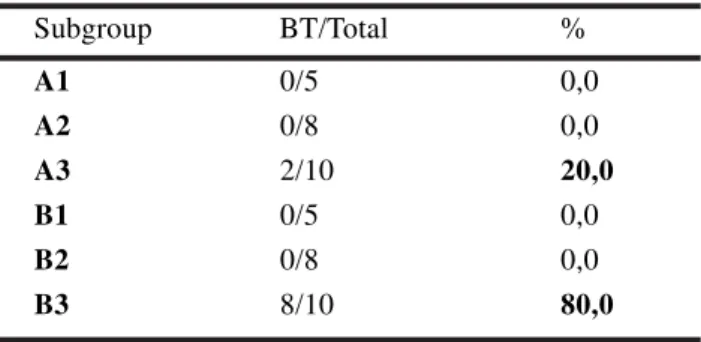
Six animals were substituted, with no loss for the research; four of them were excluded due to a suspicion of external contamination, being verified the presence of Agro bacterium tumefaciens and two were also excluded because they died during the experiment due to intestinal obstruction, secondary to colostomy stenosis. In Table 1 one can observe the descriptive statistics of the frequency for bacterial translocation, which was registered only in the animals submitted to inoculation of E. coli in a concentration of 1011 (A3 and
B3 subgroups). It is observed that 80% of the animals that were not submitted to the colostomy, presented bacterial translocation, while, the rats submitted to the procedure, the registration decreased to 20% of the analyzed animals. It can be observed, in Table 2, that the animals that underwent colostomy presented a greater frequency of bacterial recovery (PBR) in the organs and tissues analyzed, showing differences statistically significant between the groups A3 and B3 (p <0.05; ANOVA). PRB of the kidney was observed in the totality, only in the rats submitted to E. coli 1011 without
colostomy (subgroup B3). In the animals that underwent colostomy there was no indication of bacterial recovery in that organ. Escherichia coli ATTCC were positively found in the blood of 10% from the rats in subgroup A3
and in 60% from the blood of rats in subgroup B3. It was observed a greater concentration of neutral sugars
(Figure 1) and total proteins (Figure 2) in the mucous of the animals that underwent colostomy, A4 and B4, (P <0.05).
TABLE 1 - Frequency of bacterial translocation (BT) by subgroups
Subgroup BT/Total %
A1 0/5 0,0
A2 0/8 0,0
A3 2/10 20,0
B1 0/5 0,0
B2 0/8 0,0
B3 8/10 80,0
TABLE 2 - Frequency of bacterial recovery from the organs and tissues analyzed in the animals of A3 and B3 subgroups
Subgroups
A3 B3
Organs % %
Lung 20 80
Spleen 10 40
Liver 20 80
Kidney 0 40
Linphnode 20 70
FIGURE 1 - Concentration of neutral sugars for A4 and B4 subgroups (mg/ml)
Discussion
Although there has been progress in surgery, the control of infection remains a great challenge. From 593,344 surgical operations performed in the United States from 1986 to 1996, 15,525 (3%) were followed by infection. From 551 infected patients who died 77% of the deaths were due to severe infection that had led to multiple organ failure (MOF)12. Since MOF was
recognized it has been one of the most frequent causes of mortality in surgical patients, especially in those cared for in Intensive Care Unit (ICU)13. Several insults
including: trauma; liver resections; pancreatitis; and shock, can cause MOF, which once present may not respond to treatment, especially when the basic cause can no be overcome13. There has been, in the last years,
an increase in the number of publications with focus on BT9,13-19. The experimental models represent a good tool
for studying the genesis of diseases and research of new therapeutic techniques8. Among the studies, those related
to total parenteral nutrition (TPN) as cause of BT20,21. It
is known that TPN promotes atrophy and increases permeability of the bowel mucosa, which can promote impairment of the intestinal mucosal barrier, and consequently increase in the rate of infection complication. The lack of nutrients in the bowel tube, apart from promoting decrease of the enterocyte immune protection it produces changes in the normal intestinal micro biota, which together can cause BT. They also produce a decrease in the s-IgA production9,13. Rats were
chosen for the present investigation because it’s low cost and the easy management of these animals. They also present similarity of the bowel mucosa histology with that of human, as well as for being frequently used as animal model for studying BT2,6,9,13,15. Taking into
consideration the atrophic effect of nonfunctioning diverted colostomy, it points out to the importance of the bowel tube content in the genesis of derivative colitis (DC)7,8; since it is installed there are conditions for BT,
similar to those of patients in TPN. These are the reasons because the option for this research method was taken. It is known that experimental DC is associated with a decrease of the colon mucosal thickness, after ten days, in rats that underwent colostomy, and it decreases further in the 70th post-operative day8. Furthermore, it was
observed that in DC that in the nonfunctioning colon segment, the micro biota suffered quantitative and qualitative changes. According with Neut et al22, the
phenomenon is similar in humans, and the main changes is reduction of anaerobic bacteria content; interestingly the aerobic bacteria content did not undergo alterations. Since there is a decrease in anaerobic bacteria content, immune mechanism resistance is altered, and the enterocyte ability against bacteria is impaired, affecting the intestinal barrier, and facilitating BT6. Lack of bile
salts in the gastrointestinal tube can also produce similar
effect9,18,23. One can not forget that, especially in rats,
the s-IgA is almost in the bile. When derivative colitis occurs in nonfunctioning colon, there is no bile salts, and consequently decrease in s-IgA concentration affecting the immune system of this colon segment. The bowel motility involving the migratory motor complex (MMC) is implicated in the regulatory mechanism avoiding bacteria growth. Scoot et al24 observed in rats
that the high dose of morphine produces impairment of intestinal MMC promoting BT. It is also known after colostomy that there is decrease in normal peristaltic movements in the distal non functioning colon segment. These waves works as a true intestine washout, and when they are altered there is a change in the peristaltic threshold facilitating fecal stasis, and consequently increasing the probability of bacteria penetrate in the mucous barrier producing epithelial adherence13. For all
these alterations found in nonfunctioning colon segment in DC, they facilitate BT by altering the colon environment. The majority of papers about BT are based on small bowel as origin of this phenomenon. This is one of the reasons because colon in rat was chosen as subject of the present investigation. Up to the time of this study there was only one experimental investigation on this subject -, Koh et al2 experimentally studied BT
process in the large bowel. They found, using different concentrations (2x10⁵; 2x10⁸; 2x10¹¹) of E.coli R6
inoculated in rats, both in the small and large bowel, that BT could be produced. However, they stressed that BT occurred only when the concentration was 2x10¹¹.
Similar to the present study there was BT only in subgroups A3 and B3; in these subgroups the Escherichia coli concentrations were also 2x10¹¹. The results are in
accordance with the study of Cruz et al, who demonstrate in vitro that the BT process is concentration dependent, which means that the greater bacteria concentration is associated with grater possibility of BT25. In
experimental studies2,6,9,13,15, E. coli is the bacteria more
frequently found in the BT process -, up to 91% found in organs and tissues outside the bowel. The anaerobic bacteria rarely produces BT. This fact is the reason because E. colli was the choice for this investigation. There is a predisposition of bacteria adherence to the distal intestine tube, which is related to the abundance of receptors13 in the edge of colon mucosa through the
specific lecithin expression; normally present in the epithelial cells of the host20. The identification of E. coli
ATCC 25922, in the present study, was done using the Vitelí bioMérieux method, which is considered of low cost, easy and trusted with sensibility and specificity greater than 98%; in all dishes where results were similar to the inoculated bacteria the phenotypic features were tested and proved to be ATCC 25922 type. Furthermore, the absence of bacteria in tissue samples from rats that receives saline per rectum infusion (subgroups A1 and
It is important to stress that the assessment of BT using microbiology investigations can underestimated the phenomenon. Kane et al observed that the frequency of positive blood culture in patients who had undergone surgery increased from 14%, as routine blood culture was the tool for investigation, to 64% when bacteria DNA identification using Polymerase Chain Reaction (PCR) was used26. However, this is an expensive and complex
method, not available in all research institutions. Taking into account the microbiology method had interfered in the results of the present investigation, underestimating the frequency of BT, it does not undermine the analytical component of the study, since the rats from groups A and B underwent the same protocol investigation; thus, if it was the case the error would be in both groups. The significant difference between subgroups A3 and B3 -, as regard to PRB gives support to the main question f the investigation: whether the atrophy of colon mucosa evident in DC would facilitate the development of BT. From the results of this investigation one can not conclude that there was association of con mucosa atrophy and BT. The first step for BT from the bowel is bacteria adhesion to the epithelium surface. This cell property is important in determining the virulence of the majority of pathogenic bacteria9. However for adhesion
is necessary that the bacteria penetrate the intestine mucus inner part. This important defense was emphasized by Florey27, in 1933. He mentioned that the mucus layer would
act as a barrier mechanism, slowing down or avoiding that bacteria from the bowel tube could grow in the epithelial surface. There are several primary components that are part of this intestine mucus layer: mucin; neutral sugars; and total proteins, among others, which in decreasing would provide the environment for developing BT. These components increases the mucus viscosity and elasticity, facilitating the enteric bacteria union with immunoglobulin -, especially with s-IgA, thus, blocking the bacteria adhesion with the receptors of the epithelial cells, preventing mucosa colonization; and by consequence avoiding BT17. This fact
was proved by Katayama et al28, in 1997. They proved
experimentally that there were endotoxin high levels in the blood stream from rats that presented significant decrease in mucin of the bowel mucus. Neutral sugar and total protein concentrations were evaluated in the present study, either in animals with (subgroup A4) and without (subgroup B4) colostomy. The results point out for greater mucus concentration for both neutral sugars and total proteins in the rats with nonfunctioning colon (subgroup A4). It means the mucus turned out thicker and with more viscosity in the nonfunctioning colon, when compared with that from normal colon. This fact was observed when mucus was collected. There was a clear difference in mucus viscosity from the subgroups A4 and B4. It is likely that this difference was one of the factors responsible for the low BT in rats from group A when compared with animals from group B.
Conclusion
Alterations in the colon mucosa architecture from rats with colitis following fecal diversion may promote changes in bowel barrier. However they are not necessarily associated with greater bacterial translocation.
References
1. MacFie J. Bacterial translocation in surgical patients. Ann R Coll Surg Engl. 1997; 79 (3): 183 - 9.
2. Koh IHJ, Guatelli EFS, Montero, R, Keller MHG, Silva S, Goldenberg S, et al. Where is the site of bacterial translocation- smal or large bowel? Transplant Proc. 1996; 28 (5): 2661.
3. Deitch EA. Multiple organ failure. Ann Surg. 1992; 216: 127-34.
4. Sedman PC, MacFie J, Sagar P. The prevalence of gut translocation in humans. Gastroenterology. 1994; 107: 643- 9.
5. Fink MP. Effect of critical illness on microbial translocation and gastrointestinal mucosa permeability. Seps Respir Infect. 1994; 9 (3): 256-60.
6. Tamayo FG, Valdes LIT, Lopez NM. Bacterial translocation and wasting in stressed mice. Arch Med Res. 1996; 27 (2): 115-21.
7. Pinto Jr. FEL, Medeiros AC, Oliveira AJF, Medeiros KF, Ramos AMO. Colite de derivação fecal. Rev Col Bras Cir. 1999; 26 (3): 185-9.
8. Pinto Jr. FEL, Oliveira AJF, Medeiros KF, Ramos AMO, Ramos CCO, Medeiros AC. Repercussões histopatológicas da colostomia no coto colônico distal desfuncionalizado: estudo experimental em ratos. Rev Col Bras Cir. 1999; 26 (6): 327-33. 9. Rocha Sl. Recuperação em órgãos e tecidos de
Escherichia coli ATCC 25922, inoculada no íleo terminal de ratos com obstrução intestinal [Tese – Doutorado]. Paraná; 1999.
10. Yemm EW, Willis AJ. The estimation of carbohydrates in plant extracts by anthrone. Biochem J. 1954; 57: 508-15.
11. Smith PK. Measurement using bicinchoninic acid. Anal Biochem. 1985; 150: 76-85.
12. Medeiros AC, Aires Neto T, Dantas Filho AM, Pinto Jr FEL, Uchoa RAC, Carvalho MR. Infecção hospitalar em pacientes cirúrgicos de hospital universitário. Acta Cir Bras. 2003; 18(1): 15-8.
13. Castro LS. Translocação bacteriana e peroxidação lipídica na obstrução intestinal em ratos [Tese – Doutorado]. Rio de Janeiro; 1999.
Correspondence:
Francisco Edílson Leite Pinto Junior Rua: Pinto Martins, 1044/302 59014-060 Natal – RN Brazil
How to cite this article
Pinto Jr FEL, Brandt CT, Medeiros AC, Oliveira AJF, Jerônimo SM, Brito HMF. Bacterial translocation in rats nonfunctioning diverted distal colon. Acta Cir Bras. [serial on the Internet] 2007 May-June;22(3). Available from URL: http://www.scielo.br/acb
Nutr Metab Care 1999; 2 (5): 399-404
15. Alencar SSS, Medeiros AC, Brandt CT, Aguiar JLA, Rocha KBF, Silva MP. Translocação de bactérias marcadas com Tc99 na icterícia obstrutiva em ratos.
Acta Cir Bras. 2001; 17 (Suppl 1): 35-8.
16. Alexander JW, Boyce ST, Babcock GF, Gianotti L, Peck MD, Dunn DL et al. The process of microbial translocation. Ann Surg. 1990; 212 (4): 496-512.
17. Deitch EA, Rutan R, Waymack JP. Trauma, Shock and gut translocation. Am Coll of Gastroenterology. 1998; 93(2): 277-8.
18. O’Boyle CJ, MacFie J, Dave K, Sagar OS, Poon P, Mitchell CJ. Alterations in intestinal barrier function do not predispose to translocation of enteric bacteria in gastroenterologic pacients. Nutrition. 1998; 14(4): 358-62.
19. Deitch EA. Bacterial translocation or lymphatic drainage of toxic products from the gut: what is important in human beings? Surgery 2002; 131(3): 241-4.
20. MacFie J. Enteral versus parenteral nutrition: the significance of bacterial translocation and gut-barrier function. Nutrition. 2000; 16(7): 606-11.
21. Kudsk Ka, Croce MA, Fabian TC, Minard G, Tolley EA, Poret A et al. Enteral versus parenteral feeding.
Effects on septic morbidity after blunt and penetrating abdominal trauma. Ann Surg. 1983; 23(4): 503-13.
22. Neut C, Colombel JF, Guillemot F, Cortot A, Gower P, Quandalle P, et al. Impaired bacterial flora in human excluded colon. Gut. 1989; 30 (8): 1094-8.
23. Bailey ME. Endotoxin, bile salts and renal function in obstructive jaundice. Br J Surg. 1976; 63 (2): 774-8.
24. Scott LD, Cahall DL. Influence of the interdigestive myoeletric complex on enteric flora in the rat. Gastroenterology. 1982; 82: 737-45.
25. Cruz N, Lu Q, Alvarez X, Deitch EA. Bacterial translocation is bacterial species dependent: results using the human Caco-2 intestinal cell line. J Trauma. 1994; 36 (5): 612-6.
26. Kane TD, Alexander JW, Johanningman JA. The detection of microbial DNA in the blood. Ann Sur 1998, 227 (1): 1-9.
27. Florey HW. Observations on the functions of mucus and the early stages of bacterial invasion of the intestinal mucosa. J Pathol Bacteriol 1933; 37: 283-9.
28. Katayama M, Xu D, Specian R, Deitch EA. Role of bacterial adherence and the mucus barier on bacterial translocation: effects of protein malnutrition and endotoxin in rats. Ann Surg. 1977; 225 (3): 317-26.
Conflict of interest: none Financial source: none