!UNESP – UNIVERSIDADE ESTADUAL PAULISTA ! FACULDADE DE ODONTOLOGIA DE ARARAQUARA
LIVIA RODRIGUES PERUSSI VALVERDE
ARARAQUARA
2013
!
“Avaliação dos efeitos da terapia
! !!!!!!!!!!
LIVIA RODRIGUES PERUSSI VALVERDE
ARARAQUARA
2013
Tese apresentada ao Programa de Pós-Graduação em Odontologia, Área de Periodontia, da Faculdade de Odontologia de Araraquara, da Universidade Estadual Paulista, para título de Doutor em Odontologia.
Orientadora: Profª. Drª. Rosemary Adriana Chiérici Marcantonio
“Avaliação dos efeitos da terapia
AVALIAÇÃO DOS EFEITOS DA TERAPIA FOTODINÂMICA COM
HIPERICINA NO TRATAMENTO DA DOENÇA PERIODONTAL
INDUZIDA EM RATOS
COMISSÃO JULGADORA
TESE PARA OBTENÇÃO DO GRAU DE DOUTOR
Presidente e Orientador...Profa. Dra. Rosemary Adriana C. Marcantonio
2o examinador...Profa. Dra. Alessandra Nara de Souza Rastelli
3o examinador...Prof. Dr. Joni Augusto Cirelli
4o Examinador...Profa. Dra. Priscila Fernanda de Campos Menezes
5o examinador...Prof. Dr. Giuseppe Alexandre Romito
Dados Curriculares
Livia Rodrigues Perussi Valverde
Nascimento 06 de janeiro de 1983 – São Carlos/SP
Filiação Sergio Perussi Filho Janice Rodrigues Perussi
2003/2006 Curso de Graduação em Odontologia pela Universidade Estadual Paulista “Júlio de Mesquita Filho”, na Faculdade de Odontologia de Araraquara – UNESP.
2007 – 2008 Curso de Especialização em Periodontia pela APCD em convênio com a FOAr, UNESP – Araraquara/SP.
2008 – 2010 Curso de Pós-graduação em Odontologia, Área de Periodontia, Nível de Mestrado, na Faculdade de Odontologia de Araraquara – UNESP.
2010 – presente Curso de Pós-graduação em Odontologia, Área de Periodontia, Nível de Doutorado, na Faculdade de Odontologia de Araraquara – UNESP.
Dedicatória
À minha mãe Janice,
Obrigada, obrigada e obrigada!
Nada disso seria possível sem você!
Lembro de ir ao seu laboratório quando pequena e achar aqueles
aparelhos todos uma loucura! Hoje, ter tido a oportunidade de trabalhar
com você foi uma experiência única e enriquecedora. Minha admiração
por você só aumentou e me fez ver a mulher inteligente, forte, dedicada
e guerreira que você é. Seus ensinamentos e incentivo diários tem sido
estímulo para que eu continue a trilhar meus próprios passos na vida
acadêmica. Você é minha maior referência e meu melhor porto seguro.
Agradecimentos especiais
À Deus, por sempre me guiar e me confortar nos momentos mais difíceis, fazendo acreditar que tudo é possível.
Aos meus pais (Janice e Sergio), não tenho como agradecer todo amor, incentivo, confiança e conforto que sempre me ofereceram. Obrigada por estarem sempre ao meu lado, seja na alegria ou na tristeza, na saúde ou na doença! Devo à vocês tudo que sou hoje. Saibam que sempre serão meus maiores exemplos e tenho muito orgulho de ser sua filha! Amo muito vocês!
Ao meu marido (Guilherme), agradeço por todo apoio, amor, companheirismo e amizade. Depois de tantos dias de Skype (rs), estou muito feliz de finalmente estarmos juntos, caminhando perante os desafios da vida! Amo você!
Ao meu irmão (Eduardo) e à toda minha família, obrigada por estarem sempre ao meu lado torcendo por mim. Em especial à minha avó Dora, por sempre me benzer para tirar a urucubaca!!!!
O meu muito obrigada...por tudo!!!
Durante todos estes anos você foi muito mais do que uma orientadora.
Foi amiga e mãe. Esteve ao meu lado nas minhas conquistas pessoais e
soube me apoiar nos momentos mais difíceis. Depois de tantos anos
trabalhando juntas, confesso que vou sentir muita falta da convivência
do dia-a-dia. No entanto, acredito que a amizade construída ao longo
dos anos permanecerá e isso nos deixará unidas por toda a vida.
Sou eternamente grata pelos seus ensinamentos, mas acima de tudo,
sou grata por Deus ter colocado na minha vida uma pessoa a qual eu
me sinto honrada de ter convivido por tanto tempo.
Agradecimentos
À Faculdade de Odontologia de Araraquara – UNESP, em nome da Profa. Dra. Andréia Afonso Barreto Montandon e sua vice-diretora Profa. Dra. Elaine Maria Sgavioli Massucato.
Ao atual Programa de Pós-Graduação em Odontologia, área de Periodontia da Faculdade de Odontologia de Araraquara – UNESP, em nome do Prof. Mario Tanomaru Filho (coordenador) e do Prof. Dr. Carlos Rossa Junior (vice-coordenador).
À Fundação de Amparo à Pesquisa no Estado de São Paulo (FAPESP) pela concessão da bolsa de estudos (2009/17404-1) e auxílio pesquisa (2010/14932-4), sem os quais não seria possível desenvolver este trabalho.
Aos docentes da disciplina de Periodontia da Faculdade de Odontologia de Araraquara – UNESP: Profa. Dra. Silvana Regina Perez Orrico, Profa. Daniela Leal Zandim Barcelos, Prof. Dr. Joni Augusto Cirelli, Prof. Dr. José Eduardo Cezar Sampaio, Prof. Dr. Elcio Marcantonio Junior e ao Prof. Dr. Carlos Rossa Junior, agradeço por todo aprendizado, paciência, dedicação e amizade. Devo todo meu conhecimento em periodontia à vocês.
À Profa. Dra. Simone Duarte, da Universidade de Nova York (NYU) - EUA, pela oportunidade de ter desenvolvido parte do projeto de doutorado na instituição. Fica aqui minha eterna gratidão pelos conhecimentos adquiridos em seu laboratório.
computadorizada, pela ajuda com os escaneamentos dos animais. Ao Adriano, responsável pela manutenção do biotério desta faculdade, um agradecimento em reconhecimento aos seus esforços pela boa manutenção do biotério e pela sua atenção e cuidados em relação aos animais ali instalados.
À Regina Lúcia, secretária da disciplina de Periodontia desta faculdade, por toda a atenção e ajuda dispensada.
Aos demais funcionários da disciplina de Periodontia desta faculdade, meu muito obrigada. Agradecimento especial à Zezé, D. Maria e à Isabela.
À todos os funcionários da biblioteca desta faculdade, obrigada pela imensa ajuda na redação e finalização desta tese.
Aos funcionários da seção de Pós-Graduação da Faculdade de Odontologia de Araraquara – UNESP, em especial à Mara Candida Munhoz Amaral, por toda paciência, suporte e ajuda durante toda a pós-graduação.
Aos demais funcionários da Faculdade de Odontologia de Araraquara – UNESP, obrigada pelo bom funcionamento desta instituição.
À CEPOF, da USP São Carlos, pela confecção do LED (equipamento) utilizado neste trabalho.
Aos amigos brasileiros “adquiridos” na NYU, Myrella Lessio Castro, Juliana Delben e Ramiro Murata, obrigada pelo apoio e ajuda no desenvolvimento do meu projeto no laboratório da Profa. Simone.
Guilherme de Olivieira, pela ajuda nos procedimentos de micro-CT e estatística. Às grande amigas Morgana Guimarães e Sabrina Garcia de Aquino, agradeço pela paciência e ajuda durante o desenvolvimento das análises de biologia molecular. A colaboração de vocês foi fundamental e de extrema importância para a conclusão desta tese. Sem vocês, não conseguiria chegar até aqui. Muito obrigada!
Agradeço aos meus colegas de pós-graduação pelos bons momentos vividos, pelas experiências e conhecimento compartilhados. Em especial um agradecimento à amigas que se tornaram grandes amigas durante a pós-graduação: Alliny de Souza Bastos, Ana Lúcia Roselino, Fernanda Regina Godoy Rocha, Leila Santana Coimbra, Marina Montosa Belluci, Morgana Guimarães, Sabrina Garcia de Aquino, Roberta Grasselli Batitucci e Yeon Jung Kim. A convivência com vocês tornou minha vida mais colorida e divertida! Sentirei muita falta deste período que desfrutamos juntas em Araraquara. Apesar dos diferentes caminhos percorridos no futuro por cada uma, tenho certeza de que nos encontraremos pelo mundo afora! Levo no coração a amizade e bons momentos vividos com vocês.
RESUMO
imunohistoquímica por TRAP e RANKL apresentou marcação semelhante nos grupoI, III e IV. Dentre os limites deste estudo, podemos concluir que a TFD com hipericina foi capaz de reduzir a progressão da periodontite induzida in vivo de modo similar ao tratamento mecânico.
Valverde LRP. Effects of photodynamic therapy with hypericin in the treatment of periodontal diseases in rats [Tese de doutorado]. Araraquara: Faculdade de Odontologia da UNESP; 2013.
ABSTRACT
Photodynamic therapy (PDT) has been proposed as an auxiliary therapy on the treatment of peridontitis. This technique uses a non-toxic dye (photosensitizer, PS) that reacts with oxygen of the tissues in the presence of light, and is able to eliminate or reduce microorganisms on the sites of the disease. Hypericin (HY) is a very potent photoactive natural pigment. The aim of this study was evaluate the effects of HY-PDT in the treatment of periodontal disease induced in vivo. Thus, 84 rats were submitted to peridontitis induction by placing ligatures around superior 2nd molars. After 7 days, ligatures were removed and animals were randomically distributed in 4 groups and submitted to the following procedures: Group I – Control, disease without treatment; Group II – SRP, scaling and root planning with manual periodontal curettes; Group III – PDT, application of HY (10µg/ml) for 5 minutes in the periodontal pocket followed by 4 minutes of 630 nm irradiation per tooth (35,15J/cm2); Group
post-treatment. TRAP and RANKL immunohistochemistry analysis showed similar marking on groups I, III and IV. Therefore, HY-PDT was able to reduce periodontitis progression in vivo in a similar way to mechanical treatment.
LISTA DE ABREVIATURAS
HE –hematoxilin and eosin ou hematoxilina e eosina
HY – hypericin ouhipericina
LED – light emitting diode ouluz emissora de diodo
MB–methylene blue ou azul de metileno
microCT – computerized microtomography ou micro-tomografia computadorizada
OPG – osteoprotegerin ouosteoprotegerina
PDT –photodynamic therapy
RAR – raspagem e alisamento radicular
RANK – receptor activator of nuclear factor kappa B
RANKL – receptor activator of nuclear factor kappa B ligand
SRP – scaling and root planing
TBO – toluidine blue O ou azul de toulidina O
TFD – terapia fotodinâmica
SUMÁRIO
1 Introdução...18
2 Proposição...31
3 Capítulos...32
3.1 Capítulo 1...33
3.2 Capítulo 2...42
4 Conclusão...69
5 Referências...71
6 Apêndices...75
Apêndice 1 Material e método...76
7 Anexos...84
Anexo A Aprovação do Comitê de Ética...85
Anexo B Documentos comprobatórios...86
1 INTRODUÇÃO
Doença periodontal
A doença periodontal é uma desordem inflamatória à diversos microrganismos do biofilme oral os quais se acumulam sobre as superfícies supra e subgengival dos dentes1. Atualmente a periodontite afeta 10-15% da população mundial, sendo a maior causa de perdas dentárias em países desenvolvidos7. A periodontite é uma doença inflamatória multifatorial cujo fator etiológico primário são as bactérias do biofilme oral sendo este considerado um dos biofilmes mais complexos encontrados na natureza58.
Diversas bactérias periodontopatogênicas encontradas no biofilme oral são responsáveis pela indução e manutenção da doença periodontal3. A periodontite é uma doença sítio-específica, clinicamente
caracterizada por inflamação, sangramento à sondagem e pronunciada perda de inserção51.
Deste modo, o sucesso da terapia periodontal visa a completa supressão da patogenia bacteriana e a redução dos sinais clínicos da inflamação51, assim como a regeneração de tecidos que foram perdidos
físicos tem sido propostos para eliminar os microorganismos presentes no local visando à redução dos sinais clínicos de inflamação, eliminando os fatores locais que contribuem para a destruição dos tecidos periodontais3,9,10,26,37,46,48.
O mecanismo de raspagem e alisamento das superfícies radiculares (RAR) é o tratamento convencional para as doenças periodontais6,8. No entanto, anatomias dentárias complexas e
desfavoráveis como regiões de furca, grandes invaginações e concavidades, além de bolsas periodontais profundas podem dificultar o acesso de instrumentos mecânicos às superfícies dentárias, além de exigir habilidade e tempo por parte do profissional3,53.
Assim, muitos profissionais optam por associar o uso de antibióticos locais e sistêmicos ao tratamento convencional, visando uma maior descontaminação das bolsas periodontais27,37,53. Porém, o uso
regular destas substâncias pode causar diversos efeitos colaterais nos pacientes, além de uma possível resistência bacteriana ao medicamento5,21,29,31, além da dificuldade em manter concentrações
Terapia fotodinâmica
Uma das alternativas terapêuticas ao tratamento das doenças peridontais é a Terapia Fotodinâmica (TFD), do inglês Photodynamic Therapy, PDT. Esta técnica foi descrita pela primeira vez por Von Tappeiner e Raab no início do século passado, onde os autores observaram a inativação de culturas de paramécios na presença do corante laranja de acridina, quando expostas à luz solar48,58. No entanto, a
aplicabilidade desta técnica foi abandonada devido à popularização dos antibióticos na Segunda Guerra Mundial. Devido ao aumento mundial da resistência aos antibióticos23,48 tem havido um renovado interesse em terapias antimicrobianas, tendo Thomas Dougherty reportado os primeiros efeitos da TFD com uso de porfirinas13,14.
A Terapia Fotodinâmica se baseia na administração sistêmica ou tópica de um corante fotossensível não-tóxico que, estimulado por uma fonte de luz de comprimento de onda adequado, reage com o oxigênio presente nos tecidos produzindo espécies altamente reativas, como oxigênio singlete e radicais livres 5,23,31,34,48,53. Esses produtos tóxicos
formados podem modificar as estruturas das membranas plasmáticas ou até mesmo do DNA por oxidação das moléculas biológicas23,58.
acne, além de desinfecção de alimentos, de sangue para transfusão e de água para reservatórios48. Esta técnica oferece a vantagem de ser
altamente seletiva aos microorganismos ou tecido doente pois durante a TFD, apenas as células com acumulação seletiva pelo fotossensibilizador são eliminadas38,49. Além disso, a meia vida do oxigênio singlete é da ordem de nanosegundos, o que não permite que este atue longe do local em que foi produzido58. Esta terapia pode ser repetida diversas vezes,
uma vez que trata-se de um procedimento não-invasivo que não causa efeitos tóxicos cumulativos. Assim, devido ao seu baixo risco, pode ser usada em pessoas idosas ou debilitadas demais para serem submetidas a cirurgias48.
Fontes de luz na terapia fotodinâmica
Quando o laser de baixa intensidade é empregado como fonte de luz na TFD um efeito benéfico a mais pode ser observado pois além de produzir morte bacteriana, este aparato favorece a biomodulação tecidual3. Deste modo, o laser é capaz de atuar na cadeia respiratória
mitocondrial e na síntese de ATP, favorecendo o reparo39, aumentando a divisão celular e a síntese de colágeno16. No entanto, este não é um
apresenta baixo custo, fácil manuseio, além de grande acessibilidade23,42, principalmente aos dentistas.
Doença peridontal e terapia fotodinâmica
O crescente interesse na utilização da TFD como alternativa no tratamento das doenças periodontais se deve principalmente ao crescente desenvolvimento de resistência por parte dos microrganismos aos antibióticos21. Por ser altamente seletiva, esta terapia evita efeitos sistêmicos na flora bacteriana considerada “saudável” e minimiza os efeitos colaterais causados pela administração de antibióticos15.
Recentemente a TFD tem sido indicada como terapia antimicrobiana e muitos termos tem sido associados a esta técnica, tais como aPDT (antimicrobial photodynamic therapy), PACT (photodynamic
antimicrobial chemotherapy) e PDI (photodynamic
inactivation)18,22,31,42,48,50,52.
Devido à alta seletividade da técnica e ausência de resistência microbiana, a TFD tem sido estudada como terapia auxiliar do tratamento mecânico da periodontite19,48,58,23,50.
Sabe-se que bactérias gram-positivas são mais susceptíveis à fotoinativação, enquanto bactérias gram-negativas são mais resistentes à ação fotodinâmica58. Isso se deve às diferenças estruturais na membrana externa destes microrganismos, sendo que a camada externa das bactérias G- atuam como uma barreira funcional23,29,31,34,48. Nas bactérias G+, a membrana citoplasmática é circundada por uma camada relativamente porosa de peptidoglicanos e ácido lipotecoico, a qual permite a entrada do fotossensibilizador36. O envelope celular das bactérias G- consiste de uma membrana externa complexa incluindo duas bicamadas lipídicas, interna e externa, que são separadas por peptidoglicanos. Esta membrana externa produz uma barreira de permeabilidade, restringindo a ligação e penetração de diversos fotossensibilizadores34,40.
Na periodontia, a TFD tem mostrado efeitos positivos contra bactérias periodontopatogênicas como Porphyromonas gingivalis, Aggregatibacter actinomycetemcomitans, Capnocytophaga gingivalis, Fusobacterium nucleatum, Prevotella intermedia e Streptococcus sanguis12,49. Estudos utilizando a TFD como tratamento da periodontite crônica, periodontite agressiva ou mesmo em modelos de periodontite induzida por ligaduras em animais tem apresentado resultados animadores, por exemplo: diminuição estatisticamente significante de bactérias periodontopatogênicas44, redução de citocinas pró-inflamatórias
em imunossuprimidos9,18, redução de sítios com sangramento em bolsas maiores ou iguais a 5mm20 e redução da perda de tecidos periodontais3.
No entanto, recentes revisões sistemáticas e meta-análises observaram ausência de benefício adicional da TFD em relação à RAR ou benefícios apenas a curto prazo mas com resultados microbiológicos ainda contraditórios5,53. Ainda, alguns estudos observaram resultados satisfatórios aplicando a TFD como opção de tratamento para a periimplantite26,55,56. Com isso, nota-se que o desafio frente à Terapia Fotodinâmica é ajustar os parâmetros para aumentar a aplicabilidade, a eficácia e credibilidade desta técnica para que possa ser utilizada em tratamentos odontológicos num futuro próximo.
Fotossensibilizadores
O primeiro fotossensibilizador aprovado para uso clínico no tratamento de câncer foi um derivado de hematoporfirina denominado Photofrin®58. Atualmente, muitos fotossensibilizadores estão disponíveis para utilização na TFD, como porfirinas, ftalocianinas e fenotiazinas. Na área odontológica, os fotossensibilizadores mais empregados são as fenotiazinas, como azul de metileno (MB, methylene blue), azul de toluidina O (TBO, toluidine blue O) e eritrosina17,18,31,45,47,57,58,62,64.
citotóxico responsável pela foto-inativação dos microorganismos e células)23,29,34,48. Os hidroperóxidos destas reações podem levar à
formação de espécies reativas com oxigênio (ROS) e, uma vez que a reatividade desta molécula não é específica, a multiplicidade de alvos torna mais difícil para as células desenvolverem resistência celular. Ambos os mecanismos podem levar à morte celular e destruição do tecido doente48.
A fotossensitividade das bactérias tem sido relacionada à carga dos fotossensibilizadores, sendo que normalmente bactérias Gram positivas são inativadas por fotossensibilizadores neutros ou aniônicos. A afinidade de fotossensibilizadores carregados negativamente por bactérias Gram negativas pode ser aumentada pelo pré-tratamento com EDTA e ligação de moléculas catiônicas como lisina ao corante e, ainda, conjugação de anticorpos monoclonais25,29,33,34,58. Assim, o estudo de
novos fármacos que apresentem maior eficência antimicrobiana tem sido de fundamental importância para a obtenção de resultados cada vez mais promissores em bactérias G- periodontopatogênicas presentes no biofilme das doenças periodontais.
Hipericina
medicamento em quadros de distúrbios psicovegetativos, estados depressivos leves/moderados, medo e/ou agitação emocional48, sendo
um dos produtos naturais mais consumidos nos EUA41. Introduzido na TFD há quase 20 anos para tratamento antitumoral32, este pigmento tem
sido associado a uma potente ação fototóxica antitumor in vitro e in vivo2, além de apresentar propriedades antiinflamatórias, anti-sépticas, antiinfecciosas, antivirais e antimicrobianas2,48.
Figura 1 - Erva de São João e a estrutura química da hipericina
O poder fototóxico da hipericina tem sido avaliado em muitos estudos de caráter antimicrobiano15,41,63 e na eliminação de células
tumorais, induzindo tanto apoptose quanto a necrose de células tumorais43,54. Embora não tenha sido totalmente elucidado o mecanismo
sendo este radical oxidativo extremamente tóxico às bactérias. Atualmente a hipericina é considerada o fotossensibilizador mais potente encontrado na natureza30,48,59.
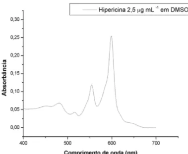
A hipericina pode ser sintetizada a partir do Emodin, um precursor da hipericina, que após sucessivas oxidações e presença de luz gera a molécula da hipericina. Este fotossensibilizador é um composto de caráter hidrofóbico e, portanto, insolúvel em água. Seu extrato deve ser dissolvido em solventes como o DMSO (dimethyl sulfoxide), a acetona, o dietil éter, etc15,32,41,54,63.
Figura 2 – Espectro de absorção da hipericina em DMSO
Terapia fotodinâmica e hipericina
De modo geral, poucos estudos de fotoinativação de bactérias tem sido realizadas. É conhecido que bactérias G+ são mais sensíveis à TFD. Alguns estudos relatam que TFD-HY apresenta resultados positivos em bactérias G+ e resultados negativos em bactérias G-41,63. Essa falha tem sido associada às diferenças nas membranas e paredes celulares das bactérias e, para contornar esta divergência e ampliar o poder de ação do fotossensibilizador, formulações catiônicas de hipericina tem sido introduzidas24. Em contrapartida, resultados recentes têm mostrado que
Diversos fatores relacionados à técnica de TFD-HY podem alterar a eficácia da eliminação de microrganismos. Sugere-se que o período de incubação é de extrema importância para a hipericina apresentar seu efeito citotóxico, assim como ambientes limitados em oxigênio podem reduzir o efeito da TFD antimicrobiana15. Estudos que ampliaram a dose de luz também revelaram maior eficiência da TFD-HY na eliminação de cepas G+ de S. aureus. Ainda, a integridade da membrana revelou ser dose-dependente em relação à concentração de hipericina, onde maiores doses apresentaram mudanças morfológicas mais expressivas e maior taxa de morte bacteriana63.
Objetivo Geral
O presente estudo teve por objetivo avaliar os efeitos da terapia fotodinâmica com uso de hipericina no tratamento da doença periodontal induzida in vivo, como terapia única ou como auxiliar do tratamento mecânico.
Objetivos Específicos
- Capítulo 1: Fazer uma revisão da literatura sobre o uso da Terapia Fotodinâmica em biofilmes orais;
CAPÍTULO 1
In: Photodynamic inactivation of biofilm: taking a lightly colored approach to stubborn infection
Wanessa CMA de Melo, Milene N de Oliveira, Pinar Avci, Asheesh Gupta, Daniela Vecchio, Magesh
Sadasivam, Rakkiyappan Chandran, Rui Yin, Livia R Perussi, George P Tegos, Janice R Perussi,
Michael R Hamblin.
Photodynamic Therapy in Dental Biofilms
∗Clinical studies and applications
Livia Rodrigues Perussi, Janice Rodrigues Perussi
Revisão submetida e aceita para o
Expert Review of Anti Infective Therapy (IF: 3.28) !!!!!!!!!!!!!!!!!!!!!!!!!!!!!!!!!!!!!!!!!!!!!!!!!!!!!!!!
∗ Este capítulo faz parte de uma reivsão mais extensa, intitulada: Photodynamic inactivation of biofilm: taking a
embedded within a polymeric matrix, typically comprising exopolysaccharide that together form a complex community. These communities of microorganisms in biofilms have been found to be responsible for a wide variety of microbial infections in different parts of the body such as: urinary tract infections, catheter infections, middle-ear infections, gingivitis, caries, periodontitis, orthopedic implants and so on. The microbial cells growing in a biofilm have properties and gene expression patterns distinct from planktonic cells. These differences include phenotypic variations in enzymatic activity, cell wall composition, and surface structure, which increase the resistance to antibiotics and other antimicrobial treatments and therefore pose a major therapeutic challenge. There is consequently an urgent need for new approaches to attack biofilm-associated microorganisms, and antimicrobial photodynamic therapy (aPDT) may be a promising candidate. aPDT involves the combination of a non-toxic dye, termed a photosensitizer, and low intensity visible light which, in the presence of oxygen, produce cytotoxic reactive oxygen species. It has been found that many biofilms are susceptible to aPDT, particularly in dental disease. This review will focus on aspects of aPDT that are designed to increase
efficacy against biofilms, including PS drug design, modalities to enhance penetration
of PS into biofilm, and combination of aPDT with biofilm-disrupting agents.
Key Words: Antimicrobial photodynamic therapy, biofilm, microbial resistance,
extracellular polysaccharide, persister cells, multidrug efflux pump, dental infection,
Clinical studies and applications
Livia Rodrigues Perussi1, Janice Rodrigues Perussi2
1 Faculdade de Odontologia de Araraquara – UNESP, Brazil 2 Instituto de Química de São Carlos – USP, Brazil
Periodontal diseases are among the most common oral diseases in the world, leading to tooth loss. Upon poor oral hygiene, constituents of the diet interact with oral bacteria leading to changes in the local microflora resulting in an upsurge of oral biofilms that adhere in teeth and soft tissues of the mouth. Thus, bleeding of the gingiva (gingivitis) can advance and involve supporting tissues of the teeth such as bone, creating the periodontal pocket (periodontitis). An adequate treatment must be done in order to stop the advance of the disease. However, due to the complex biofilm composition, systemic therapy may be associated with conventional therapy of periodontal diseases.
Chronic use of antibiotics has led resistance to many bacterial strains. Thus, the development of alternatives therapies is necessary in order to control anaerobic gram-negative microorganisms in periodontal infections. The large number of reviews articles on the association of low-power lasers with photosensitizers in periodontal disease conditions [1-7] reflects the increasing use of PDT in dentistry, which represents a novel therapeutic approach in the management of oral biofilms.
as the nutrient source, S. sanguis was killed using an aluminium disulphonated phthalocyanine (AlPcS2) as a light-activated antimicrobial agent [8].
In 2002, O’Neill et al used multi-species biofilms of oral bacteria irradiated with light from a helium/neon laser. The biofilms consisted of extremely large numbers of bacteria (9.109) and 97.4% were killed following irradiation with 31.5 J in the
presence of 25 mg/ml toluidine blue O (TBO), concluding that this may be useful in the treatment of dental plaque-related diseases [9].
Fontana et al, 2009 investigated the photodynamic ability to reduce the number of dental bacteria in the planktonic phase and in biofilms. The microorganisms were sensitized with 25 µg/ml of methylene blue (MB) and exposed to red light for 5 minutes. As results, they observed 63% reduction of bacteria in the suspension form and only 32% in the biofilms. Thus, the conclusion was that PDT is less effective in biofilm conditions than in bacteria planktonic phase [10].
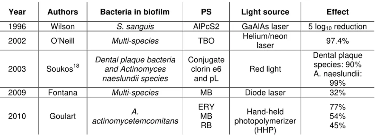
summarizes some important outcomes of each study.
Table 1.In vitro studies outcomes.
Year Authors Bacteria in biofilm PS Light source Effect
1996 Wilson S. sanguis AlPcS2 GaAlAs laser 5 log10 reduction
2002 O’Neill Multi-species TBO Helium/neon laser 97.4%
2003 Soukos18
Dental plaque bacteria and Actinomyces naeslundii species
Conjugate clorin e6
and pL Red light
Dental plaque species: 90% A. naeslundii:
99% 2009 Fontana Multi-species MB Diode laser 32%
2010 Goulart actinomycetemcomitans A.
ERY MB RB Hand-held photopolymerizer (HHP) 77% 54% 45%
As it can be observed, antimicrobial PDT is very effective in periodontal studies in vitro. However, when biofilms studies are evaluated and even more, when clinical studies are conducted, PDT results are controversial.
An in vivo study evaluated PDT as a treatment for induced periodontitis in diabetic rats. PDT was applied once with toluidine blue O (TBO) 100 µg/ml and red source laser for 133 seconds. Results showed that PDT groups had less bone loss than group given only the SRP treatment. Even though the animals had a systemic disease, the therapy was able to decrease the signals of periodontal destruction [13].
A clinical study conducted in 10 patients with diagnosis of aggressive periodontitis on which they were submitted to SRP or PDT and evaluated in terms of clinical outcomes. After 3 months, both treatments led to improvements in reducing the signals of disease, but with no significant statistical differences among them [14].
decreased in the groups with the associated therapies [15].
In 2011, a statement released by the American Academy of Periodontology (AAP) reported that lacks evidence of PDT effectiveness based on the clinical trials, where only two trials observed great bacterial reduction from out of ten already published. Therefore, AAP states that PDT is unpredictable and inconsistent in the ability to reduce bacterial loads compared to SRP alone [16]. However, to our knowledge, comparisons between PDT clinical studies are difficult to eliminate all bias. There are PDT applications in many different diseases, such as chronic periodontitis and aggressive periodontitis. As it is know, the nature and the pathogenesis of the diseases are different, making difficult to compare the therapies outcomes. Moreover, the PDT technique has many parameters that can alter between studies methodologies, such as photosensitizers used, concentrations of the dyes, time of pre-irradiation and dose of light.
Even though PDT has still a long way to roam in studies related to dental biofilms, it is already clear that this therapy can be interesting in many clinical situations due to its unique characteristics [13,14]. PDT applications related to caries, endodontic infections and dental implants have also been evaluated and promising results have been achieved [17].
1. Wood S, Metcalf D, Devine D, Robinson C. Erythrosine is a potential photosensitizer for the photodynamic therapy of oral plaque biofilms. J Antimicrob Chemother. 57, 680-4 (2006).
2. Shivakumar V, Shanmugam M, Sudhir G, Pavithra S. Priyadarshoni Scope of photodynamic therapy in periodontics and other fields of dentistry. J Interdiscip Dentistry. 2 (2), 78-83 (2012).
3. Sudhakara Reddy R, Ramya Kotha, Ramesh Tatapudi, Subbarayudu Gudapati, Sai Madhavai N, Sai Kiran CH. Photo Dynamic Therapy in Oral Diseases. Int J Biol Med Res. 3(2), 1875-83 (2012).
4. Malik R, Manocha A, Suresh DK. Photodynamic therapy - A strategic review. Indian J Dent Res. 21, 285-91 (2010).
5. Bains VK, Gupta S, Bains R. Lasers in Periodontics: An overview. J Oral Health Comm Dent. 4, 29-34 (2010).
6. Azarpazhooh A, Shah PS, Tenenbaum HC, Goldberg MB. The effect of photodynamic therapy for periodontitis: A systematic review and meta-analysis. J Periodontol. 81 (1) 4-14 (2010).
7. Aoki A, Sasaki KM, Watanabe H, Ishikawa I. Lasers in nonsurgical periodontal therapy. Periodontol 2000. 36, 59-97 (2006).
8. Wilson M, Burns T, Pratten J. Killing of Streptococcus sanguis in biofilms using a light-activated antimicrobial agent. J Antimicrob Chemother. 37 (2), 377-81 (1996).
9. O’Neill JF, Hope CK, Wilson M. Oral bacteria in multi-species biofilms can be killed by red light in the presence of toluidine blue. Lasers Surg Med. 31 (2), 86-90 (2002).
10. Fontana CR, Abernethy AD, Som S, Ruggiero K, Doucette S, Marcantonio RC, et al. J Periodontal Res. 44 (6), 751-9 (2009).
11. Goulart R de C, Bolean M, Paulino T de P, Thedei G Jr, Souza SL, Tedesco AC, et al. Photodynamic therapy in planktonic and biofilm cultures of Aggregatibacter actinomycetemcomitans. Photomed Laser Surg. 28 (1), 53-60 (2010).
Aggregatibacter actinomycetemcomitans. Photomed Laser Surg. 28 (1), 85-90 (2010).
13. De Almeida JM, Theodoro LH, Bosco AF, Nagata MJ, Bonfante S, Garcia VG. Treatment of experimental periodontal disease by photodynamic therapy in rats with diabetes. J Periodontol. 79 (11), 2156-65 (2008).
14. De Oliveira RR, Schwartz-Filho HO, Novaes AB Jr, Taba M Jr. Antimicrobial photodynamic therapy in the non-surgical treatment of aggressive periodontitis: a preliminary randomized controlled clinical study. J Periodontol. 78 (6), 965-73 (2007).
15. Ge L, Shu R, Li Y, Luo L, Song Z, Xie Y, et al. Adjunctive effect of photodynamic therapy to scaling and root planning in the treatment of chronic periodontitis. Photomed Laser Surg. 29 (1), 33-7 (2011).
16. American Academy of Periodontology statement on the efficacy of lasers in the non-surgical treatment of inflammatory disease. J Periodontol. 82 (4), 513-4 (2011).
17. Gursoy H, Ozcakir-Tomruk C, Tanalp J, Yilmaz S. Photodynamic therapy in dentistry: a literature review. Clin Oral Investig. 27, [Epub ahead of print] (2012).
18. Soukos NS, Mulholland SE, Socransky SS, Doukas AG. Photodestruction of Human Dental Plaque Bacteria: Enhancement of the Photodynamic Effect by Photomechanical Waves in an Oral Biofilm Model. Lasers Surg Med. 33 (3), 161-8. (2002).
!
CAPÍTULO 2
Hypericin- PDT Treatment of Experimental
Periodontitis
In Vivo.
Livia Rodrigues Perussi-Valverde, Rafael Scaf de Molon, Guilherme José Pimentel Lopes de Oliveira, Anderson Orzari Ribeiro, Janice Rodrigues Perussi, Rosemary Adriana Chierici Marcantonio.
Livia Rodrigues Perussi-Valverde, MS, PhD student1; Rafael Scaf de Molon, MS,
PhD student1; Guilherme José Pimentel Lopes de Oliveira, MS, PhD student1;
Anderson Orzari Ribeiro, PhD, Adjunct Professor2; Janice Rodrigues Perussi, PhD, Adjunct Professor3; Rosemary Adriana Chierici Marcantonio, PhD, Titular Professor1
1Departamento de Diagnostico e Cirurgia, Faculdade de Odontologia de Araraquara,
Universidade Estadual Paulista – UNESP, Araraquara, SP, Brazil
2 Centro de Ciências Naturais e Humanas, Universidade Federal do ABC - UFABC,
Santo André, SP, Brazil
3Instituto de Química de São Carlos, Universidade de São Paulo, São Carlos, SP,
Brazil
Corresponding author:
Profa. Rosemary Adriana Chierici Marcantonio
Department of Diagnosis and Surgery, Araraquara Dental School Universidade Estadual Paulista – UNESP; Rua Humaitá, 1680 Araraquara, São Paulo, Brazil, Zip Code 14801-903
Background: Hypericin has been quoted as a powerful photosensitizer but no reports of HY-PDT (Hypericin – Photodynamic Therapy) for periodontal diseases to date have been observed. The aim of this study was to evaluate the HY-PDT effects on experimental periodontitis in vivo as a monotherapy or as an adjunct to mechanical periodontal treatment.
Methods: Eighty-four rats had periodontitis induced by cotton ligatures that were inserted in the subgingival area of maxillary second molars. After seven days, the ligatures were removed and the animals were randomly divided into the following treatments groups: Control (C); Scaling and root planning (SRP) with mini-curettes; PDT (HY 10µg/ml, LED 590nm) and SRP+PDT. Seven, 15 and 30 days post-treatment the animals were euthanized. The furcation area was evaluated by immunohistochemical analysis for TRAP and RANKL in the 7-days samples; histometric and micro-computerized tomography (microCT) analysis for bone loss observations were performed at all periods samples.
Results: MicroCT and histometric analysis of bone at 7 and 15-days periods showed significant differences between SRP, PDT and SRP+PDT groups to Control group, whereas treatments groups revealed more bone repair. At 30-days period, no differences were found among all groups. PDT and SRP+PDT immunohistochemistry analysis showed TRAP-positive cells and RANKL-staining similar to Control group. Conclusion: In this study conditions, HY-PDT as a single therapy or as an auxiliary treatment of periodontitis in vivo showed similar results to SRP regarding bone loss at all time points. Therefore, more studies should be performed in optimize the periodontal outcomes.
Periodontitis is characterized by loss of periodontal tissues due to accumulation of bacterial biofilm around the structure of the teeth. If untreated, the biological response leads to inflammation and bone loss.1 In order to investigate host bacteria interactions, the ligature model in rats is a feasible way to evaluate periodontal therapies in vivo.2-4
!
The conventional treatment of periodontal diseases includes scaling and root planning (SRP), which is a mechanical procedure indicated to eliminate bacterial deposits on tooth surfaces in order to diminish signs of inflammation.4,5 However, the pattern of the disease, anatomical variations, involvement of systemic diseases and different therapeutic approaches can make mechanical treatment not sufficient as a monotherapy.4,6-9 Thus, chemical support in association to SRP is commonly used to amplify the bacterial reduction and help eliminate clinical signs of periodontitis. Although antibiotic is commonly an ally, a long-term prescription can lead to microorganism resistance and many other side effects to the patient.9-14
New therapies have been proposed as auxiliary to periodontal mechanical treatment, such as photodynamic therapy (PDT). This technique requires a non-toxic light-sensitive dye (known as photosensitizer) that interacts with a specific target cell in the presence of oxygen and is activated by a visible light of adequate wavelength. This reaction produces singlet oxygen and other reactive oxygen compounds (ROS) that are toxic to bacteria.15-18
!
Although many studies in vitro and in vivo have already evaluated the efficacy of PDT in the treatment of periodontitis, results are still contradictory.17,19,20
make it difficult to achieve a protocol with 100% efficiency. !
It is also important to consider that most of the PDT studies are conducted with lasers to activate the PS.6, 21-23 Although they have many advantages as a light source, this technology is very expensive, limiting the applicability of this technique. An alternative is the light emitting diode (LED),24 which is reasonably less expensive
and may be easily found in a dental office.25,26!
Other aspect to emphasize is that most studies associating PDT and periodontitis uses mainly phenothiazines dyes, like methylene blue (MB) and toluidine blue O (TBO), which have poor effects against G- microorganisms and can also stain the teeth as well as implants and restorations.17,23,25,27 Hypericin (HY) is a
natural photosensitive pigment that is extracted from various species of the Hypericum genus, the herbaceous plant known as St. John’s Wort and is on the market for many years as an anti-depression herbal agent.28-31 HY is considered one of the most powerful photosensitizers found in nature due to the high generation of superoxide anion and singlet oxygen.15,30,32 Although the phototoxicity of HY on
several tumor cells has been reported, the data concerning its photoactivity on microorganisms are scarce.33,34 A detailed study to obtain the experimental
parameters to achieve satisfactory selective photoinactivation of C. albicans and C. dubliniensis with hypericin has been achieved.35 In another study, HY was showed to
kill E. coli on a similar amount as Zn-phthalocyanine since 12 J cm-2 leads to a 7 log
reduction, almost twice the inactivation obtained with rose Bengal and methylene blue at high concentration and at the same light dose.36 A recent study evaluating
to date.28,31!
During progression of periodontal disease, bone reabsorption occurs in the presence of cytokines that are released in the inflammatory sites. Receptor activator of nuclear factor-kappa B ligant (RANKL) and multinuclear tartrate-resistant acid-phosphatase (TRAP)-positive cells are related to osteoclast differentiation and activation, leading to osteoclatogenesis and bone loss. Both cytokines play an important role in the alveolar bone during periodontitis.23,37-39 To evaluate bone
destruction, micro-computerized tomography (microCT) is a very sensitive technique providing three-dimensional images such as bone height, bone density and bone structures40-42 and has been indicated to evaluate periodontal bone modifications.
Regarding previous PDT in vivo studies, there are no reports of bone repair by means of micro-CT.
A great amount of questions still needs to be answered regarding PDT as a treatment for periodontal diseases. To our knowledge, there are no reports using HY for this purpose. Thus, the aim of this study was to investigate the effects of HY-PDT as a monotherapy or as an adjunct treatment to experimental periodontitis in a rat model.
!
Material and Methods ∗
!
Sample
Experimental group comprised eighty-four male Holtzman rats (Rattus Novergicus), weighting 200-250g, maintained in the animal facilities of the Araraquara Dental School, with controlled temperature (23±2°C), humidity (65-75%), !!!!!!!!!!!!!!!!!!!!!!!!!!!!!!!!!!!!!!!!!!!!!!!!!!!!!!!!
housed in plastic cages, fed by a standard laboratory diet and given water ad libitum. The study protocol was conducted according to the recommendations of the National Council for Control of Animal Experimentation (CONCEA) and was approved by the Local Institutional Experimentation Committee for Animal Care and Use (Protocol 21/2009). #
Experimental Periodontal Disease Induction
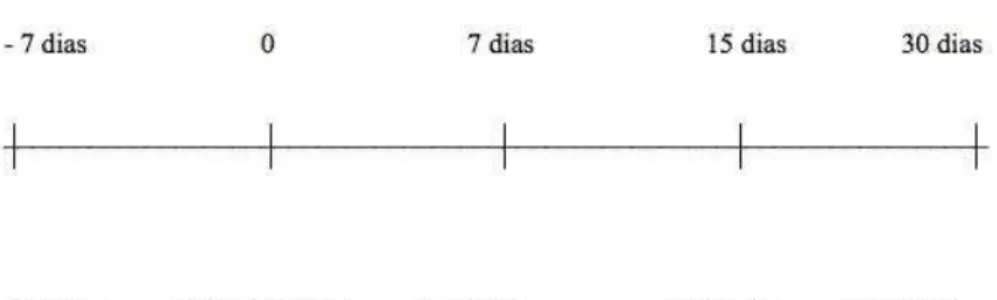
The animals underwent general anesthesia (0.08 ml of ketamine hydrochloride and 0.04 ml of xylazine per 100g body weight) administered via intramuscular injection. The ligature model was obtained by placing a cotton thread around the second maxillary molars at the level of the gingival sulcus. The ligatures were placed bilaterally and kept for 7 days in order to induce periodontitis. After the 1-week induction period, the ligatures were removed and the treatments were performed.
Experimental Protocol
At the day of ligature removal, all treatments were performed. The animals were randomly∝ assigned into four groups: Control (C, n=21) in which no treatment was performed; Scaling and root planning (SRP, n=21); Photodynamic therapy (PDT, n=21); and SRP associated with PDT (SRP+PDT, n=21). On each group, the animals were divided into three experimental periods: seven, 15 and 30 days post treatment, with seven animals per period on each group.
!!!!!!!!!!!!!!!!!!!!!!!!!!!!!!!!!!!!!!!!!!!!!!!!!!!!!!!! #!Aprovação do Comitê de Ética: Anexo A!
∝
The SRP treatment was conducted with mini-five curettes (#7-8, #11-12, #13-14)∗ through distal-mesial traction movements in both vestibular and palatine surfaces of the second maxillary molars. The furcation and interproximal areas were scaled through cervico-occlusal traction movements with the same curettes.
Photosensitizer
Hypericin was synthesized from emodin according to the literature43 and the product was checked by 1H nuclear magnetic resonance (NMR). The stock solution 1
mg/mL was prepared in dimethyl sulfoxide (DMSO 1%). In order to achieve the established concentration (10 µg/mL), the stock was diluted in phosphate buffered saline (PBS, pH 7.4) and kept in the dark to avoid photodegradation30.
Light source
The maximum absorption band of HY is 590 nm and therefore the light source used to activate the photosensitizer was a yellow LED (light emitting diode) operating at 590 ± 10 nm at an intensity of 115 mW.cm-2. The LED apparatus (tip: 1 mm
diameter) was developed by the Optics Group (Research Center for Optics and Photonics) of the Physics Institute, University of São Paulo - USP, São Carlos, SP - Brazil.
PDT Treatment
Hypericin was inserted into the gingival sulcus at a volume of 0.2 mL using a syringe (1 mL) and an insulin needle (13 x 0.45 mm), without a bevel, through all !!!!!!!!!!!!!!!!!!!!!!!!!!!!!!!!!!!!!!!!!!!!!!!!!!!!!!!!
periodontal pocket for five minutes (pre-irradiation time). The irradiation time was 4 minutes per tooth, where the LED was applied in a parallel position to the teeth occlusal surface. The fluence delivered was 35.15 J/cm2 per tooth.
Experimental Periods
The animals were sacrificed at 7, 15 and 30 days post treatments, by an overdose of anesthesia administration. After euthanasia, the maxillary jaws were hemisected. Half of the blocks was separated for histology and immunohistochemical processing, whereas the hemi-maxillas were fixed in 4% buffered paraformaldehyde (pH 7.2) for 48 hours and decalcified for 2 months in 7% EDTA (0.1M, pH 7.2) and embedded in paraffin. The remaining blocks were placed in H2O2 for 48 hours in
order to facilitate removal of the soft tissues and, subsequently, stored in 70% ethanol for alveolar bone analysis by micro-computerized tomography.
Histometric analysis
After decalcification, the specimens were embedded in paraffin, cut into 6µm sections of longitudinal serial slices and stained with hematoxilin and eosin.
For the histometric analysis, a calibrated blinded examiner randomly selected three histological images from each animal corresponding to the beginning, middle and end of each sample. The proportion of bone tissue in the furcation area was assessed using an image analyzer program#. The measures of each section were performed three times by the same examiner within a week interval. The area analyzed was delimited from the furcation roof to a distance of 1000 µm apically and !!!!!!!!!!!!!!!!!!!!!!!!!!!!!!!!!!!!!!!!!!!!!!!!!!!!!!!!
by César-Neto et al, 2006.44
MicroCT Analysis
The hemi-maxilla samples of seven rats per group and period were scanned with MicroCT system∞. The x-ray generator was operated at 50 kVp, beam current at 500µA, 0.5mm aluminum filter at an image resolution of 12.45µm. The images were reconstructed with specific softwareξ in all three spatial dimensions and then, all the images were orientated and saved in sagittal slices (2000 x 1336) with Skyscan software℘. The volumetric measurements were performed after the selection of a 3-D ROI (region of interest) with CTAn program≈. 3-During the drawing of the ROI the examiner was guided by morphological landmarks. The ROI was delimited from the most mesial root of third molar to the most distal root of first molarserved as endpoint landmark borders, because the experimental periodontal disease and the maximum bone loss was evident on the second molar around the furcation and interproximal area. After the delimited landmarks borders, the contours of the ROI were drawn at regular intervals with a slice-based method, every 10 planes, resulting in minimum inclusion of the teeth roots and maximum quantification of alveolar bone. All contours were drawn beginning immediately below the cemento-enamel junction (CEJ) and moving 1,5 mm in the apical direction. Thus, the entire bone area of the interproximal region and the furcation area of the second molar were involved in the ROI. Finally,
!!!!!!!!!!!!!!!!!!!!!!!!!!!!!!!!!!!!!!!!!!!!!!!!!!!!!!!!
∞ Skyscan, 1176, Aartselaar, Belgium
ξ NRecon 1.6.1.5 – Skyscan, Belgium!
℘ Data Viewer 1.4.3.1 - Skyscan, Belgium
binarized ROI. The architectural parameter evaluated was bone volume fraction (%) that represents the volume of the mineralized tissue.
Immunohistochemical analysis
Serial longitudinal sections (4µm) were mounted on silanized slidesς and incubated for 2 hours at 58oC. Briefly, tissue sections were deparaffinized and rehydrated. No antigen retrieval was performed. The sections were treated with 3% hydrogen peroxide for 30 minutes at room temperature (RT). Afterwards, sections were incubated with 3% bovine serum albumin for 2 hours. Therafter, the sections were incubated with rat TRAP or RANKL polyclonal antibodies raised in rabbit (1:200 dilution for both proteins), in a humid chamber at RT overnight. As negative control, primary antibody was omitted and incubated with 1% PBS. Following the incubation period, the sections were washed with 1% PBS and incubated with biotinylated immunoglobulin for 30 minutes at RT. After washing, sections were incubated by biotin-streptavidin peroxidaseΦ complex for 30 at RT. Diaminobenzidine/hydrogen peroxideψ was used as a chromogen substrate. All sections were counter-stained with Carrazi Hematoxylin for 45 seconds and mounted in Permount.
Standardized photomicrographs of the most coronal portion of the furcation region were taken using a light microscope (20x, original magnification). The area of the furcation for immunohistochemical analysis was selected as the same methodology described for histometric analysis.
!!!!!!!!!!!!!!!!!!!!!!!!!!!!!!!!!!!!!!!!!!!!!!!!!!!!!!!!
ς Dako, Glostrup, Denmark
Φ DAKO-LSAB!
cells around bone tissue were assessed whereas osteoclasts with more than two nuclei were considered TRAP-positive multinuclear cells. The RANKL marker was analyzed in the same furcation area by percentages: no marking (0%), weak marking (<25% of cells), moderate marking (<50% cells) and strong intensity (>50% cells), according to Garcia et al.45
Data Analysis
The histometric and microCT data distribution were statistically analyzedusing the test of Shapiro-Wilk to assess whether the data were distributed according to the central theorem of distribution. All groups showed normal distribution of data, and then the parametric test ANOVA (One Way) supplemented with Tukey test was used for inter-group analysis of the data. For the histometric data, the paired t-test was used to intra-groups for comparison taking into account different evaluation periods. Statistical analysis was performed using GraphPad Prism 5.0°. All tests in this study were applied with a 95% confidence level (p <0.05).
Results
Histometric analysis
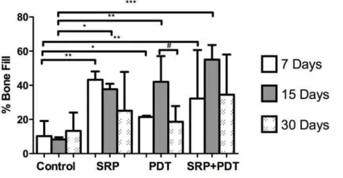
The histometric data showed that on the 7-days analysis, all the treatment groups (SRP, PDT and SRP+PDT) presented a higher percentage of bone fill in furcation area when compared to Control group (p< 0.05; p< 0.001). The same observation was verified at 15-days analysis; all the treatment groups presented a higher percentage of bone fill in furcation area when compared to Control group (p< 0.05; !!!!!!!!!!!!!!!!!!!!!!!!!!!!!!!!!!!!!!!!!!!!!!!!!!!!!!!!
treated groups.
The intragroup analysis showed that there was statistically significant difference in the PDT group, between the periods of 15 and 30-days (p<0.05), which presented greater bone fill in the furcation area at 15-days samples.
Figure 1. Percentage of bone fill in all groups at the 7, 15 and 30 days experimental periods.
* Significant differences between Control and PDT group at 7-days and between Control and SRP group at 15-days (p<0.05). Tukey’s test.
** Significant differences between Control and SRP and SRP+PDT groups at 7-days and between Control and PDt group at 15-days (p<0.01). Tukey’s test.
*** Significant difference between Control and SRP+PDT group at 15-days (p<0.001). Tukey’s test.
# Significant difference between 15 and 30-days period at PDT group (p<0.05). Paired t-test.
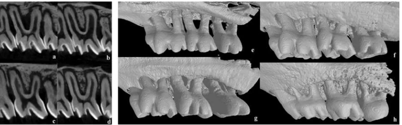
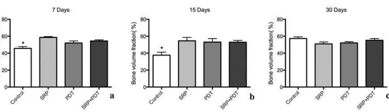
The results of bi-dimensional and three-dimensional sagittal micro-computed tomography views of maxillary molars from each group at 7 days are shown in figure 2a to 2d and 2e to 2h, respectively. Graphs of all experimental periods with the results of bone fraction volume (BVF) are shown in figure 3a to 3c. Relevant effect on bone repair, represented by BVF, was evidenced in the control group compared to SRP, PDT and SRP+PDT groups at 7 and 15-day periods. No significant difference was evident at 30-day period evaluation between groups (p<0.05).
Figure 3. Figure 3a to 3c – Bone volume fraction in all experimental periods of 7 (a), 15 (b) and 30 (c) days, respectively.
* Significant difference between treatment groups (SRP, PDT and SRP+PDT) and Control group (p<0.05). Tukey’s test.
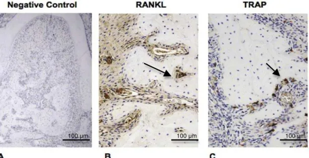
Immunohistochemical analysis
Figure 4. TRAP and RANKL immunostaining (IS) in the furcation area, 7-days post treatment in sections from the Control group. Negative control (A) of IS, original magnification: 5x; arrow indicating RANKL IS (B) and TRAP-positive cell (C), original magnification: 20x.
Table 1. RANKL Immunostaining and TRAP-positive cells staining on the groups analyzed.
Control SRP PDT SRP+PDT RANKL
Immunostaining
Weak (<25%) to moderate marking (<50%)
No marking (0%) - weak marking
(<25%)
Moderate marking (<50%)
Moderate marking (<50%)
TRAP-positive cells
8 3 7 9
Discussion
In this study, clinical signs of inflammation were evident and bone loss was observed by the tridimensional images after the 7-days induction period of
100 µm 100 µm
accordance to previous in vivo studies performed in rodents with the same experimental period for periodontites.1,2,5,9,41,45
Photodynamic therapy, initially indicated for treatment of malignant tumors, recently has been proposed to inactivate bacteria and may also be cited as antimicrobial or antibacterial-PDT (a-PDT), photodynamic antimicrobial chemotherapy (PACT) or even photodynamic inactivation (PDI).9,15,26,33,45-47 Since we did not evaluate bacterial load, the PDT term was used. This therapy has advantages such as selectivity and lack of resistance development, being a very promising treatment in association to periodontal mechanical therapy.9,15,17,18
Periodontitis is an inflammatory disease formed by accumulation of oral biofilm, mainly formed by periodontopathogenic bacteria, mostly G-. This type of bacteria possesses an outer membrane that restricts binding and penetration of many photosensitizers, especially when arranged in a biofilm.15,33,48 Thus, one of the challenges to PDT is to find a suitable drug that can penetrate in bacteria walls appropriately so that the efficiency of the therapy is maximized. This may explain why PDT for periodontal diseases have less efficient outcomes than reports on other diseases mainly constituted by G+ bacteria.25,33 Yow et al.49 observed that
when arranged in biofilms. Furthermore, Zanin et al.25 reported that mature biofilms are less susceptible to PDT. The microCT and histometric results at 7 and 15-days (Figures 1 and 3, respectively) showed significant differences between Control and treatment groups (SRP, PDT and SRP+PDT), but without differences among them. PDT alone or combined to SRP did not reveal a significant greater bone repair compared to SRP group. The lack of expressive outcomes in the PDT and SRP+PDT groups may be related to deficiencies on the PDT parameters and as well as on the hypericin’s characteristics. Some explanations can be cited, such as: hypericin may have had difficulty in binding to the G- bacteria walls arranged in a complex biofilm in periodontitis; the pre-irradiation time could have been short; the dose used could not be sufficient to photosensitize HY on a biofilm in vivo and lastly, the concentration of HY may have been low. Therefore, the PDT outcomes in this study were not significant compared to SRP and some modifications in the parameters of HY-PDT should be made in further studies, such as utilization of nanoparticules to improve hypericin’s delivery to its targets.
In this study, PDT as a monotherapy or as adjunct to SRP showed similar results to SRP alone, considered the gold- therapy for periodontitis. To our knowledge, SRP will never be an option but a sine qua non treatment to diminish progression of periodontitis and thus, PDT should never be indicated as a monotherapy.50 SRP should always be performed in order to disrupt the biofilm
as adjunct to SRP should be a treatment option but not a substitute to conventional therapy by SRP.
Beyond that, the inflammatory character of periodontitis leads to bleeding, which can alter the concentration and efficiency of the photosensitizer, once hemoglobin is present on gingival crevicular fluid, reducing the effectiveness of the PDT.19,26 In this study, in order to avoid this problem, once the cotton ligature was removed, the bleeding was controleed using a soft gauze. Since all the treatments were performed immediately after ligature removal, we cannot affirm that blood was totally removed and thus, did not interfere with the absorbance of the photosensitizer and efficiency of the therapy. Thus, bleeding and saliva may have interfered in our PDT outcomes, even though the treatments were carefully conducted. Even more, our PDT treatments were performed in a single session. Recent studies in carcinoma cells observed that more applications of PDT can lead to more effectiveness of the therapy and can also produce more reactive oxygen species (ROS).51
Photodynamic therapy has two pathways of action and production of ROS: type I and type II reactions. Hypericin is mostly related to a high yield of singlet oxygen formation (type II reaction) even in low concentrations (<0.1 µg/mL), a characteristic that makes its photosensitizing effect so powerful49. Since singlet oxygen’s action time is very short, this reactive oxygen is only toxic to the organism in a small range of seconds and in a small radious of action (20 nm), avoiding interactions with the surrounding tissues.15,17,29 Therefore, those peculiar characteristics made HY-PDT a challenge for periodontal treatment.
concentrations of photosensitizers may lead to aggregation in aqueous medium.26,30 Therefore, HY is considered potentially more effective in lower concentrations so 10 µg/mL seemed to be a feasible initial parameter in this in vivo study.
To improve even more the HY-PDT outcomes, changes in the settings such as pre-incubation time, irradiation time, dose and HY concentration should be evaluated. Nevertheless, future studies should evaluate PDT outcomes in multiple sessions. With exception of PDT group at 15 and 30-days (Figure 1) which had significant less bone filling at 30-days compared to 15-days period, the 30-day period observation between all groups had no statistical significant difference within the bone repair (Figure 3). One of the explanations may be the re-colonization of the periodontal pockets.53 Prates et al.9 evaluated the short-term of bacterial reduction after PDT and reported no difference between the initial bacterial load (analyzed immediately after ligature removal) and 7-days post treatment. The authors concluded that re-growth or re-colonization may have occurred. Other aspect to emphasize is that the ligature model involves trauma of the periodontal tissues, like reported by Li, Amar.40 Although without statistical significant difference, these observations could explain the tendency of equal bone filling and repair in all 4 groups at the 30-day period compared to groups at 7 and 15-days. Thus, we can presume that more applications of the therapy could lead to an augmentation of the bone repair at long term, as well as changes in the parameters of the PDT and on the delivery system of HY to G- bacteria.
this apparatus is available in almost every dental office since it is used in other dental procedures such as curing composites and dental bleaching. Thus, these features can make PDT a technique more achievable for dentists and financially more reliable to the patient as well.
Li, Amar40 comparing three different methods to evaluate bone loss during
Within the limitations of this study, hypericin-PDT as a monotherapy or as adjunct to SRP diminished periodontitis progression in vivo, similarly to conventional mechanical treatment. Thus, hypericin can be considered a promising photosensitizing agent. However, more studies are required in order to provide a more efficient protocol to justify the use of HY-PDT in the treatment of periodontal diseases.
!
ACKNOWLEDGEMENTS
! This study was financially supported by the São Paulo State Research
Support Foundation (Process no. 2009/17404-1 and no. 2010/14932-4). There were no conflicts of interest related to this study.
Sources of support
São Paulo State Research Support Foundation - FAPESP, São Paulo-SP, Brazil (process no. 2009/17404-1 and no. 2010/14932-4).
Conflict of interest and declaration of financial support
The authors declare no conflicts of interest.
REFERENCES
1. Bottura PE, Milanezi J, Fernandes LA, Caldas HC, Abbud-Filho M, Garcia VG, et al. Nonsurgical periodontal therapy combined with laser and photodynamic therapies for periodontal disease in immunosuppressed rats. Transplantation proceedings. 2011 Jun;43(5):2009-16.