478
Srp Arh Celok Lek. 2016 Sep-Oct;144(9-10):478-484 DOI: 10.2298/SARH1610478R
ОРИГИНАЛНИ РАД / ORIGINAL ARTICLE UDC: 616.314-089-06; 615.46:616.314
Correspondence to:
Dragana RAKAŠEVIĆ Faculty of Dental Medicine University of Belgrade Dr Subotića 4, 11000 Belgrade Serbia
SUMMARY
Introduction Peri-implantitis is an inflammatory lesion of peri-implant tissues. Eradication of the caus-ative bacteria and decontamination of the implant surface is essential in achieving predictable and stabile clinical results. Photodynamic therapy (PDT) is non-invasive adjuvant therapeutic method to surgery in the treatment of bacterial infection.
Objective The aim of this study was to evaluate early clinical and microbiological outcomes of peri-implantitis after surgical therapy with adjuvant PDT.
Methods Fifty-two diagnosed peri-implantitis sites were divided into two groups. PDT was used for decontamination of implant surface in the study group; in the control group, chlorhexidine gel (CHX) followed by saline irrigation was applied. Several clinical parameters were recorded before the treatment (baseline values) and three months after surgical treatment. Samples for microbiological identification were collected before therapy, during the surgical therapy (before and after decontamination of im-plant surface), and three months thereafter, and analyzed with identification systems using biochemical analysis.
Results The use of PDT resulted in significant decrease of bleeding on probing in comparison to CHX (p < 0.001). It showed significant decontamination of implant surfaces with complete elimination of anaerobic bacteria immediately after surgical procedure and three months later.
Conclusion The results indicate that PDT can be used as an adjuvant therapy to surgery for decontami-nation of implant surface and surrounding peri-implant tissues within the treatment of peri-implantitis.
keywords: peri-implantitis; decontamination; photodynamic therapy
Efficiency of photodynamic therapy in the treatment
of peri-implantitis – A three-month randomized
controlled clinical trial
Dragana Rakašević1, Zoran Lazić2, Bojan Rakonjac3, Ivan Soldatović4, Saša Janković1, Marko Magić5,
Zoran Aleksić1
1University of Belgrade, Faculty of Dental Medicine, Department of Periodontology, Belgrade, Serbia; 2Medical Military Academy, Department of Oral Implantology, Belgrade, Serbia;
3Medical Military Academy, Institute of Microbiology, Belgrade, Serbia;
4University of Belgrade, School of Medicine, Institute of Medical Statistics and Informatics, Belgrade, Serbia; 5University of Belgrade, Faculty of Dental Medicine, Department of Oral Surgery, Belgrade, Serbia
INTRODUCTION
Peri-implantitis has been defined as an inflam-matory process that affects the supporting mar-ginal bone around an implant in function and results in bone resorption [1, 2]. It has been shown that the biofilm formed around osseoin-tegrated implant has an important role in initi-ation and progression of peri-implant diseases [3, 4]. Most common microorganisms that are related to peri-implantitis are anaerobic bacte-ria, such as Prevotella intermedia, Porphyromo-nas gingivalis, Aggregatibacter actinomycetem-comitans, Bacteroides forsythia, Treponema den-ticola, Prevotella nigrescens, Peptostreptococcus
spp., Fusobacterium nucleatum. Some authors suggested that Staphylococcus aureus and Can-dida albicans may be connected with initiation of peri-implantitis [3, 5]. Excessive mechani-cal stress, residual cement, poor plaque control could be among risk factors in the onset and development of peri-implantitis [2].
Peri-implantitis is a complex disease; there-fore, the therapy continues to be a challenge. Surgical therapy of peri-implantitis has been suggested to be superior to non-surgical
ther-apy [6]. Decontamination of implant surface is one of the most important and difficult steps because of the screw-shaped design and rough-ness where microorganism and their products are incorporated. Many methods have been suggested, such as mechanical (dental curettes, ultrasonic scalers, air-powder abrasive), physical and/or chemical methods (citric acid, chlorhex-idine, EDTA), usually combined with local or systematic antibiotics [7–12]. No single protocol has been suggested for solving this problem.
an excited singlet state. Subsequently, it may decay back to its ground state, transition high-energy triplet state, which can react with biomolecules such as endogenous oxygen to produce very reactive products, like singlet oxygen. Singlet oxygen has cytotoxic effect through damage of cell mem-brane and cell wall. Using this therapy, the target bacteria can be destroyed without adverse effect on the implant surface and surrounding peri-implant tissue [14, 15].
Some articles showed that using chlorhexidine (CHX) as adjunctive chemical agent to decontamination, reduced inflammation and microorganisms on the implant surface. Bioadhesive gels with higher concentrations of CHX have shown greater effectiveness in various clinical situations [16, 17]. However, ideal treatment for peri-implantitis is not defined yet. There are no clinical trials showing effects of PDT during surgical therapy of peri-implantitis.
OBjECTIvE
The aim of this study was to compare early clinical and microbiological effects of the adjuvant use of PDT to sur-gical treatment of peri-implantitis, comparing the effects of decontamination of the implant surface to those when 1% CHX gel was used.
METHODS
Patients were selected from two dental clinics: Department of Periodontology, University of Belgrade, and Department of Implantology, Military Medical Academy, from January 2014 to February 2015. The research protocol was submitted to and approved by the Ethical Committee, University of Belgrade, Serbia (number 36/28). The study was carried out in accordance with the ethical principles of the World Medi-cal Association Declaration of Helsinki. Before the proce-dure, all participants were informed about the study and signed a written consent. All participants had to meet the following inclusion criteria: age >18 years, no periodontal or peri-implant treatment three months prior to the study, presence of minimum one implant in function, early or moderate type of peri-implantitis classified by Froum and Rosen [18]. Exclusion criteria were the following: uncon-trolled medical conditions, use of systemic antibiotics in the previous three months, use of anti-inflammatory drugs in the previous six months, pregnancy and lactation, therapy of peri-implantitis in the last three months previously.
Measurements and recording of clinical parameters
The measurements were done and recorded before any treatment. All measurements were made at the following six sites of the implant with signs of peri-implantitis: me-siobuccal, mid-buccal, distobuccal, mesio-oral, mid-oral, disto-oral by one examiner (DR), using a graduated probe (PCPUNC 15, Hu Friedy, Chicago, IL, USA). The applied probing force was standardized force of 0.25 N. The
im-plant shoulder was used as landmark for calculation of mucosal recession and clinical attachment level. The fol-lowing clinical parameters were registered:
• Peri-implant probing depth (PPD) – measured in mil
-limeters from the mucosal margin to the bottom of the peri-implant pocket;
• Clinical attachment level (CAL) – measured in mil
-limeters from the implant shoulder to the bottom of the peri-implant pocket;
• Mucosal recession (MR) – calculated as the difference
between the CAL and PPD;
• Bleeding on probing (BOP) – evaluated as being
pres-ent if bleeding was evident within 30 seconds after probing, or absent, if no bleeding was observed;
• Suppuration (SUP) – present or absent.
Clinical parameters were measured and recorded be-fore (baseline values) and three months after therapy. All clinical examinations were performed after removal of the abutments attached to the implants. Fifteen days after therapy the provisional restoration was performed on the treated implants.
Microbiological samples and analysis
The first samples for microbiological analysis were taken before any measurements from the deepest peri-implant pockets. After the removal of subgingival plaque, sample sites were isolated with cotton rolls and gently air dried to avoid contamination with saliva. Fine sterile paper points were inserted into the peri-implant pocket until mild re-sistance and left in place for 30 seconds. The paper points were immediately transferred to the substrate used as mul-tipurpose transport system (ESWAB LQ Amies, COPAN Diagnostics Inc., Murrieta, CA, USA).
The second and third samples were taken separately during the surgical therapy from the implant surfaces. Af-ter opening the flap and removing granulation tissue, the second sample was obtained from the implant surface by a transport swab. The swab was immediately transferred to the substrate used as multipurpose transport system (ESWAB LQ Amies, COPAN Diagnostics Inc.). After decontamination using chemical/PDT therapy, the third swab was taken from the implant surface applying the aforementioned procedure.
480
Rakašević D. et al. Eiciency of photodynamic therapy in the treatment of peri-implantitis – A three-month randomized controlled clinical trial
Treatment procedure
After clinical parameters were recorded and samples were taken, all the patients underwent a single episode of non-surgical therapy. It implied mechanical method for debridement of implants and remaining dentition in or-der to reduce signs of inflammation. Instructions for oral hygiene were proposed during the same visit.
Surgical treatment
During the surgical procedure, all the patients were ran-domly divided into two groups: the study group (PDT group) and the control group (CHX group). Similar meth-odology procedure was described in a study by De Waal et al. [17].
Surgical treatment of peri-implantitis was performed by one experienced oral surgeon (ZL) two weeks after non-surgical therapy (in accordance with Schwarz et al. [19]). Mucoperiosteal buccal and lingual incisions were made with surgical blade No. 15 under local anesthesia (2% li-docaine with epinephrine, 1:100,000). Flaps were designed to permit optimal access to the peri-implant bone defect for granulation tissue removal and decontamination of the implant surface. Full thickness mucoperiosteal flaps were elevated buccally and lingually. Removal of granula-tion tissue and mechanical implant surface cleaning were done using graphite curettes (Straumann® Dental Implant System; Straumann AG, Basel, Switzerland).
Decontamination of implant surfaces
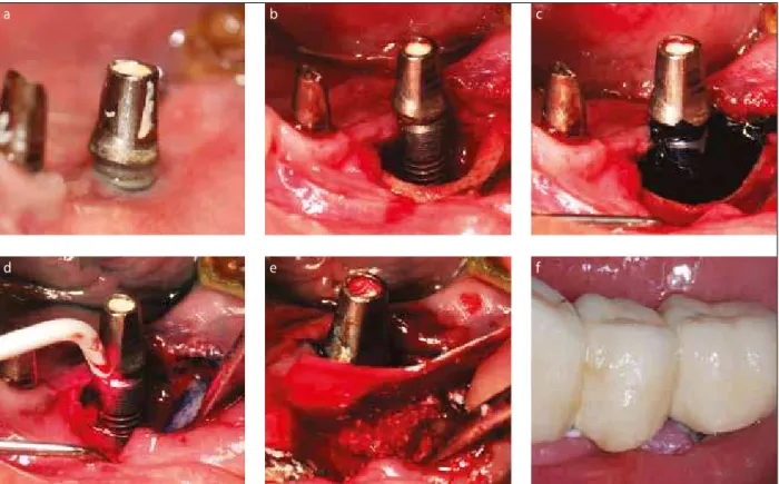
In the study group, after careful removal of granulation tissue and mechanical debridement of implant surface, decontamination of implant surfaces and peri-implant tis-sues was performed using PDT (HELBO, Photodynamic Systems GmbH, Wels, Austria). Photosensitizer, pheno-thiazine chloride (HELBO® Blue Photosensitizer, bredent medical GmbH & Co. KG), was applied onto implant surface, bone and peri-implant soft tissue, for 3 minutes. Irrigation of photosensitizer was performed with saline, according to instructions of the manufacturer. Implant surface and the surrounding tissue were exposed to the laser light by means of fibers (HELBO® TheraLite Laser HELBO® 2D Spot Probe; Bredent medical GmbH & Co. KG) for 30 seconds/spot, which operates on wave length of 660 nm and irradiance of 100 mW (Figure 1a–f).
In the control group, after removal of granulation tis-sue, 1% gel of chlorhexidine (Chlorhexamed® – Direkt; GlaxoSmithKline, GmbH & Co. KG, München, Germany) was put on implant surface. One minute after exposing implant surface with CHX, it was irrigated for 1 minute by saline.
Bone augmentation and bio-resorbable membrane were applied in peri-implant defects using artificial bone of bovine origin (Bio-Oss and Bio-Gide; Geistlich Pharma, Wolhusen, Switzerland). The mucoperiosteal flaps were repositioned and sutured [17, 19].
All the patients from both groups were prescribed an-tibiotics over a five-day period (amoxicillin caps. 500 mg,
a b c
d e f
three per day). It was recommended that patients don’t use mouthwash during the postoperative period.
Followed up period
Three months after therapy samples for microbiological analysis were taken from the reduced peri-implant pockets in the same way described above. In addition, measure-ments of clinical parameters were recorded as well.
Statistical analysis
Data are presented as mean ± standard deviation or n (%) depending on data type. Chi-squared test, Mann–Whitney U-test, and Student’st-test were used to assess the differences between the groups. Cochran’s Q-test and Wilcoxon signed-rank test were used to assess significant differences within the groups. All analyses were performed in SPSS Statistics for Windows 20.0 (IBM Corp., Armonk, NY, USA). All p-values less than 0.05 were considered statistically significant.
RESULTS
There were 52 peri-implantitis sites diagnosed in 40 sys-temically healthy patients, which were treated and re-ex-amined in a three-month period. Demography and clinical description of the study population are shown in Table 1. No adverse events and side effects were reported during and after therapy.
Clinical parameters and outcomes
From a total of 52 treated implantitis sites, 12 peri-implantitis sites belonged to the category of moderate, and 31 to the category of early peri-implantitis, classified by Froum and Rosen [18]. The rest of peri-implantitis in-stances had depths of ≥ 5 mm in only one peri-implant pocket, as one of the inclusion criteria.
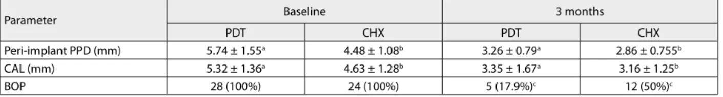
The mean value and standard deviation of PPD and CAL are shown in Table 2. Both groups showed statis-tically significant reduction of peri-implant pockets (p < 0.001). It has been shown that the greater (but not statistically significant) reduction of PPD was in the study group compared to the controls (p = 0.07). The results of our study showed that there was no statistically significant difference in CAL between the two tested groups three months after the therapy (p = 0.883). Also, there were no statistically significant differences in marginal recession.
In the study group, there was a statistically significant reduction of BOP at all six points compared with the control group three months after the therapy (p < 0.001). Changes in percentage of BOP are shown in Table 2. PDT has shown statistically significant reduction of suppuration compared to CHX treatment (p < 0.002).
Microbiological outcomes
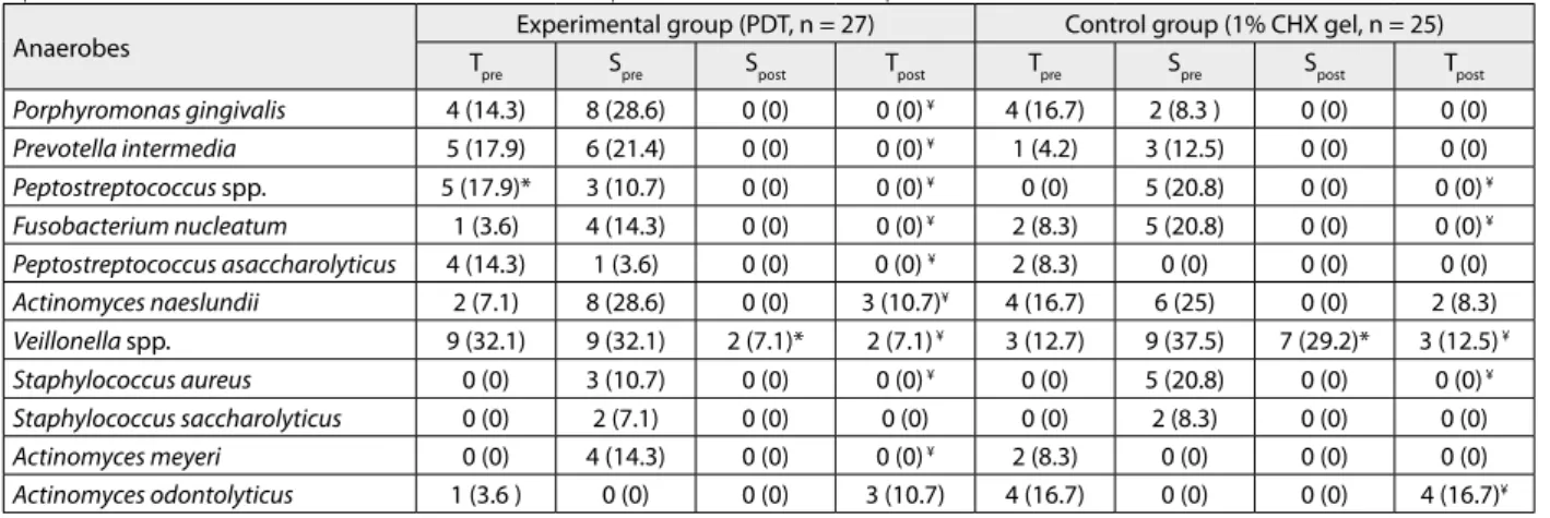
All cultivated anaerobic microorganisms which were isolated from deepest peri-implant pockets and implant surfaces are shown in Table 3. It was shown that some of these bacteria were isolated only from implant surfaces. Table 1. Demographic and clinical description of the study population
Characteristics Study group Control group p-value
Number of subjects 21 (52.5%) 19 (47.5%)
Gender Male 12 (57.14%) 12 (63.15%) 0.703
Mean age (years) (mean ±SD) 57.59 60.00 0.408
Mean time (years) after implant placement (mean ±SD) 7.68 ± 3.76 6.21 ± 3.064 0.236
Subjects with a history of treated periodontitis; n (%) 8 (29.6%) 10 (40%) 0.432
Localization of implants, n (%) Maxilla 8 (29.6%) 6 (24) 0.647
Mandible 19 (70.4%) 19 (76%)
Type of restoration, n (%)
Cement retained fixed partial denature 13 (46.4%) 19 (79.2%)
0.010a
Screw retained fixed partial denture 6 (21.4%) 2 (8.3%)
Cement retained single crown 4 (14.3%) 3 (12.5%)
Overdenture on implants 5 (17.9%) 0 (0)
a Signiicant statistical diference in type of prosthetic restauration among the groups at baseline by Pearson’s χ2 test (p < 0.01)
SD – standard deviation
No signiicant diferences were observed among the groups at baseline by Pearson’s χ2 or Independent Samples Test (T-test).
Table 2. Mean pocket probing depth (PPD) ±SD, mean clinical attachment level (CAL) ±SD, mean number of marginal recession (%), mean bleeding on probing (BOP) – positive sites (%) at each implant at baseline and three months later
Parameter Baseline 3 months
PDT CHX PDT CHX
Peri-implant PPD (mm) 5.74 ± 1.55a 4.48 ± 1.08b 3.26 ± 0.79a 2.86 ± 0.755b
CAL (mm) 5.32 ± 1.36a 4.63 ± 1.28b 3.35 ± 1.67a 3.16 ± 1.25b
BOP 28 (100%) 24 (100%) 5 (17.9%)c 12 (50%)c
a Signiicant statistical diference measured before and three months after PDT by T-test (p < 0.001); b Signiicant statistical diference measured before and three months after CHX by T-test (p < 0.001); c Signiicant statistical diference between the groups by Pearson’s χ2 test (p < 0. 001)
482
Candida spp. was also isolated from the deepest peri-implant pockets.
The results showed that the amount of anaerobic mi-croorganisms was significantly reduced and in most cases eliminated, after using both anti-infection therapeutic methods during surgery therapy as well as three months after the therapy procedure. It showed significant reduc-tion of anaerobic microorganisms in the study group in comparison with the control group, immediately after decontamination of the implant surface and also three months after (Veillonella spp.,p < 0.010; Staphylococcus aureus,p < 0.002; Peptostreptococcus spp.,p < 0.002; Pep-tostreptococcus asaccharolyticus,p < 0.035; Actinomyces naeslundii,p < 0.014; Prevotella intermedia,p < 0.011;
Porphyromonas gingivalis,p < 0.001; Actinomyces mey-eri, p < 0.007). Three months after therapy, Actinomyces naeslundii was only isolated in peri-implant pockets in the control group.
The results of the study show that using chlorhexidine leads to significant reduction of Actinomyces odontolyticus
(p < 0.046), Fusobacterium nucleatum (p = 0.23) compared with using photodynamic therapy. The presence of Ag-gregatibacter actinomycetemcomitans, Tannerella forsythia,
and Treponema denticola was not identified.
DISCUSSION
The short-term results of the present study show that both examined methods for debridement and decontamination of implant surfaces during surgical therapy (PDT and 1% chlorhexidine gel), significantly improve clinical and mi-crobiological outcomes.
In the current literature, most attention is given to sys-temic and/or local use of antibiotics or implantoplasty for the decontamination of implant surfaces during surgical procedures [3, 10, 20]. However, these methods showed side effects, as well as bacterial resistance to some drugs
and allergic reactions during and after their consumption [10]. It has been recorded that implantoplasty leads to marginal recession, which is disadvantageous in terms of function and aesthetics [2, 20].
Some authors consider PDT to be less harmful and ef-fective solution for the decontamination of the implant surface [13, 14]. This therapy was widely used in non-surgical therapy of peri-implantitis [15, 21, 22]. It has been suggested that using only photodynamic therapy several times may lead to total recovery [22]. Schwarz et al. [6] suggested that surgical therapy of peri-implantitis has been superior to non-surgical therapy. The explanation lies in the open access and controlled removal of granulation tis-sue and decontamination of the exposed implant surfaces. The results of the present study show that there are statistically significant changes in peri-implant probing depth and clinical attachment level measured at baseline and three months after therapy. The depth of peri-implant pockets in both tested groups was reduced approximately 2 mm. Higher reduction of peri-implant pocket depth was in the study group, but without statistical significance. This could be explained by larger baseline levels of peri-im-plant pockets depth recorded in the study group. It seems that PDT can promote re-osseointegration more rapidly than CHX, without side effects on the bone. The results show significant reduction of BOP and suppuration three months after using PDT. It is known that BOP is one of the most essential and important signs of peri-implantitis; therefore, the use of PDT can achieve elimination of BOP and promote better healing. In the study by De Waal et al. [17], similar methodology was used, but they didn’t achieve the significant difference in clinical parameters using two concentration of chlorhexidine solutions (0.12% and 2%) in the examined follow-up periods. These results differ from ours due to improvements of clinical parame-ters in our study. In the study by Heitz-Mayfield et al. [10], after surgical debridement and implant surface decontami-nation following by saline irrigation, systemic amoxicillin Table 3. Number (n) of culture-positive implants at baseline and culture-negative after decontamination of selected anaerobes; mean number (%) of total anaerobic bacteria load on culture-positive implants; before any treatment (Tpre); before decontamination – during surgical therapy (Spre); after decontamination – during surgical therapy (Spost); three months later (Tpost )
Anaerobes Experimental group (PDT, n = 27) Control group (1% CHX gel, n = 25)
Tpre Spre Spost Tpost Tpre Spre Spost Tpost
Porphyromonas gingivalis 4 (14.3) 8 (28.6) 0 (0) 0 (0) ¥ 4 (16.7) 2 (8.3 ) 0 (0) 0 (0) Prevotella intermedia 5 (17.9) 6 (21.4) 0 (0) 0 (0) ¥ 1 (4.2) 3 (12.5) 0 (0) 0 (0) Peptostreptococcus spp. 5 (17.9)* 3 (10.7) 0 (0) 0 (0) ¥ 0 (0) 5 (20.8) 0 (0) 0 (0) ¥ Fusobacterium nucleatum 1 (3.6) 4 (14.3) 0 (0) 0 (0) ¥ 2 (8.3) 5 (20.8) 0 (0) 0 (0) ¥ Peptostreptococcus asaccharolyticus 4 (14.3) 1 (3.6) 0 (0) 0 (0) ¥ 2 (8.3) 0 (0) 0 (0) 0 (0) Actinomyces naeslundii 2 (7.1) 8 (28.6) 0 (0) 3 (10.7)¥ 4 (16.7) 6 (25) 0 (0) 2 (8.3) Veillonella spp. 9 (32.1) 9 (32.1) 2 (7.1)* 2 (7.1) ¥ 3 (12.7) 9 (37.5) 7 (29.2)* 3 (12.5) ¥ Staphylococcus aureus 0 (0) 3 (10.7) 0 (0) 0 (0) ¥ 0 (0) 5 (20.8) 0 (0) 0 (0) ¥ Staphylococcus saccharolyticus 0 (0) 2 (7.1) 0 (0) 0 (0) 0 (0) 2 (8.3) 0 (0) 0 (0) Actinomyces meyeri 0 (0) 4 (14.3) 0 (0) 0 (0) ¥ 2 (8.3) 0 (0) 0 (0) 0 (0) Actinomyces odontolyticus 1 (3.6 ) 0 (0) 0 (0) 3 (10.7) 4 (16.7) 0 (0) 0 (0) 4 (16.7)¥
N = 52;
* Statistically signiicant change from baseline to three months after therapy, as well as before and after decontamination of implant surface between the two groups according to Pearson’s χ2 test, p < 0.05;
¥ Statistically signiicant change from baseline to three months after therapy, as well as before and after decontamination of implant surface during surgical
procedure within the two tested groups according to Cochran’s Q-test, p < 0.05
and metronidazole were prescribed. Although clinical re-sults of their study were similar to ours, using adjunctive PDT could be efficient, as it leads to lower consumption of antibiotics and antiseptics, without side effects.
Cultivation of anaerobic microorganisms showed the presence of various anaerobes isolated from peri-implant pocket before any treatment, as well as on implant surface before decontamination. Most of these bacteria belonged to the pathogenic microorganisms from red and orange complex described by Socransky et al. [23]. In our study, bacteria of yellow, purple, and green complex were also found, which is assumed to be the first phase of coloniza-tion of implant surface. This can also represent a possible bridge for the adherence of bacteria from orange and red complexes in a later phase of colonization and maturation of dental biofilm. The results also showed the presence of
Staphylococcus aureus. It was mentioned that it can be one of the possible pathogenic microorganisms that promote initiation and progression of peri-implantitis. This bacte-ria was isolated only from the implant surface, so it can be assumed that this can be a very virulent strain, which can enable the progression of peri-implantitis [24]. The presence of Candida albicans could be due to inappropri-ate oral hygiene. Therefore, Candida couldn’t have had an influence on initiation of peri-implantitis, as mentioned in some early papers [2].
In both examined groups, statistically significant reduc-tion of anaerobic microorganisms from the peri-implant pockets was evident in the follow-up period and also from implant surface immediately after decontamination. The results show that application of PDT significantly reduced the amount of bacteria, especially those from the red and orange complex. De Waal et al. [17] achieved reduction of microorganisms from implant surface between groups during a surgical procedure, but results were not as statisti-cally significant as in our study. Our study showed signifi-cant reduction of microorganisms after using both anti-infection therapeutic methods of decontamination during surgical therapy. Invitro study of Marotti et al. [14] showed significant reduction of anaerobic microorganisms after application of PDT and 0.12% solution of chlorhexidine on anodized implants, which are results similar to ours. Another study showed that using PDT with toluidine blue as a dye can reduce but not eliminate pathogenic
micro-organisms from implant surfaces (Prevotella intermedia,
Porphyromonas gingivalis) [15]. Presence of these bacteria is one of the most important factors in peri-implantitis progression. In contrast, our study has shown elimination of these bacteria. The reason for these differences could be the composition of photosensitizer. Phenothiazine chlo-ride might have greater ability to bind different microor-ganisms compared to toluidine blue.
In the control group, chlorhexidine gel was chosen for decontamination because of its widely spread use as an antiseptic in different therapeutic procedures. It might be capable of adhering to implant surface. The results show statistically significant reduction of Fusobacterium nuclea-tum and Actinomyces odontolyticus after using 1% of CHX in adhesive gel, which can be explained by high concen-tration and viscosity of CHX in the gel. Although earlier studies described that CHX has a bactericidal effect on mi-croorganisms regardless of the concentration [17], it seems that CHX cannot remove dental biofilm, which can lead to renewed adherence of microorganism and reappearance of inflammation. Similarly, in the control group, the results of the present study showed the reduction of BOP, as one of the signs of inflammation, of only 50%, which confirmed the earlier assumption.
CONCLUSION
Even with limitations of the present study, such as short observation period, it can be concluded that photody-namic therapy achieves significant reduction of micro-organisms from implant surface and peri-implant tissue when compared with chlorhexidine application, without adverse and side effects on implants and surrounding tis-sue. Therefore, PDT could be suggested as a new strategy for decontamination of implant surface during surgical treatment of peri-implantitis.
ACkNOWLEDGEMENT
This study was partly supported by the Ministry of Educa-tion, Science and Technological Development of the Re-public of Serbia (project No. 41008).
REFERENCES
1. Lindhe J, Meyle J. Peri-implant diseases: Consensus Report of the Sixth European Workshop on Periodontology. J Clin Periodontol. 2008; 35(8 Suppl):282–5.
[DOI: 10.1111/j.1600-051X.2008.01283.x] [PMID:18724855] 2. Smeets R, Henningsen A, Jung O, Heiland M, Hammacher C,
Stein JM. Definition, etiology, prevention and treatment of peri-implantitis-a review. Head Face Med. 2014; 10:34.
[DOI: 10.1186/1746-160X-10-34] [PMID:25185675] 3. Mombelli A. Microbiology and antimicrobial therapy of
peri-implantitis. Periodontol. 2000. 2002; 28:177–89. [PMID: 12013341] 4. Dabdoub SM, Tsigarida AA, Kumar PS. Patient-specific analysis of periodontal and peri-implant microbiomes. J Dent Res. 2013; 92(12 Suppl):168s–75s.
[DOI: 10.1177/0022034513504950] [PMID: 24158341]
5. Heitz-Mayfield LJ, Lang NP. Comparative biology of chronic and aggressive periodontitis vs. peri-implantitis. Periodontol 2000. 2010; 53:167–81.
[DOI: 10.1111/j.1600-0757.2010.00348.x] [PMID: 20403112] 6. Schwarz F, Jepsen S, Herten M, Sager M, Rothamel D, Becker J.
Influence of different treatment approaches on non-submerged and submerged healing of ligature induced peri-implantitis lesions: an experimental study in dogs. J Clin Periodontol. 2006; 33(8):584– 95. [PMID: 16899102]
7. Levin L, Frankenthal S, Joseph L, Rozitsky D, Levi G, Machtei EE. Water jet with adjunct chlorhexidine gel for nonsurgical treatment of peri-implantitis. Quintessence Int. 2015; 46(2):133–7.
[DOI: 10.3290/j.qi.a32819] [PMID: 25262677]
484
outcome following combined surgical therapy of peri-implantitis: a randomized controlled clinical study. J Clin Periodontol. 2011; 38(3):276–84.
[DOI: 10.1111/j.1600-051X.2010.01690.x] [PMID: 21219392] 9. Carcuac O, Abrahamsson I, Charalampakis G, Berglundh T. The
effect of the local use of chlorhexidine in surgical treatment of experimental peri-implantitis in dogs. J Clin Periodontol. 2015; 42(2):196–203. [DOI: 10.1111/jcpe.12332] [PMID: 25385434] 10. Heitz-Mayfield LJ, Salvi GE, Mombelli A, Faddy M, Lang NP.
Anti-infective surgical therapy of peri-implantitis. A 12-month prospective clinical study. Clin Oral Implants Res. 2012; 23(2):205–10.
[DOI: 10.1111/j.1600-0501.2011.02276.x] [PMID: 22092831] 11. Valderrama P, Wilson TG Jr. Detoxification of implant surfaces
affected by peri-implant disease: an overview of surgical methods. Int J Dent. 2013; 2013:740680.
[DOI: 10.1155/2013/740680] [PMID: 23983691]
12. Romanos GE, Gupta B, Yunker M, Romanos EB, Malmstrom H. Lasers use in dental implantology. Implant Dent. 2013; 22(3):282–8. [DOI: 10.1097/ID.0b013e3182885fcc] [PMID: 23571715]
13. Rajesh S, Koshi E, Philip K, Mohan A. Antimicrobial photodynamic therapy: An overview. J Indian Soc Periodontol. 2011; 15(4):323–7. [DOI: 10.4103/0972-124X.92563] [PMID: 22368354]
14. Marotti J, Tortamano P, Cai S, Ribeiro MS, Franco JE, de Campos TT. Decontamination of dental implant surfaces by means of photodynamic therapy. Lasers Med Sci. 2013; 28(1):303–9. [DOI: 10.1007/s10103-012-1148-6] [PMID: 22790655] 15. Dortbudak O, Haas R, Bernhart T, Mailath-Pokorny G. Lethal
photosensitization for decontamination of implant surfaces in the treatment of peri-implantitis. Clin Oral Implants Res. 2001; 12(2):104–8. [PMID: 11251658]
16. Rodriguez-Perez M, Bravo-Perez M, Sanchez-Lopez JD, Muñoz-Soto E, Romero-Olid MN, Baca-García P. Effectiveness of 1% versus 0.2% chlorhexidine gels in reducing alveolar osteitis from mandibular third molar surgery: a randomized, double-blind clinical trial. Med Oral Patol Oral Cir Bucal. 2013; 18(4):e693–700. [PMID: 23722126]
17. de Waal YC, Raghoebar GM, Meijer HJ, Winkel EG, van Winkelhoff AJ. Implant decontamination with 2% chlorhexidine during surgical peri-implantitis treatment: a randomized, double-blind, controlled trial. Clin Oral Implants Res. 2015; 26(9):1015–23.
[DOI: 10.1111/clr.12419] [PMID: 24861411]
18. Froum SJ, Rosen PS. A proposed classification for peri-implantitis. Int J Periodontics Restorative Dent. 2012; 32(5):533–40. [PMID: 22754901]
19. Schwarz F, John G, Sahm N, Becker J. Combined surgical resective and regenerative therapy for advanced peri-implantitis with concomitant soft tissue volume augmentation: a case report. Int J Periodontics Restorative Dent. 2014; 34(4):489–95.
[DOI: 10.11607/prd.1794] [PMID: 25006766]
20. Romanos GE, Javed F, Delgado-Ruiz RA, Calvo-Guirado JL. Peri-implant Diseases: A Review of Treatment Interventions. Dent Clin North Am. 2015; 59(1):157–78.
[DOI: 10.1016/j.cden.2014.08.002] [PMID: 25434564] 21. Javed F, Romanos GE. Does photodynamic therapy enhance
standard antibacterial therapy in dentistry? Photomed Laser Surg. 2013; 31(11):512–8.
[DOI: 10.1089/pho.2012.3329] [PMID: 24138192]
22. Schar D, Ramseier CA, Eick S, Arweiler NB, Sculean A, Salvi GE. Anti-infective therapy of peri-implantitis with adjunctive local drug delivery or photodynamic therapy: six-month outcomes of a prospective randomized clinical trial. Clin Oral Implants Res. 2013; 24(1):104–10.
[DOI: 10.1111/j.1600-0501.2012.02494.x] [PMID: 22568744]
23. Socransky SS, Haffajee AD, Cugini MA, Smith C, Kent RL, Jr. Microbial complexes in subgingival plaque. J Clin Periodontol. 1998; 25(2):134–44. [PMID: 9495612]
24. Lee A, Wang HL. Biofilm related to dental implants. Implant Dent. 2010; 19(5):387–93.
[DOI: 10.1097/ID.0b013e3181effa53] [PMID: 20881809]
КРАТАК САДРжАЈ
Увод Периимплантитис је инфламаторни процес који зах-вата мека ткива и потпорну кост око осеоинтегрисаног имплантата. Елиминација патогених микроорганизама и деконтаминација имплантне површине представљу нај-битнији корак у постизању стабилних клиничких резултата. Фотодинамска терапија (ФДТ) представља додатни неинва-зивни метод у терапији бактеријских инфекција.
Циљ рада Циљ рада била је процена клиничких и микро-биолошких параметара након хируршке терапије периим-плантитиса уз додатну примену ФДТ.
Методе рада Сва дијагностикована места периимплан-титиса (n = 52) била су подељена у две групе: у студијској групи, за деконтаминацију имплантне површине током хируршке процедуре коришћена је ФДТ; у контролној гру-пи, за деконтаминацију имплантне површине коришћен је хлорхексидин у гелу (CHX). Клинички параметри праћени су пре терапијске процедуре и три месеца после терапије.
Узорци за микробиолошку анализу узимани су пре и три месеца после терапије, као и током хируршке процедуре, пре и после деконтаминације имплантне површине. За идентификацију изолованих анаероба коришћен је систем који ради по принципу биохемијске анализе изолованих микробиолошких сојева.
Резултати Резултати студије су показали да применом ФДТ долази до знатне редукције крварења на провокацију у поређењу са применом CHX (p < 0,001). Примена ФДТ, као помоћног терапијског средства, омогућава потпуну елими-нацију анаеробних бактерија са имплантне површине.
Закључак Резултати показују да ФДТ може да се користи као помоћно терапијско средство за деконтаминацију им-плантне површине и периимплантног ткива у оквиру тера-пије периимплантитиса.
Кључне речи: периимплантитис; деконтаминација; фото-динамска терапија
Процена ефикасности фотодинамске терапије у терапији периимплантитиса
после три месеца: рандомизирана контролисана клиничка студија
Драгана Ракашевић1, Зоран Лазић2, Бојан Ракоњац3, Иван Солдатовић4, Саша Јанковић1, Марко Магић5, Зоран Алексић1
1Универзитет у Београду, Стоматолошки факултет, Клиника за пародонтологију и оралну медицину, Београд, Србија;
2Војномедицинска академија, Клиника за оралну имплантологију, Београд, Србија;
3Војномедицинска академија, Институт за микробиологију, Београд, Србија;
4Универзитет у Београду, Медицински факултет, Институт за медицинску статистику и информатику, Београд, Србија;
5Универзитет у Београду, Стоматолошки факултет, Клиника за оралну хирургију, Београд, Србија