Anatomical studies on twelve clones of
Camellia species with reference to their
taxonomic significance
Rajanna L.1 and M. Ramakrishnan2*
1
Department of Botany, Bangalore University, Jnana Bharathi Campus, Bangalore - 560056, India 2
PG Department of Biotechnology, CMR Institute of Management Studies, 6th ‘A’ Main, HRBR Lay out, 2nd Block, Kalyan Nagar, Bangalore – 560043, India
Abstract
Anatomical studies of leaf and stem of twelve clones of Camellia were investigated. Cross sections of the stem of all the clones exhibited a typical pattern of arrangement of tissues characteristics of woody plants. Two types of idioblastic sclereids were found in the medullary parenchyma of the taxa studied. While astrosclereids were present in 10 of the twelve clones, the vesciculose sclereids were found only in the four clones belonging to C.
sinensis. Leaves of the clones show variations in the number of palisade layers. Astro sclereids, brachy
sclereids, and dendritic forms were observed in the leaves, their distribution varying in the different clones. A few other micromorphological features are also recorded. Our study forms a basis for answering uncertainties in taxonomic revision in the genus Camellia.
Keyword: Camellia sp., micromorphology, multiple epidermis, mesophyll layers, sclereids
Introduction
On the basis of phenological characteristics south Indian cultivated tea varieties were grouped into three categories Camellia sinensis, C. assamica and C. assamica subsp. lasiocalyx1,2. Several taxonomists evaluated and revised the genus by employing mainly macromorphological characters3-5. Free-hand sections of living plant tissues often provide adequate information for rapid and inexpensive microscopic observation of their internal structure. Leaf micromorphological features do have considerable systematic value6,7. Epidermal characters have been considered to be of great use in studying relationships between taxa8. Multiple epidermises, palisade layers and foliar sclereids provide useful information for taxonomy9. Stem and leaf sclereids are commonly found in Theaceae and are considered to be pleisomorphic10. Taxonomic importance of foliar sclereids have been emphasised by many botanists11-13. In south Indian tea germplasm, anatomical variations in leaf were reported14-16. More comprehensive investigations on leaf and stem anatomy is required in south Indian tea clones to provide a foundation for future taxonomic revision and scientific identification of commercial tea species and varieties. Leaf and stem micromorphology will establish a better systematic construction in this genus. In the present study, anatomical features of stem and leaf of twleve clones of
Camellia are reported and their taxonomic significances are discussed.
Materials and Methods
Tea shoots of twelve clones of Camellia species were collected from UPASI-TRI germplasm collection centre, Valparai, India (Table 1). Fully expanded, sun-exposed mature leaf and internodes were selected for the study in each clone. The samples were thoroughly washed with water, and sectioned without prior fixation. Sections were taken by following standard freehand sectioning method17. Later, the sections were dehydrated in a graded alcohol series, double stained with safranin and aniline blue combination and mounted in 50% glycerol18. The sections were examined with Nikon Labophot-2 light microscope, and photomicrographs were obtained using Canon S5 IS camera.
Table1 List of evaluated Camellia species and their clones
Camellia species Clones
Camellia assamica UPASI-2, UPASI-3, Assam Seedling
C. sinensis UPASI-9, UPASI-10, TRF-2, SA-6
Results
The Cross sections of stem of all the clones of Camellia, showed circular to ovate shape with uniseriate epidermis and unicellular trichomes. The epidermis with a thick cuticle was observed only in certain clones of
C. sinensis (UPASI 10, UPASI 9 and SA 6) in the present investigation (Fig. 1 b). Three to four layers of
collenchymatous sub epidermal cells and about four layers of continuous sheath of parenchymatous cortex were observed in the clones studied (Table 2). A complete cylinder of two to three layered sclerenchymatous pericycle was observed surrounding the vascular system. Vascular system exhibited an indistinct cambial zone. The establishment of the vascular system, as seen in stem transections, does not occur uniformly around the circumference of the axis (Fig. 1 a). The vascular system at a given level of a stem exhibited both primary and secondary state of developments. Hence, some regions of the vascular system have shown the secondary-growth stage earlier than others (Fig. 1 a). The xylem and phloem are arranged as continuous cylinders with the differentiation of primary and secondary tissue (Fig. 1 a). Secondary phloem and xylem are arranged in the form of a continuous cylinder traversed by narrow medullary rays (Fig. 1 b). Vessels of primary xylem are comparatively smaller and arranged in radial rows. The pith represents approximately 40% of stem volume and found parenchyma cells containing sclereids (Fig. 1 b). The cross sections of stem of the diverse clones examined do not show much variation. However, regarding the idioblastic sclereids in the clones studied shown differences (Table 2). Astroslereids are present in all clones of Camellia sinensis, C. assamica and only in two of the five clones of C. assamica subsp. lasiocalyx. Vesiculose sclereids were found only in the four clones of C.
sinensis but not in any of the other eight clones belonging to C. assamica (3 clones) and C. assamica subsp. lasiocalyx (5 clones). All the clones of Camellia have bifacial leaves, unilayered epidermis, and vascular tissue
(Fig. 1 c). The size and shape of epidermal cells varied between species and between the adaxial and abaxial epidermis of the same species. Unicellular trichomes on abaxial side of leaves are present in all the clones as is true in the entire genus Camellia; however in TRI 2025 of C. assamica subsp. lasiocalyx are restricted to midrib region (Fig. 1 i). Well differentiated palisade parenchyma on the adaxial side of the leaf was observed. In the clones studied, the mesophyll comprised of one to three layers of palisade parenchyma and loosely arranged spongy parenchyma (Fig, 1 d-g). Palisade tissue was one-layered in four clones, 2 layered in six of the clones, and 3-layered in two of the clones (Table 2). All the clones of three Camellia taxa examined have shared similar vascular anatomy in leaves (Fig. 1 c). The midrib bundle was slightly flattened and surrounded by thick-walled cell layers. Veins in the section were embedded and surrounded by parenchymatous bundle sheaths (Fig. 1 e). The type and distribution patterns of foliar sclereids clearly distinguish the three groups of clones belonging to
Camellia. Astrosclereids are present in all the twelve clones. Brachysclereids are restricted to the clones of C. assamica while dendritic sclereids are restricted to the clones of C. assamica subsp. lasiocalyx. Camellia sinensis clones have only astrosclereids while the other two groups have each two types of sclereids (Table 2).
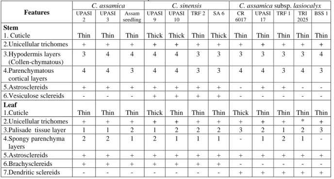
Table 2 Anatomical variation in tea clones of three Camellia species
Features
C. assamica C. sinensis C. assamica subsp. lasiocalyx
UPASI 2 UPASI 3 Assam seedling UPASI 9 UPASI 10
TRF 2 SA 6 CR
6017
UPASI 17
TRF 1 TRI
2025 BSS 1
Stem
1. Cuticle Thin Thin Thin Thick Thick Thin Thick Thin Thin Thin Thin Thin
2.Unicellular trichomes + + + + + + + + + + + +
3.Hypodermis layers (Collen-chymatous)
3 4 4 4 4 3 3 3 3 3 3 4
4.Parenchymatous cortical layers
4 4 3 4 4 3 3 4 4 3 4 3
5.Astrosclereids + + + + + + + - + + -
-6.Vesiculose sclereids - - - + + + + - - - - -
Leaf
1.Cuticle Thin Thin Thin Thick Thin Thin Thin Thick Thin Thin Thin Thin
2.Unicellular trichomes + + + + + + + + + + * +
3.Palisade tissue layer 1 1 2 1 2 2 2 3 2 1 2 3
4.Spongy parenchyma layers
2 2 1 2 1 1 1 - 1 2 1 -
5.Astrosclereids + + + + + + + + + + + +
6.Brachysclereids + + + + + + + - - -
7.Dendritic sclereids - - - + + + + +
Discussion
Thick cuticle has been considered as a reliable character indicating drought resistance14,16. Markedly thick cuticle is present in stems of only some of the clones of C. sinensis but not in the other two groups of clones (Table 2, Fig. 1 b). In the leaves, on the other hand show thick cuticle in one of C. sinensis and C.
assmica subsp. lasiocalyx. In this context, it becomes difficult to assort the thickness of cuticle as an essential
feature of some significance, either from logical point of view or from the point of view of taxonomic importance. However, a very thick cuticle found in stems and leaves of UPASI 9 (present study) seems to have a bearing on its relationship to its inherent drought tolerant nature, as emphasized by Balasubramanian et al., (2010)19. Similar significance of cuticle has been emphasized in bread wheat variety20 and also in xeric grasses21. The present findings of micromorphological features are in consonance with those of earlier study on
Camellia sinensis from Brazil22. However, sclereids reported from pith in stems have not been found by others. Lu et al., (2008)23 reported single layered epidermis in C. rhytidophylla, and two layered in C. parvimuricata, C.
hupehensis and C. ilicifolia. Pi et al., (2009)24 reported discontinuous multiple epidermis in 43 species of C.
japonica.
Conclusion
Therefore, discriminating tea plants based on the number of mesophyll layer are disputable although earlier studies classified certain wild and ornamental Camellia species. It is not usually considered natural to determine their taxonomic status based on only one or two characters. Our reports on stem sclereids in Camellia
sinensis and C. assamica were first of its kind and a highly discriminating descriptor for certain clones of C. assamica subsp. lasiocalyx as they are absent. Our findings on sclereids, type and distribution pattern are
distinguishing features among the clones studied, not particularly influenced by environmental factors. We conclude that anatomical variations in the genus Camellia are relatively stable, their diversity and regularity are of some taxonomic value. Our study provides useful data for addressing taxonomic problems and discrimination of commercial teas using tea descriptors.
Acknowledgements
One of the authors (MR) would like to acknowledge the support and encouragement of the Chairman
and Principal of CMR IMS, Bangalore, India. The authors are thankful to UPASI-Tea Research Institute,
Valparai, Tamil Nadu, India for providing the plant material. We are also grateful to Dr. D. Sivaramakrishna, Retired Professor, Department of Botany, Bangalore University, Bangalore, India for going through this manuscript.
References
[1] Venkataramani, K.S.; Sharma, V. S. (1974). The tea clone: ‘Sundaram’, Planters’ Chronicle, 69, pp. 353- 355.
[2] Mohanan, M.; Sharma, V. S. (1981). Morphology and systematics of some tea (Camellia species) cultivars. Proceedings of Annual
Symposium on Plantation Crops, PLACROSYM IV, Central Coffee Research Institute, Chikmagalur, India, pp. 391-400.
[3] Sealy, J. R. (1958). A revision of the genus Camellia. London, The Royal Hort. Soc., pp. 1–239.
[4] Zhang, H. D.; Ren, S. X. (1998). Flora Reipublicae Popularis Sinicae, Vol. 49 (3), Science Press, Beijing.
[5] Ming, T. L. (1999). A systematic synopsis of the genus Camellia. Acta Bot. Yunnanica, 21, pp. 149–159.
[6] Haron, N. W.; Moore D. M. (1996). The taxonomic significance of leaf micromorphology in the genus Eugenia L. (Myrtaceae). Bot. J.
Linn. Soc., 120, pp. 265–277.
[7] Lux, A.; Morita, S.; Abe, J.; Ito, K. (2005). An Improved Method for Clearing and Staining Free-hand Sections and Whole-mount
Samples. Annals of Botany, 96, pp. 989–996.
[8] Ao, C. Q; Ye, C. X.; Zhang, H. D. (2007). A systematic investigation of leaf epidermis in Camellia using light microscopy. Biologia,
62, pp. 157–162.
[9] Baranova, M. (1972). Systematic anatomy of the leaf epidermis in the Magnoliaceae and some related families. Taxon, 21, pp. 447–
469.
[10] Luna, I.; Ochoterena, H. (2004). Phylogenetic relationships of the genera of theaceae based on morphology. Cladistics, 20, pp. 223-270.
[11] Rao, T. A.; Bhattacharya, J. (1978). A review on foliar sclereids in angiosperms. Bulletin of the Botanical survey of India, 20, pp. 91-98.
[12] Trift, I.; Anderberg, A. A. (2006). Foliar sclereids in Dionysia (Primulaceae) from a phylogenetic perspective. Edinburgh Journal of
Botany, 63, pp. 21-48.
[13] Zhang, W.; Hu, Y.; Li, Z.; Wang, P.; M. Xu. (2009). Foliar sclereids in tea and its wild allies, with reference to their taxonomy.
Australian Systemic Botany, 22, pp. 286-295.
[14] Ramon, K.; Chang, P. C. (1982). Comparative foliar anatomical studies of clonal tea. Proc.4th. Int. Symp. Plant. Crop. UPASI Tea.
Inst. Cinchona. Tamil Nadu. India. pp. 413-424.
[15] Manivel, L. (1998). Tea Botany and Horticulture. Horticultural Reviews, 22, pp. 267-295.
[16] Balasubramanian, S.; Muraleedharan, N. (2000). Anatomical variations in the leaf of tea clones. Planters Chronicle, 96 (2), pp. 85-86.
[17] Ruzin, S.E. (1999). Plant microtechnique and microscopy. New York: Oxford University Press.
[19] Balasubramanian, S.; Parathiraj, S.; Netto, L. A. (2010). Unique graft combination of tea Cr-6017 / UPASI-9. Current science. 98 (11), pp. 1508-1517.
[20] Mehrun-Nisa, K. Y. (2007). Evaluation on anatomical and morphological traits in relation to low water requirement conditions of
bread wheat (Triticum aestivum l.). Pak. J. Bot., 39(7), pp. 2725-2731.
[21] Ubeda, J.A. (1993). Morpho-anatomy of drought resistance in different ecotypes of Cenchrus cilians L., from Cholistan. M. Phil
thesis, Dept. Bot. Univ. Agric, Faisalabad, Pakistan, pp. 121.
[22] Marcia, R. D.; Daniele, O. M. (2006). Morpho diagnosis of leaf and stem anatomy of Camellia sinensis (L.) Kuntze, Theaceae.
Brazilian jounal of Pharmacognosy, 16 (4), pp. 545-551.
[23] Lu, H. F.; Jiang, H. F.; Shen, Z. G.; Shen, J. B.; Peng, Q. F; Cheng, C. G. (2008). Comparative leaf anatomy, FTIR discrimination and biogeographical analysis of Camellia section Tuberculata (Theaceae) with a discussion of its taxonomic treatments. Plant Syst. Evol.,
274, pp. 223–23.
[24] Pi-Erxu; Peng-Qiufa; Lu-Hongfei; Shen-Jingbo; Du-Yueqiang; Huang-Feilai; HU-Hui. (2009). Leaf morphology and anatomy of