Submitted23 March 2016
Accepted 24 May 2016
Published6 July 2016
Corresponding author
Frank J. Varriale, frankvarriale@kings.edu
Academic editor
Mathew Wedel
Additional Information and Declarations can be found on page 15
DOI10.7717/peerj.2132
Copyright
2016 Varriale
Distributed under
Creative Commons CC-BY 4.0
OPEN ACCESS
Dental microwear reveals mammal-like
chewing in the neoceratopsian dinosaur
Leptoceratops gracilis
Frank J. Varriale
Department of Biology, King’s College, Wilkes-Barre, PA, United States
ABSTRACT
Extensive oral processing of food through dental occlusion and orbital mandibular movement is often cited as a uniquely mammalian trait that contributed to their evolu-tionary success. Save for mandibular translation, these adaptations are not seen in extant archosaurs or lepidosaurs. In contrast, some ornithischian dinosaurs show evidence of precise dental occlusion, habitual intraoral trituration and complex jaw motion. To date, however, a robust understanding of the diversity of jaw mechanics within non-avian dinosaurs, and its comparison with other vertebrates, remains unrealized. Large dental batteries, well-developed dental wear facets, and robust jaws suggests that neoceratopsian (horned) dinosaurs were capable chewers. But, biomechanical analyses have assumed a relatively simple, scissor-like (orthal) jaw mechanism for these animals. New analyses of dental microwear, presented here, show curvilinear striations on the teeth ofLeptoceratops. These features indicate a rostral to caudal orbital motion of the mandible during chewing. A rostrocaudal mandibular orbit is seen in multituberculates, haramiyid allotherians, and some rodents, and its identification inLeptoceratops gracilis is the first evidence of complex, mammal-like chewing in a ceratopsian dinosaur. The term circumpalinal is here proposed to distinguish this new style of chewing from other models of ceratopsian mastication that also involve a palinal component. This previously unrecognized complexity in dinosaurian jaw mechanics indicates that some neoceratopsian dinosaurs achieved a mammalian level of masticatory efficiency through novel adaptive solutions.
SubjectsEvolutionary Studies, Paleontology
Keywords Dental microwear, Jaw action, Mastication, Chewing, Ornithischia, Dinosauria, Ceratopsia, Jaw mechanics
INTRODUCTION
Ösi & Weishampel,2009;O’connor et al.,2010). Among non-avian dinosaurs, mastication is well supported in hadrosaurid ornithopods. Hadrosaurs possess numerous cranio-dental specializations for mastication, including a jaw joint depressed below the dental arcade, an elongated coronoid process, and most strikingly a dental battery consisting of many closely packed teeth with dentine of differing hardness (Ostrom,1961;Weishampel,1984;Norman & Weishampel,1985;Williams, Barrett & Purnell,2009;Erickson et al.,2012;Cuthbertson et al.,2012). Many neoceratopsians exhibit similar adaptations, providing support for an identical level of masticatory sophistication (Ostrom,1964;Ostrom, 1966;Tanoue et al., 2009;Erickson et al.,2015). Because neoceratopsian skulls lack intracranial joints with large gaps (Holliday & Witmer, 2008), and have a simple hinge-like jaw mechanism, biomechanical analyses of their chewing infer an unsophisticated orthal motion that resulted in a scissor-like (orthal) adduction of the lower jaw (Ostrom,1964;Ostrom, 1966; Tanoue et al.,2009).
Food often leaves microscopic traces known as microwear, in the form of pits and scratches on the occlusal surfaces of teeth (Teaford,1988a;Teaford,1991). Comparison of microwear within and among taxa and analyses of scratch orientation have been successfully used to test hypotheses of jaw motion in numerous mammalian taxa (Teaford & Byrd,1989; Teaford,1991;Charles et al.,2007), as well as various dinosaur groups (Weishampel,1984; Upchurch & Barrett, 2000;Rybczynski & Vickaryous,2001;Williams, Barrett & Purnell, 2009;Whitlock,2011;Mallon & Anderson,2014;Ösi et al.,2014). Unlike mammalian teeth, each neoceratopsian tooth possessed a single, relatively flat, near vertical occlusal surface resulting from shear between the maxillary and mandibular dentitions (Hatcher et al.,1907; Dodson, Forster & Sampson,2004). Occlusal microwear on ceratopsian teeth is the result of the direction and magnitude of the jaw closing power stroke of mastication, and an exceptional record of jaw action is recorded on teeth as a result of the planar nature of the occlusal surface. Given a strictly orthal model, predicted microwear should be composed of striations that are rectilinear and limited to a single modality at or near the apicobasal axis of the tooth. To test this hypothesis, dental microwear was examined in the non-ceratopsid neoceratopsianLeptoceratops gracilis, B. Brown (Brown,1914).
MATERIALS & METHODS
Dental microwear was examined in a nearly complete, articulated skull of the neoceratopsianLeptoceratops gracilis(CMN 8889; Canadian Museum of Nature, Ottawa, Ontario, Canada) as well as the holotype material (AMNH FR 5205; American Museum of Natural History, New York, USA) and isolated teeth (n=2) assigned to the taxon (YPM VPPU 018133; Yale Peabody Museum of Natural History, New Haven, Connecticut, USA). The conclusions in this analysis are derived primarily from examination of CMN 8889. AMNH FR 5205 and YPM VPPU 018133 were examined to provide qualitative support for conclusions drawn from CMN 8889; they were not quantitatively analysed because preserved microwear was either of slightly lesser quality and did not provide a large sample size (AMNH FR 5205) or represented isolated teeth (YPM VPPU 018133).
Jet regular (product number C6012) was used to obtain peels of occlusal surfaces, whereas the President impression putty (product number C4843) was used to create walls around the edge of peels, transforming them into cups capable of receiving epoxy resin. Prior to molding, teeth were screened for alterations and artifacts that can occur due to taphonomic, post depositional, and museum conservation processes. Teeth from all specimens included here were deemed suitable for analysis because they met a number of criteria suggested by other workers as indicative of genuine microwear. Microwear on theLeptoceratopssample is relatively uniform, having a regular pattern and showing no evidence of multiple abrupt shifts or large gouges indicative of preparation marks (Teaford,1988b). Striations are well defined and show none of the obliteration and dulling characteristic of taphonomic particulate abrasion or acid etching (Teaford,1988b;King, Andrews & Boz,1999). When preservative obscuring occlusal surfaces was encountered, it was removed using a gentle scrubbing action with cotton swabs and acetone or ethanol as the solvent.
Casts were poured using a two-part epoxy resin (EPO-TECH #301) designed to cure slowly at room temperature with minimal exothermal heat production. Casts were allowed to cure for 14–21 days at ≈23◦C. Upon solidification, casts were removed from their
molds and secured to SEM specimen mounts (SPI SuppliesR Aluminum Pin-Type Mounts
#1507L-MB) using a nonconductive modified nitrocellulose solution (Duco CementR). Casts were then gold-palladium coated for 180 s using a Denton Vacuum Desk III set to 40 milliamps and a 50% Argon gas mixture. Silver paint (SPI SuppliesR, Ag Colloidal Suspension #05001-AB) was used to create a conductive connection between the coated specimen and SEM mount, ensuring electron transmission.
Microscopic examination was conducted using an Amray 1810 scanning electron microscope set to a working distance of 11 mm, and a 20-keV electron beam in secondary emission mode. The occlusal surface was oriented orthogonal to the electron beam to provide a controlled position. This, coupled with the secondary emission mode, helped to minimize the extinction of features that can occur with tilting and use of backscatter electrons (Galbany, Martinez & Perez-Perez,2004). Specimens were photographed at 100X using PolaroidR, Polapan 55 film (ISO 50/18◦20 s/Sek/s), and micrographs were scanned
as bit-map images at a resolution of 300 dpi for computer analysis.
Wear features were digitized using Microware, Version 4.02, a semi-automated image analysis system for the quantification of dental microwear (Ungar,2002). Microware 4.02 records features as four points (x,y coordinates) on a Cartesian grid as the user defines the length and width of a feature by clicking on its ends with a computer mouse. These Cartesian data were imported into a Microsoft Excel spreadsheet, and a macro (Articles S1 andS2) was written to trigonometrically calculate the angle of microwear striations within a 180◦arc progressing from apical to basal on the distal side of the tooth.
micrographs were flipped horizontally using Adobe Photoshop prior to digitization with Microware 4.02 to obtain angular data directly comparable and easily visualized for all teeth in all quadrants of the dentition. Micrographs from teeth in the right dentary quadrant were designated as the standard and all other quadrants manipulated to provide data comparable with this quadrant. Left dentary and right maxillary micrographs were flipped horizontally. Left maxillary teeth did not require manipulation because angles measured from this quadrant are the same as those in the right dentary. This protocol produced comparable angles without the need for mathematical transformation via the trigonometric principle that alternate interior angles are equal. Analyses of angular data (Data S1) were conducted using Oriana, Version 2.0 (Kovach,2003), a circular statistics and rose diagram software package. Oriana was used to generate rose diagrams, calculate mean angle, Rao’s spacing test, and length of the mean vector (r). Rose diagrams depict angular data on a unit circle with increasing angle as a function of clockwise rotation. As such, all rose diagrams herein depict angles relative to wear on a left dentary tooth of a left facing skull, facilitating direct visual comparison among dental quadrants. In all rose diagrams 90◦ is equivalent to the
caudal direction and 180◦ is ventral.
r and mean angle
The homogeneity of scratch orientations can be measured by the valuer, which is the length of the mean vector of circularly-distributed data on a unit circle with an imaginary radius of 1.0 (Zar,1998;Mardia & Jupp,2000). The mean vector is inversely proportional to uniformity, with values ranging from 0 to 1.0. Values ofrapproaching 0 indicate striations whose angles are uniformly dispersed around the circle, whereas those approaching 1.0 indicate angles with high homogeneity confined to a relatively small angular arc. The following formula describesr:
r=1
n v u u t n X i=1
cosθi
!2 +
n
X
i=1
sinθi
!2
wherenis the sample size andθis the angle of theith striation. Because a single axis can be described by two different angles 180◦apart, using these data in the above equation without
transformation would yield anr value lower than expected. For example, two striations with angles 10◦and 190◦are parallel to each other and striking in the same direction, thus
anr value of 1.0 is expected. However, these values yield anr=0 when inserted into the equation because they are on opposite sides of the unit circle and thus evenly distributed. For axial data, all angles are doubled (multiplied by 2), and any angle greater than or equal to 360◦is subtracted by 360◦. This transformation has the effect of rotating any angle of θ that is in the 180◦–359◦hemisphere back into the 0◦–179◦hemisphere so that it is equal to its counterparts in that half of the circle, and yielding a correctrvalue. The mean angle can then be found using the mean sine and cosine values calculated above for the length of the mean vector, using the inverse tangent function:
θ=tan−1 n
X
i=1
sinθi
! n X
i=1
cosθi
!
Because of the doubling of angles for thercalculation above, the angle resulting from this equation should be halved to arrive at the correct mean angle (Zar,1998;Mardia & Jupp, 2000).
Rao’s spacingL-test
Rao’s spacing test evaluates the null hypothesis that a sample of angles is uniformly distributed around a circle (Mardia & Jupp,2000). A value less than the predetermined significance level rejects the null hypothesis and supports the alternative, that the angles show a preferred direction. This test was chosen because it is more powerful when confronted with bimodal data separated by 180◦, compared to other similar tests such as
the Rayleigh test of uniformity. The Rayleigh test incorporatesr in calculating its test statistic Z (Z=nr2). Because bimodal data on opposite sides of the circle yield lowr
values, theZ value would be proportionately small. SmallZ values fail to reach the given level of significance (p=0.05), increasing the likelihood of a type II error, which would fail to reject the null hypothesis. Rao’s spacing does not user, but instead examines the spacing between points on the unit circle and compares their deviation from the uniform case where the spacing should be 2π /nradians (360◦/n). TheLstatistic is calculated:
L=1
2 n
X
i=1
Ti−2
π
n
wherenis the sample size, andTiis the spacing or difference between the observed angles given the following:
Ti=θ(i)−θ(i−1), i=1,...,n−1,Tn=2π−(θ(n)−θ(1)).
BecauseTi is the distance betweennobserved points and 2π /nis the expected distance betweennpoints then the difference between them should be large if the observed points are clustered, thus yielding a large value ofL(Mardia & Jupp,2000).
RESULTS
Microwear striations on teeth of Leptoceratops(CMN 8889) conform to predictions of orthal chewing by being unimodal in distribution (Fig. 1). However, they differ greatly in other parameters. Striations are not rectilinear, as would be expected of a simple scissor-like closure of the mandible, but are curvilinear (Figs. 2A–2B and3). Striations begin at the apicodistal border on dentary teeth and curve though an arc of nearly 50◦before ending in
either a horizontal or apicomesial orientation near the mesial border (Figs. 2Aand3A–3B). Maxillary teeth show a counterpart curvature, with striations beginning apicomesially and ending distal to apicodistally (Figs. 2Band3C–3D).
Figure 1 Histogram of striation orientations pooled from all teeth examined inLeptoceratops gra-cilis(CMN 8889).Classes are in 10◦increments. A single mode is present, with the greatest frequency of
scratches in an arc from 30◦to 60◦. Line intersecting largest bar is the pooled sample mean angle (44.1◦).
N=1,504.
teeth is composed of the enamel, hard mantle dentine, and orthodentine materials discussed byEricksonand others (2015). Microwear is formed in, and traverses all of these tissues, but only a thin rim of enamel encapsulates the edge (cutting edge) of the occlusal surface, As such, most of the occlusal surface is hard mantle dentine and orthodentine. As mentioned above, micrographs were taken near the apicodistal and apicomesial edges of teeth, resulting in capture of microwear formed primarily in orthodentine and some hard mantle dentine. Microwear can be seen to traverse materials of differing hardness without appreciable shift in direction (Figs. 2Aand2C).
A Rao’s spacing test was performed on angular measurements from each tooth (Table 1), and in all cases a uniform distribution of measurements about a 360◦circle is rejected
(p<0.01), supporting a preference for the mean direction. Furthermore, striations are not oriented with or near the apicobasal axis of the tooth but are inclined distally relative to it. This distal inclination is consistent across all teeth examined with means from individual teeth ranging from 26◦–55◦(Table 1andFigs. 2C–2Fand4).
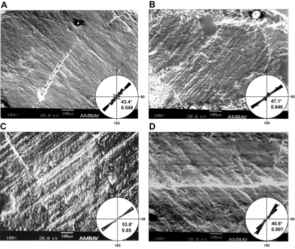
Figure 2 Dental microwear on representative teeth ofLeptoceratops gracilis(CMN 8889). (A) Light microscope images of the seventh left maxillary, and (B) sixth left dentary teeth showing curvilinear mi-crowear traversing the entire occlusal surface. Arrows indicate the direction of initiation and exit of the power stroke. Microwear near the site of initiation is oriented caudodorsally whereas the same wear near the end of the stroke is oriented rostrocaudally, a≈50◦shift in orientation. Dotted lines in (A) and (C)
de-marcate approximate junction of hard mantle dentine (HMD) with orthodentine (O). (C, D) SEM micro-graphs imaged from rectangles indicated in (A) and (B) respectively. At this magnification (100×) wear appears uniform with a striation dominated texture. (E, F) Rose diagrams of angular data from striations in (C) and (D). Rose diagrams are on a unit circle and summarize angular orientation relative to wear on a left dentary tooth. 90◦=caudal and 180◦=ventral directions. Values in lower right quadrant of rose
di-agrams are the mean angle and the length of the mean vector (r). Arrows in rose diagrams indicates direc-tion and magnitude ofr.
that summarizes the parallelism of scratches. If all scratches are nearly parallel, or have a similar angular orientation, then the value ofrapproaches one, if they are more randomly distributed then this value approaches zero (Zar,1998;Mardia & Jupp,2000).
Microwear preserved on additional specimens ofLeptoceratops(AMNH FR 5205, YPM
VPPU 018133) corroborates qualitative observations made on CMN 8889. An isolated right
maxillary tooth assigned toLeptoceratops(YPM VPPU 018133) shows dental microwear
with an apicomesial (rostroventral) orientation near the site of powerstroke initiation, and striations curve through a similar arc to end in a mesiodistal (rostrocaudal) orientation
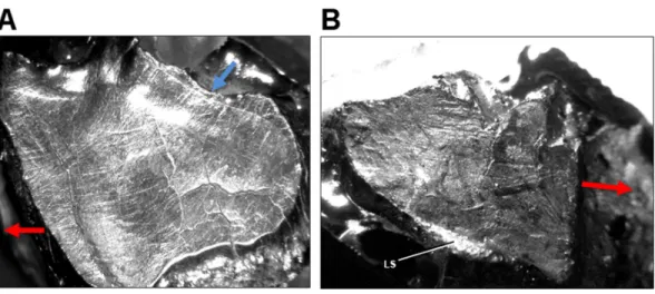
(Compare Fig. 5AwithFig. 2A). Teeth from AMNH FR 5205 show multiple stages of
Figure 3 Light microscope images of additional teeth fromLeptoceratops gracilis(CMN 8889) show-ing semicircular dental microwear. (A) Eighth right dentary tooth. (B) 12th right dentary tooth. (C) 10th right maxillary tooth. (D) 11th right maxillary tooth. Colored arrows correspond to those inFig. 6and in-dicate (where visible) the overall orientation of microwear at the initiation (blue) and ending (red) of the power stroke. Arrow (black) in (D) indicates step between two facets (f1, f2) formed via differential wear. These facets were likely caused by the occlusion of this tooth against two opposing right dentary teeth at different stages of eruption. Images not to scale.
the mesiodistal axis, and showing a slight change in curvature. The wear also reflects a similar orientation as the nearby labial shelf (LS). Considering this structure as a result of differential wear via incomplete shear of maxillary teeth against their dentary counterparts, its orientation should be similar to nearby microwear on the occlusal surface.
DISCUSSION & CONCLUSIONS
Table 1 Summary and inferential statistics for angular data from teeth examined inLeptoceratops gracilis(CMN 8889). Alveolar position of each tooth from rostral is indicated by number.
Maxillary teeth LM5* LM6 LM7 LM13* RM2* RM3* RM4 RM10 RM11 RM13*
N 142 100 130 104 37 71 84 103 39 8
Meanθ◦ 43.56 47.15 26.85 39.63 31.23 38.66 40.63 35.3 34.87 46.6
Length ofθ◦(r) 0.686 0.646 0.533 0.833 0.98 0.867 0.897 0.665 0.9 0.993
Rao’s spacing (U) 198.208 203 191.123 246.954 308.541 243.248 264.029 216.012 260.246 292 Rao’s spacing (p) <0.01 <0.01 <0.01 <0.01 <0.01 <0.01 <0.01 <0.01 <0.01 <0.01
Dentary teeth LD4* LD6 LD7* LD13 RD8 RD9* RD10* RD12 RD14*
N 30 129 67 175 61 59 58 76 31
Meanθ◦ 53.65 54.44 55.09 43.44 53.82 51.29 52.96 43.61 40.78
Length ofθ◦(r) 0.905 0.884 0.886 0.548 0.85 0.976 0.987 0.9 0.958
Rao’s spacing (U) 256 259.712 252.642 213.686 288.39 301.197 311.779 267.116 280.574 Rao’s spacing (p) <0.01 <0.01 <0.01 <0.01 <0.01 <0.01 <0.01 <0.01 <0.01
Notes.
*Data for additional teeth not figured in text.
LD, left dentary; LM, left maxillary; RD, right dentary; RM, right maxillary.
along the mean striation angle. To traverse an unbroken arc, the mandible must have undergone uninterrupted precise motion as the dentary teeth slid past the maxillary teeth during the power stroke. Indeed, the high degree of striation parallelism (highr) indicates a power stroke event that was performed under precise muscular action and one that must have been stereotyped due to the homogenous nature of curvilinear striations. (Figs. 2–5).
Refutation of the orthal model of mastication in neoceratopsians requires an alternative with increased explanatory power. Unlike mammals, ceratopsians retained the plesiomorphic organization of jaw adductor muscles present in their amniote ancestors. Them. adductor mandibulae externusgroup and them. adductor mandibulae posteriorare the major jaw closing muscles (Haas,1955;Holliday,2009). These muscles have caudodorsal vectors, with the former having a stronger dorsal component and the latter a more caudal orientation. Them. pterygoideusgroup is also involved in jaw closure, having a rostrodorsal vector. However, its role is more pronounced during the beginning of adduction (Carroll, 1969) rather than the power stroke, and a correlate in microwear is undetectable. Microwear at the apicodistal limit of dentary teeth is oriented in the direction of the supratemporal fenestrae, suggesting that the power stroke was initiated by the m. adductor mandibulae externusgroup. Although them. adductor mandibulae externusgroup would have assisted in retraction of the jaw due to its caudodorsal vector, the muscle of primacy for producing the mesiodistally oriented microwear at the basomesial edge of dentary teeth was the m. adductor mandibulae posterior. Production of the uniform arc seen on these teeth must have involved a precise yet smooth transition between actions of them. adductor mandibulae externusgroup and them. adductor mandibulae posterior (Fig. 6).
Figure 4 Micrographs of selected teeth from each of the four dental quadrants inLeptoceratops gracilis(CMN 8889).(A) 13th left dentary tooth, (B) sixth left maxillary tooth, (C) eighth right dentary tooth, and (D) fourth right maxillary tooth. Orientations of micrographs are as follows; apical at left and basal at right. In left dentary and left maxillary micrographs distal is at top and mesial is at bottom. In right dentary and right maxillary images distal at bottom and mesial is at top. Rose diagrams summarize angular orientation relative to wear on a left dentary tooth. Description of rose diagram orientation and data are the same as inFig. 2.
Figure 5 Light microscope images of dental microwear in additional specimens ofLeptoceratops. (A) Isolated right maxillary tooth assigned toLeptoceratops(YPM VPPU 018133) showing semicircular dental microwear. (B) Fifth right dentary tooth fromLeptoceratops gracilis(AMNH FR 5205) showing a predominance of mesiodistally oriented wear near the base of the facet, and in a similar orientation as the labial shelf (LS). Colored arrows correspond to those inFig. 6and indicate the overall orientation of microwear at the initiation (blue) and ending (red) of the power stroke. Images not to scale.
Figure 6 Model of circumpalinal mastication inLeptoceratops. (A) Adduction of the lower jaw dom-inated by action (blue arrow) of them. adductor mandibulae externusgroup (mAME) and beginning of the power stroke of mastication. (B) Progression of the power stroke into a palinal (retraction) phase dominated by transition from the mAME to action (red arrow) of them. addcutor mandibulae posterior
been demonstrated in the basal ceratopsianPsittacosaurus. Previously,Sereno(1987) and Norman & Weishampel(1991) suggested a propalinal mechanism forPsittacosaurus, with Sereno(1987) forwarding a palinal powerstroke. However, recent work has clarified a more detailed mechanism where inclined dental facets combined with diverging tooth rows and an orthopalinal motion yielded continuous occlusion during the power stroke (Sereno, Xijin & Lin,2010). The term clinolineal was coined to encompass the jaw mechanism of Psittacosaurus, because occlusion occurred over an inclined linear direction (Sereno, Xijin & Lin,2010). However, microwear produced by clinolineal and orthopalinal chewing is rectilinear and not strongly curved, as that demonstrated here in Leptoceratops. Unlike the condition inPsittacosaurus and derived ceratopsids, the semicircular microwear of Leptoceratops supports a power stroke that progressed smoothly into a palinal phase (Fig. 6). The term circumpalinal is proposed here to describe the semicircular orbit that is accomplished during the overall palinal (front-to-back) jaw action inLeptoceratops, and to distinguish this style of chewing from the clinolineal and orthopalinal mastication of other ceratopsians.
Among dinosaurs, complex mastication may have been achieved through intracranial joints, in a manner very different from that seen in mammals. Euhadrosaurs (duck-billed dinosaurs) have been reconstructed as masticating using pleurokinesis, a unique motion in which the maxillae and associated teeth swung laterally as the mandibular dentition occluded with them (Weishampel,1984;Norman & Weishampel,1985). However, the existence of pleurokinesis is currently contested. The work ofHolliday & Witmer(2008) indicates that despite having intracranial synovial joints, hadrosaurids were only partially kinetically competent. Cuthbertson and others (2012) further examined the masticatory apparatus inBrachylophosaurus andEdmontosaurusby scrutinizing dental microwear, arthrology, and kinematic models. They concluded that the facial skeleton of these taxa were akinetic. Still, microwear from the teeth of euhadrosaurs indicates that the jaws may have slid past one another in a pleurokinetic mechanism (Williams, Barrett & Purnell, 2009). However, it now seems that mandibular long-axis rotation, combined with an orthopalinal powerstroke and accessory propalinal motion, has the greatest explanatory power for interpreting observed microwear (Cuthbertson et al.,2012;Nabavizadeh,2014; Mallon & Anderson,2014).
What little is known of their mastication indicates that each of the major ankylosaur clades may have had their own unique and complex jaw actions.Rybczynski & Vickaryous (2001) showed that mastication in the derived ankylosaurid Euoplocephalus was accomplished by swinging the caudal portion of the dentary bones laterally as a result of translation within the jaw joint. The predentary and dentary bones forming a condyloid joint, permitting medial rotation of the anterior dentary. The combined effect created a propalinal arcing of the mandible with both shearing and crushing abilities (Rybczynski & Vickaryous,2001). Ősi and others (2014) examined microwear, jaw architecture, and myology in the nodosauridHungarosaurus, reconstructing a different action than that inEuoplocephalus. Microwear on dentary teeth ofHungarosaurusis bimodal with a near apicobasally oriented class that traverses most of the occlusal surface, and a mesiodistal class that is principally located near the base of occlusal facets. They propose that jaw action was initiated with an orthal motion followed by transition into a palinal powerstroke, these actions being assisted by mandibular rotation or medial flexion.
The action seen here inLeptoceratops bears some resemblance to that proposed for Hungarosaurus (Ősi et al.,2014); however, there are also some notable departures. The apicobasally/apicodistally oriented microwear onHungarosaurusdentary teeth does not show a consistent and continuous transition to a mesiodistal orientation. Save for a few scratches showing a curved transition, the two classes are disconnected, indicating that transition to a palinal stroke was not as smooth as inLeptoceratops, but a discrete event. The proposed chewing cycle forHungarosaurusbears this out, with orthal and palinal phases depicted as discreet and rapid changes in vector (Ősi et al.,2014). The transition in Leptoceratopswas not swift and abrupt, but one spread over the entire powerstroke with the possibility of one muscle group trading action to another. The relatively flat glenoid of Hungarosauruscompared to the deeply cupped joint inLeptoceratopsalso influences the path a powerstroke can take (see below) and is an additional reason for the departure of action in these two taxa. Nevertheless, the basic jaw action of Hungarosaurustraces a similar, albeit, disjointed path.
The semicircular character of dental microwear striations inLeptoceratopsresulted from a combination of the aforementioned muscular actions directing motion at the jaw joint. As the chewing cycle began, the quadrate started at the caudal rim of the mandibular glenoid, causing apicodistally oriented striations on dentary teeth. When the quadrate reached the base of the glenoid, microwear on teeth transitioned to a mesiodistal orientation, and then to an apicomesial orientation as further adduction caused the quadrate to ride up the rostral wall of the glenoid. An analogous rostrocaudal masticatory orbit within an akinetic skull occurs in several mammalian groups, including multituberculates (Wall & Krause,1992), haramiyid allotherians (Butler,2000), and some rodents (Rose,2006). Palinal occlusion also occured in cynodonts, but microwear and tooth morphology show no evidence of orbital motion of the mandible (Crompton,1972;Rybczynski & Reisz,2001). The presence of mammal-like orbital motion inLeptoceratopsis striking because of the structural differences in the jaw joint between dinosaurs and mammals. In mammals, the condyle is on the dentary bone and the glenoid depression is on the underside of the skull in the squamosal. Dinosaurs are essentially the reverse, as they retain the ancestral amniote condition of a condyle on the quadrate of the skull and the glenoid depression in the articular of the lower jaw (Norman & Weishampel,1991). This retention of the ancestral condition suggests that complex motion of the lower jaw, involving multiple vectors in an orbital motion, need not require a mammalian jaw architecture for its development, and it broadens our understanding of comparative biomechanics.
The greatest biodiversity of ceratopsians occurs during the latter half of the Cretaceous, a time known for the diversification and revolution of terrestrial communities by angiosperm plants (Lloyd et al.,2008). However, recent investigations have questioned or shown no correlation in diversification between dinosaur groups and angiosperms (Weishampel & Jianu,2000;Barrett & Willis,2001;Lloyd et al.,2008;Butler et al.,2010). The recognition of a novel chewing adaptation in a ceratopsian dinosaur suggests that previous assignments (Weishampel & Norman,1989;Weishampel & Jianu,2000) of taxa to masticatory groupings (orthal pulper, orthal slicer, etc.) may have been too gross.
The disparate chewing mechanisms ofEuoplocephalusandHungarosauruscoupled with
a depauperate understanding of chewing in other ankylosaurs demonstrates the need for refining the details of jaw action in herbivorous dinosaurs. This refinement, in addition to other areas of inquiry (sampling bias, spatio-temporal diversity and abundance) may bring much needed resolution to the ongoing question of angiosperm-dinosaur coevolution.
material, at a minimum it corroborates circumpalinal mastication inLeptoceratops gracilis, and at most hints at a wider distribution of this chewing style within Leptoceratopsidae.
Recent work also addresses the question of wider distribution, and indicates that circumpalinal mastication was not present in derived ceratopsids (Varriale, 2011; Mallon & Anderson,2014). However, this leaves a large paraphyletic portion of Ceratopsia for which jaw action is unknown, and as such the ancestral action for all ceratopsians is also unknown. Clinolineal mastication could extend to the base of Ceratopsia or be limited to Psittacosauridae. If limited, then circumpalinal or orthopalinal mastication may be the ancestral conditions. A fourth, as-yet undiscovered action may be present there, or the orthal mechanism that seems to be present in the ceratopsian outgroup Pachycephalosauria (Sues & Galton,1987;Varriale, 2015) and many other ornithischians (Thulborn,1971; Weishampel,1984;Crompton & Attridge,1986;Norman & Weishampel,1991;Barrett,1998; Barrett,2001), could have been retained at the ceratopsian base. To further support the results here forLeptoceratops, and answer the aforementioned questions, a much wider range of taxa will need to be sampled, including other non-ceratopsid neoceratopsians as well as basal ceratopsians outside of Psittacosauridae. This expanded analysis is currently underway by F Varriale.
Recognition of circumpalinal chewing within Leptoceratopsadds a distinct style of mastication to the dinosaurian repertoire, one that is fundamentally different from previously understood mechanisms involving cranial or mandibular kinesis. This suggests that some dinosaurs may have possessed a mammalian level of masticatory prowess and biomechanical diversity, achieving this convergence through novel independent adaptations of the masticatory apparatus.
ACKNOWLEDGEMENTS
Thanks to Andrew Farke, Jordan Mallon, David Weishampel, and Mark Teaford for comments and discussion that improved initial drafts of this manuscript. Paul Barrett and Jeremy Green provided thorough and insightful reviews that enhanced the quality of the narrative. Walter Joyce (YPM), Carl Mehling (AMNH), Natalia Rybczynski (CMN), and Kieran Shephard (CMN) provided access to specimens.
ADDITIONAL INFORMATION AND DECLARATIONS
Funding
Funding for this research was provided by grants from the Jurassic Foundation, Sigma Xi: Grants in Aid, The Geological Society of America, and a Stephen J. Gould Award from the Paleontological Society. The funders had no role in study design, data collection and analysis, decision to publish, or preparation of the manuscript.
Grant Disclosures
Sigma Xi.
The Geological Society of America.
Stephen J. Gould Award from the Paleontological Society.
Competing Interests
The author declares there are no competing interests.
Author Contributions
• Frank J. Varriale conceived and designed the experiments, performed the experiments, analyzed the data, contributed reagents/materials/analysis tools, wrote the paper, prepared figures and/or tables, reviewed drafts of the paper.
Data Availability
The following information was supplied regarding data availability: The raw data has been supplied asData S1.
Supplemental Information
Supplemental information for this article can be found online athttp://dx.doi.org/10.7717/ peerj.2132#supplemental-information.
REFERENCES
Angielczyk KD. 2004.Phylogenetic evidence for and implications of a dual origin of propaliny in anomodont therapsids (Synapsida).Paleobiology 30:268–296 DOI 10.1666/0094-8373(2004)030<0268:PEFAIO>2.0.CO;2.
Barrett PM. 1998.Herbivory in the non-avian dinosauria. PhD Dissertation Thesis, University of Cambridge.
Barrett PM. 2001. Tooth wear and possible jaw action ofScelidosaurus harrisoniiOwen and a review of feeding mechanisms in other thyreophoran dinosaurs. In: Carpenter K, ed.The armored dinosaurs. Bloomington: Indiana University Press, 25–52. Barrett PM, Upchurch P. 1994.Feeding mechanisms ofDiplodocus.Gaia10:195–203. Barrett PM, Willis KJ. 2001.Did dinosaurs invent flowers? Dinosaur—angiosperm
co-evolution revisited.Biological Reviews76:411–447DOI 10.1017/S1464793101005735. Brown B. 1914.Leptoceratops, a new genus of Ceratopsia from the Edmonton Cretaceous
of Alberta.Bulletin of the American Museum of Natural History33:567–580.
Butler PM. 2000.Review of the early allotherian mammals.Acta Palaeontologica Polonica 45:317–342.
Butler RJ, Barrett PM, Penn MG, Kenrick P. 2010.Testing coevolutionary hypotheses over geological timescales: interactions between Cretaceous dinosaurs and plants. Bi-ological Journal of the Linnean Society 100:1–15DOI 10.1111/j.1095-8312.2010.01401.x. Carroll RL. 1969.Problems of the origin of reptiles.Biological Reviews44:393–431
DOI 10.1111/j.1469-185X.1969.tb01218.x.
Charles C, Jaeger J-J, Michaux J, Viriot L. 2007.Dental microwear in relation to changes in the direction of mastication during the evolution of Myodonta (Rodentia,
Chinnery B. 2004.Description ofPrenoceratops pieganensisgen. et sp. nov. (Dinosauria: Neoceratopsia) from the two medicine formation of montana.Journal of Vertebrate Paleontology24:572–590
DOI 10.1671/0272-4634(2004)024[0572:DOPPGE]2.0.CO;2.
Cox CB. 1998.The jaw function and adaptive radiation of the dicynodont mammal-like reptiles of the Karoo basin of South Africa.Zoological Journal of the Linnean Society 122:349–384DOI 10.1111/j.1096-3642.1998.tb02534.x.
Crompton AW. 1972. The evolution of the jaw articulation in cynodonts. In: Joysey KA, Kemp TS, eds.Studies in vertebrate evolution. Edinburgh: Oliver and Boyd, 231–251. Crompton AW, Attridge J. 1986. Masticatory apparatus of the larger herbivores during
Late Triassic and Early Jurassic times. In: Padian K, ed.The beginning of the age of dinosaurs: faunal change across the Triassic–Jurassic boundary. New York: Cambridge University Press, 223–236.
Crompton AW, Hotton NI. 1967.Functional morphology of the masticatory apparatus of two dicynodonts (Reptilia, Therapsida).Postilla109:1–51.
Cuthbertson RS, Tirabasso A, Rybczynski N, Holmes RB. 2012.Kinetic limitations of intracranial joints inBrachylophosaurus canadensisandEdmontosaurus regalis (Dinosauria: Hadrosauridae), and their implications for the chewing mechanics of hadrosaurids.The Anatomical Record: Advances in Integrative Anatomy and Evolutionary Biology295:968–979 DOI 10.1002/ar.22458.
Dodson P. 1993.Comparative craniology of the Ceratopsia.American Journal of Science 293:200–234DOI 10.2475/ajs.293.A.200.
Dodson P, Forster CA, Sampson SD. 2004. Ceratopsidae. In: Weishampel DB, Dodson P, Osmólska H, eds.The dinosauria. Berkeley: University of California Press, 494–513.
Erickson GM, Krick BA, Hamilton M, Bourne GR, Norell MA, Lilleodden E, Sawyer WG. 2012.Complex dental structure and wear biomechanics in hadrosaurid dinosaurs.Science338:98–101DOI 10.1126/science.1224495.
Erickson GM, Sidebottom MA, Kay DI, Turner KT, Ip N, Norell MA, Sawyer WG, Krick BA. 2015.Wear biomechanics in the slicing dentition of the giant horned dinosaur Triceratops.Science Advances1DOI 10.1126/sciadv.1500055.
Galbany J, Martinez LM, Perez-Perez A. 2004.Tooth replication techniques, SEM imag-ing and microwear analysis in Primates: methodological obstacles.Anthropologie 42:5–12.
Haas G. 1955.The jaw musculature inProtoceratopsand in other ceratopsians.American Museum Novitates1729:1–24.
Hatcher JB, Marsh OC, Lull RS, Osborn HF. 1907.The ceratopsia.Monographs of the United States Geological Survey49:1–300.
Hiiemae KM. 2000. Feeding in mammals. In: Schwenk K, ed.Feeding: form, function and evolution in tetrapod vertebrates. San Diego: Academic Press, 411–448.
Holliday CM, Witmer LM. 2008.Cranial kinesis in dinosaurs: intracranial joints, pro-tractor muscles, and their significance for cranial evolution and function in diapsids. Journal of Vertebrate Paleontology 28:1073–1088DOI 10.1671/0272-4634-28.4.1073. Hopson JA. 1980.Tooth function and replacement in early Mesozoic ornithischian
di-nosaurs: implications for aestivation.Lethaia13:93–105 DOI 10.1111/j.1502-3931.1980.tb01035.x.
King GM. 1990.Dicynodonts: a study in palaeobiology. London: Chapman and Hall. King GM. 1996.Reptiles and herbivory. London: Chapman and Hall.
King T, Andrews P, Boz B. 1999.Effect of taphonomic processes on dental microwear. American Journal of Physical Anthropology 108:359–373
DOI 10.1002/(SICI)1096-8644(199903)108:3<359::AID-AJPA10>3.0.CO;2-9. King GM, Oelofsen BW, Rubidge BS. 1989.The evolution of the dicynodont feeding
sys-tem.Zoological Journal of the Linnean Society96:185–211 DOI 10.1111/j.1096-3642.1989.tb01826.x.
Kovach WL. 2003.Oriana—circular statistics for Windows. Pentraeth: Kovach Comput-ing Services.
Lloyd GT, Davis KE, Pisani D, Tarver JE, Ruta M, Sakamoto M, Hone DW, Jen-nings R, Benton MJ. 2008.Dinosaurs and the cretaceous terrestrial revolution. Proceedings of the Royal Society of London B: Biological Sciences275:2483–2490 DOI 10.1098/rspb.2008.0715.
Mallon JC, Anderson JS. 2014.The functional and palaeoecological implications of tooth morphology and wear for the megaherbivorous dinosaurs from the Dinosaur Park
Formation (Upper Campanian) of Alberta, Canada.PLoS ONE9:e98605–e98605
DOI 10.1371/journal.pone.0098605.
Mardia KV, Jupp PE. 2000.Directional statistics. Chichester: John Wiley and Sons. Nabavizadeh A. 2014. Hadrosauroid jaw mechanics and the functional significance of
the predentary bone. In: Eberth DA, Evans DC, eds.Hadrosaurs. Bloomington: Indiana University Press, 467–482.
Norman DB, Crompton AW, Butler RJ, Porro LB, Charig AJ. 2011.The Lower Jurassic ornithischian dinosaurHeterodontosaurus tuckiCrompton & Charig, 1962: cranial anatomy, functional morphology, taxonomy, and relationships.Zoological Journal of the Linnean Society 163:182–276DOI 10.1111/j.1096-3642.2011.00697.x.
Norman DB, Weishampel DB. 1985.Ornithopod feeding mechanisms: their bear-ing on the evolution of herbivory.The American Naturalist 126:151–164 DOI 10.1086/284406.
Norman DB, Weishampel DB. 1991. Feeding mechanisms in some small herbivorous dinosaurs: processes and patterns. In: Rayner JMV, Wootton RJ, eds.Biomechanics in evolution. Cambridge: Cambridge University Press, 161–181.
Ősi A, Barrett PM, Földes T, Tokai R. 2014.Wear pattern, dental function, and jaw mechanism in the Late Cretaceous ankylosaurHungarosaurus.The Anatomical Record 297:1165–1180DOI 10.1002/ar.22910.
Ősi A, Weishampel DB. 2009.Jaw mechanism and dental function in the Late Cre-taceous basal eusuchianIharkutosuchus.Journal of Morphology270:903–920 DOI 10.1002/jmor.10726.
Ostrom JH. 1961.Cranial morphology of the hadrosaurian dinosaurs of North America. Bulletin of the American Museum of Natural History122:33–186.
Ostrom JH. 1964.A functional analysis of jaw mechanics in the dinosaurTriceratops. Postilla88:1–35.
Ostrom JH. 1966.Functional morphology and evolution of the ceratopsian dinosaurs. Evolution20:290–308DOI 10.2307/2406631.
Ostrom JH. 1978.Leptoceratops gracilisfrom the ‘‘lance’’ formation of wyoming.Journal of Paleontology52:697–704 DOI 10.2307/1303974.
Rose KD. 2006.The beginning of the age of mammals. Baltimore: Johns Hopkins University Press.
Rybczynski N, Reisz RR. 2001.Earliest evidence for efficient oral processing in a terrestrial herbivore.Nature411:684–687DOI 10.1038/35079567.
Rybczynski N, Vickaryous MK. 2001. Evidence of complex jaw movement in the Late Cretaceous ankylosauridEuoplocephalus tutus(Dinosauria: Thyreophora). In: Carpenter K, ed.The armored dinosaurs. Bloomington: Indiana University Press, 299–317.
Sampson SD. 1993.Cranial ornamentations in ceratopsid dinosaurs: systematic, behavioural, and evolutionary implications. PhD Dissertation Thesis, Toronto: University of Toronto.
Sereno PC. 1987.The ornithischian dinosaurPsittacosaurusfrom the lower cretaceous of Asia and the relationships of the ceratopsia. PhD Dissertation Thesis, New York: Columbia University.
Sereno PC. 2012.Taxonomy, morphology, masticatory function and phylogeny of heterodontosaurid dinosaurs.ZooKeys226:1–225DOI 10.3897/zookeys.226.2840. Sereno PC, Xijin Z, Lin T. 2010.A new psittacosaur from Inner Mongolia and the
parrot-like structure and function of the psittacosaur skull.Proceedings of the Royal Society, Series B: Biological Sciences277:199–209DOI 10.1098/rspb.2009.0691. Sues H-D, Galton PM. 1987.Anatomy and classification of the North American
Pachy-cephalosauria (Dinosauria: Omithischia).Palaeontographica, Abteilung A198:1–40. Tanoue K, Grandstaff BS, You H-L, Dodson P. 2009.Jaw mechanics in basal
ceratopsia (Ornithischia, Dinosauria).Anatomical Record292:1352–1369 DOI 10.1002/ar.20979.
Teaford MF. 1988a.A review of dental microwear and diet in modern mammals. Scanning Microscopy 2:1149–1166.
Teaford MF. 1988b.Scanning electron microscope diagnosis of wear patterns versus artifacts on fossil teeth.Scanning Microscopy2:1167–1175.
Teaford MF. 1991. Dental microwear: what can it tell us about diet and dental function? In: Kelley MA, Larsen CS, eds.Andvances in dental anthropology. New York: Wiley, 341–356.
Teaford MF, Byrd KE. 1989.Differences in tooth wear as an indicator of changes in jaw movement in the guinea pigCavia porcellus.Archives of Oral Biology34:929–936 DOI 10.1016/0003-9969(89)90048-4.
Thulborn RA. 1971.Tooth wear and jaw action in the Triassic ornithischian dinosaur Fabrosaurus.Journal of Zoology 164:165–179
DOI 10.1111/j.1469-7998.1971.tb01303.x.
Ungar PS. 2002.Microware software. A semi-automated image analysis system for the quantification of dental microwear. Version 4.02. Fayetteville: University of Arkansas. Available athttp:// microware1.software.informer.com/.
Upchurch P, Barrett PM. 2000. The evolution of sauropod feeding mechanisms. In: Sues H-D, ed.Evolution of herbivory in terrestrial vertebrates: perspectives from the fossil record. Cambridge: Cambridge University Press, 79–122.
Varriale FJ. 2011.Dental microwear and the evolution of mastication in the ceratopsian dinosaurs. PhD Dissertation Thesis, Baltimore: Johns Hopkins University: School of Medicine.
Varriale FJ. 2015.Dental microwear inPachycephalosaurusandStegocerassupports orthal mastication in Pachycephalosauria (Ornithischia) [Abstract].Journal of Vertebrate Paleontology, Program and Abstracts35:230.
Wall CE, Krause DW. 1992.A biomechanical analysis of the masticatory apparatus ofPtilodus(Multituberculata).Journal of Vertebrate Paleontology12:172–187 DOI 10.1080/02724634.1992.10011448.
Weishampel DB. 1984.Evolution of jaw mechanisms in ornithopod dinosaurs.Advances in Anatomy, Embryology, and Cell Biology87:1–109
DOI 10.1007/978-3-642-69533-9_1.
Weishampel DB, Jianu C-M. 2000. Plant-eaters and ghost lineages: dinosaurian
herbivory revisited. In: Sues H-D, ed.Evolution of herbivory in terrestrial vertebrates: perspectives from the fossil record. Cambridge: Cambridge University Press, 123–143. Weishampel DB, Norman DB. 1989.Vertebrate herbivory in the Mesozoic; jaws, plants,
and evolutionary metrics.Geological Society of America Special Papers238:87–101 DOI 10.1130/SPE238-p87.
Whitlock JA. 2011.Inferences of diplodocoid (Sauropoda: Dinosauria) feeding behavior from snout shape and microwear analyses.PLoS ONE6:e18304 DOI 10.1371/journal.pone.0018304.
Williams VS, Barrett PM, Purnell MA. 2009.Quantitative analysis of dental microwear in hadrosaurid dinosaurs, and the implications for hypotheses of jaw mechanics and feeding.Proceedings of the National Academy of Sciences of the United States of America106:11194–11199DOI 10.1073/pnas.0812631106.