Loss of RhoB Expression Enhances the Myelodysplastic
Phenotype of Mammalian Diaphanous-Related Formin
mDia1 Knockout Mice
Aaron D. DeWard1,3, Kellie Leali1,2, Richard A. West1,2, George C. Prendergast4, Arthur S. Alberts1*
1Laboratory of Cell Structure and Signal Integration, Van Andel Research Institute, Grand Rapids, Michigan, United States of America,2Flow Cytometry Core Facility, Van Andel Research Institute, Grand Rapids, Michigan, United States of America,3Program in Cell and Molecular Biology, Michigan State University, East Lansing, Michigan, United States of America,4Lankenau Institute for Medical Research, Wynnewood, Pennsylvania, United States of America
Abstract
Myelodysplastic syndrome (MDS) is characterized by ineffective hematopoiesis and hyperplastic bone marrow. Complete loss or interstitial deletions of the long arm of chromosome 5 occur frequently in MDS. One candidate tumor suppressor on 5q is the mammalian Diaphanous (mDia)-related formin mDia1, encoded byDIAPH1(5q31.3). mDia-family formins act as effectors for Rho-family small GTP-binding proteins including RhoB, which has also been shown to possess tumor suppressor activity. Mice lacking theDrf1gene that encodes mDia1 develop age-dependent myelodysplastic features. We crossed mDia1 and RhoB knockout mice to test whether the additional loss of RhoB expression would compound the myelodysplastic phenotype. Drf12/2RhoB2/2 mice are fertile and develop normally. Relative to age-matched Drf12/2RhoB+/2 mice, the age of myelodysplasia onset was earlier inDrf12/2RhoB2/2animals—including abnormally shaped erythrocytes, splenomegaly, and extramedullary hematopoiesis. In addition, we observed a statistically significant increase in the number of activated monocytes/macrophages in both the spleen and bone marrow ofDrf12/2RhoB2/2 mice relative toDrf12/2RhoB+/2mice. These data suggest a role for RhoB-regulated mDia1 in the regulation of hematopoietic progenitor cells.
Citation:DeWard AD, Leali K, West RA, Prendergast GC, Alberts AS (2009) Loss of RhoB Expression Enhances the Myelodysplastic Phenotype of Mammalian Diaphanous-Related Formin mDia1 Knockout Mice. PLoS ONE 4(9): e7102. doi:10.1371/journal.pone.0007102
Editor:Mikhail V. Blagosklonny, Roswell Park Cancer Institute, United States of America
ReceivedAugust 1, 2009;AcceptedAugust 18, 2009;PublishedSeptember 21, 2009
Copyright:ß2009 DeWard et al. This is an open-access article distributed under the terms of the Creative Commons Attribution License, which permits unrestricted use, distribution, and reproduction in any medium, provided the original author and source are credited.
Funding:Supported by the Van Andel Foundation and American Cancer Society RSG-05-033-01-CSM. The funders had no role in study design, data collection and analysis, decision to publish, or preparation of the manuscript.
Competing Interests:The authors have declared that no competing interests exist.
* E-mail: Art.Alberts@vai.org
Introduction
mDia-family formins assemble linear actin filaments and modulate microtubule dynamics in response to adhesive and proliferative stimuli [1]. They are regulated by Rho-family small GTP-binding proteins such as RhoB [2,3]. Rho GTPases activate formins through direct binding and disruption of an autoinhibitory mechanism mediated by regulatory domains that flank the actin/ microtubule-binding formin homology-2 (FH2) domain [1]. While the roles of mDia formins in directed cell migration, cell division, and development are well established [4], only recently have gene-targeting experiments in mice revealed roles for mDia family formins in immune and myeloid cell proliferation [5,6,7].
TheDIAPH1gene encoding human mDia1 (located at 5q31.3) lies between the two commonly deleted regions mapped by conventional cytogenetics in myelodysplastic syndrome (MDS) patient samples [8]. 5q- MDS is characterized by peripheral cytopenias and ineffective hematopoiesis [9]. How defects in one or more 5q genes trigger MDS or contribute to malignant progression in conjunction with additional chromosomal abnor-malities remains unknown [10]. Gene targeting experiments on the murine DIAPH1 homolog Drf1 show that loss of mDia1 expression impairs the growth control of hematopoietic progen-itors [6]. However, the specific mechanism by which loss of mDia1 expression triggers the MDS-like phenotype is currently under investigation.
The small GTPase RhoB binds to both mDia1 and mDia2 on endosomes and has a role in endosome trafficking [2,3]. RhoB also plays an important role in the apoptotic response to DNA damage [11], and loss of RhoB expression has been shown to correlate with late-stage malignancy [12,13]. Mice lacking RhoB alone do not show any signs of myelodysplasia or any developmental or fertility defects, but Ras-transformed mouse embryonic fibroblasts (MEFs) from these mice are resistant to apoptosis in the presence of farnesyltransferase inhibitors (FTIs), doxorubicin, or taxol [14,15,16]. Together, these studies suggest RhoB possesses tumor suppressor activity.
In this study, we hypothesized that the myelodysplasia observed in Drf1-null mice would be enhanced by the additional loss of one of its regulators, RhoB. After examination of the peripheral blood, bone marrow, and spleen hematopoietic progenitor cells, we show that Drf12/2RhoB2/2mice develop age-dependent myelodysplasia before
Drf12/2RhoB+/2mice. These results are consistent with a model for
disease progression in MDS that includes the alteration of multiple tumor suppressors in hematopoietic stem or progenitor cells.
Materials and Methods
Gene targeting
experiments, data was acquired from sixDrf12/2RhoB+/2and six
Drf12/2RhoB2/2mice at 100 days of age, and from eightDrf12/2 RhoB+/2and nineDrf12/2RhoB2/2mice at 400 days of age.
Ethics Statement
All experiments performed were approved in advance by the Van Andel Research Institute Institutional Animal Care and Use Committee.
Flow cytometry analysis
Peripheral blood, bone marrow, and splenic single-cell suspen-sions were characterized by flow cytometric analysis. Peripheral blood was extracted from the heart using a syringe equipped with a fine gauge needle. Bone marrow was flushed from femurs using a syringe with a fine gauge needle and 3 mL of PBS. Single-cell suspensions of the spleen were obtained by mincing tissue with glass slides and subsequent passage and scraping of tissue in a ThermoShandon biopsy bag (Thermo Fisher Scientific).
Cells were incubated for 15 min at 20uC in the dark. Incubation was followed by addition of 16 FACSLyse reagent (Becton Dickinson) for 15 min at 20uC in the dark. After RBC lysis, the remaining cells were washed in 2 mL PBS with 0.1% sodium azide. Cells were fixed in 1.0% methanol-free formaldehyde (Polysciences, Inc.) in PBS containing 0.1% bovine serum albumin and refrigerated at 4.0uC until acquisition. Appropriate subclass and negative controls were used to detect nonspecific binding of antibody and autofluorescence. A minimum of 10,000 events for fresh mononuclear cells and 5,000 events for splenic cells were acquired. Flow cytometric analyses were conducted using either a FACSCalibur 4-color or a FACSAria 12-color flow cytometer (Becton Dickinson). Data were analyzed using Becton Dickinson CellQuest Pro and FACSDiVa software.
Monoclonal antibodies
The following monoclonal antibodies were used: CD29APC from BioLegend; CD45PerCP (30-F11), anti-CD41FITC (MWReg30), anti-CD71FITC (C2), anti-CD74FITC (In-1), anti-TER-119PE (Ly-76, Ter-119), anti-CD13PE (R3-242), anti-CD19PE (1D3), and anti-CD11bAPC (D12) from BD PharMingen; and F4/80 (BM8), CD8aPE (5H10), anti-CD4APC (RM4-5), anti-CD34APC (MEC14.7), and anti-CD3FITC (500A2) from Invitrogen/Caltag Laboratories.
Cell cycle analysis, complete blood count (CBC), and statistics
Cell cycle analyses used propidium iodide (Sigma) in a modified Vindelov’s preparation. A minimum of 10,000 events were collected by flow cytometry. Data were analyzed using Becton Dickinson CellQUEST Pro and Verity House ModFIT LT software. CBC analysis was performed using a VetScan HM2 Hematology System (Abaxis). All statistical analysis was performed using the one-tailed Mann Whitney test for significance. Each point on scatter plots represent a single mouse, and each plot includes a horizontal line to indicate the median of the data. Scatter plots and statistics were performed using GraphPad Prism 5.0 software.
Results
We previously reported thatDrf12/2andDrf1+/2mice develop
age-dependent myelodysplasia, typically around 450 days of age [6]. We asked whether the additional homozygous loss of RhoB would enhance disease progression relative toDrf1-null mice that still contain a functional allele of RhoB. Previous reports have
shown thatRhoB+/2cells are indistinguishable fromRhoB+/+cells
in their apoptotic response to DNA damage [16]. We character-ized multiple mice of each genotype at 100 and 400 days of age to assess the presence of myelodysplastic features.
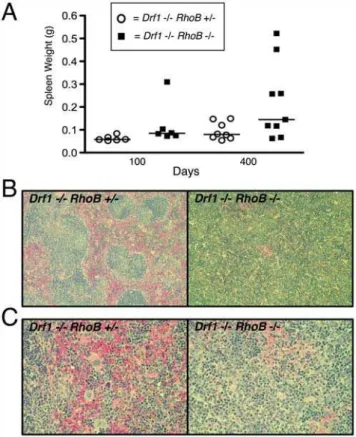
Upon necropsy, we found that several 400-day-old Drf12/2
RhoB2/2mice had splenomegaly, as determined by whole spleen
weight compared to that of Drf12/2RhoB+/2 mice (Fig. 1A).
Pathological examination of H&E-stained splenic sections showed significant dysplasia, and there was often atrophy of the white pulp with poorly formed germinal centers (Fig. 1B). Splenic sections from Drf12/2RhoB2/2 mice also revealed abnormal ratios of
myeloid and erythroid composition (frequently a myeloid:ery-throid ratio greater than 2.0) and an increased presence of extramedullary hematopoiesis (EMH) (Fig. 1C).
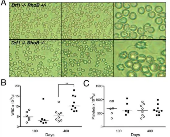
Based on our previous work inDrf1-null mice [6] and the recent finding that the mDia2 formin contributes to erythroblast enucleation [18], we examined the peripheral blood to determine if there was morphological evidence of erythrocyte dysplasia. Peripheral blood smears from Drf12/2RhoB2/2 mice showed a
marked elevation in abnormally shaped erythrocytes compared with blood from Drf12/2RhoB+/2
mice. The erythrocytes were often spiked (echinocyte) or teardrop in appearance (Fig. 2A), consistent with dysplastic features observed in patients with MDS. Drf12/2RhoB+/2 mice did show signs of dysplasia at 400 days,
with several teardrop-shaped erythrocytes, but to a lesser extent than Drf12/2RhoB2/2 mice. CBC analysis of peripheral blood
Figure 1. Mice lackingDrf1 andRhoB develop splenomegaly
and splenic disorganization.Drf12/2RhoB2/2andDrf12/2RhoB+/2
mice were necropsied at 100 and 400 days of age.A. Spleens were removed from mice and weighed immediately. Each point on scatter plot represents data from a single mouse.B. Formalin-fixed spleens from 400-day-old mice were paraffin-embedded and stained with H&E. Sections shown are at 106magnification.C. Splenic sections fromB, but at 406magnification.
revealed a significant increase in the WBC count of Drf12/2
RhoB2/2 mice relative to that of Drf12/2RhoB+/2 mice at 400
days of age (Fig. 2B). Abnormal platelet counts are sometimes observed in certain subsets of MDS, but we did not observe any significant differences in these mice (Fig. 2C).
We then focused our analysis specifically on the bone marrow and spleen to examine myelopoiesis in these compartments. Flow cytometry was used to determine the percentage of lymphocytes, monocytes, and granulocytes based on the pan-leukocyte marker CD45. Using the gating strategy outlined previously [6], we found that 400-day-old Drf12/2RhoB2/2 mice had an increased
percentage of granulocytes and a concomitant decrease in lymphocytes in the bone marrow compartment (Fig. 3A). Analysis of splenic single-cell suspensions isolated from 400-day-old mice showed a similar increase of granulocytes in Drf12/2RhoB2/2
mice (Fig. 3B).
To further examine potential differences in myelopoiesis between Drf12/2RhoB+/2
mice and Drf12/2RhoB2/2 mice, we performed flow cytometry on cells from the bone marrow and spleen to detect levels of F4/80 (a pan macrophage marker) and CD11b (integrin aM; monocyte development marker). By 400 days of age,Drf12/2RhoB2/2mice had an increased percentage of
F4/80+ cells in the spleen, but we observed no significant
difference in the bone marrow (Fig. 3C). On the other hand, CD11b+cells were significantly elevated in both the marrow and
spleen by 400 days of age. We also examined cellular expression of the marker CD29 (b1 integrin; important for homing and retention in lymphoid organs). We found an increased percentage
of CD29+cells in the marrow, with an even more pronounced
increase in the spleen ofDrf12/2RhoB2/2mice relative toDrf12/2 RhoB+/2 mice (Fig. 3C). These results are consistent with the
observed increase in myeloid cell proportion found by histopathol-ogy (Fig. 1).
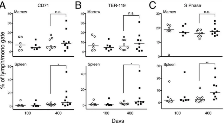
Finally, we analyzed the levels of TER-119 (erythroid-specific marker) and CD71 (transferrin receptor; a marker of proliferating erythroid precursors) in the marrow and spleen ofDrf12/2RhoB2/2
and Drf12/2RhoB+/2 mice. While there were no differences in
the bone marrow, the levels of CD71+and TER-119+cells were
elevated in the spleens of 400-day-old Drf12/2RhoB2/2 mice
(Fig. 4,A and B). An increase in erythroid precursors was also evident in H&E-stained splenic sections (data not shown). Consistent with this observation and the presence of splenic EMH, we also found a substantial increase in splenic cells undergoing S phase in 400-day-old Drf12/2RhoB2/2 mice relative to the number in Drf12/2RhoB+/2 mice (Fig. 4C).
Discussion
MDS is thought to arise because of multiple alterations in a hematopoietic stem cell [10]. We previously found that knocking out mDia1 expression in mice leads to the age-dependent development of myelodysplasia [6]. Here, we show that the additional knockout of RhoB expression in Drf1-null mice accelerates the progression to myelodysplasia. Several candidate tumor suppressor genes in humans reside on the long arm of chromosome 5 [8]. One of these genes isDIAPH1, which encodes the actin assembly protein mDia1. While genetic ablation of
Figure 2. Peripheral blood fromDrf12/2RhoB2/2mice show age-dependent abnormalities.A. Peripheral blood smears stained with
Wright-Giemsa from 400-day-old mice. Left and center panels are images at 40x; right panels are 60x.B. Total WBC count from peripheral blood.C. Platelet numbers from peripheral blood CBC analysis. In bothBandC, each point on scatter plot represents data from a single mouse (o =Drf12/2RhoB+/2;
mDia1 expression alone in mice resembles 5q- MDS, it is likely that mDia1 acts in concert with multiple other genes on the same commonly deleted region to suppress malignancy [8].
MDS can progress to a more advanced stage and ultimately develop into leukemia. The transformation events involved in disease progression are thought to include genes that contribute to cell-cycle control, apoptosis, and differentiation [10]. The small GTPase RhoB is required for apoptosis in response to DNA-damaging agents and farnesyltransferase inhibitors [14,16]. Previous reports have shown that RhoB negatively regulates Akt survival signaling and mediates apoptosis in a p53-dependent manner [19,20]. Given its role in apoptosis and its known interactions with the mDia formins, we hypothesized that deletion ofRhoBwould enhance the progression of MDS in mice lacking
mDia1 expression. Our results support this hypothesis and further substantiate our mouse model of age-associated myelodysplasia. It is also interesting to speculate that loss of RhoB expression may interfere with hematopoietic stem cell maintenance, since recent work has highlighted a role forRhoBin stem cell self-renewal [21]. Together, these findings suggest that multiple mechanisms may contribute to the myelodysplastic phenotype in our mice.
Our mouse model uniquely positions us to test some important questions related to thein vivorole of RhoB in the anti-neoplastic affects of FTIs. FTIs have been tested clinically to treat various myelodysplasias, with varying degrees of efficacy [22]. But whether FTIs can alleviate the disease phenotype in Drf1-null or heterozygous mice and whether RhoB is required in vivo to mediate this response remains to be determined.
Figure 3. Flow cytometry analysis of mouse bone marrow and splenic cells.A. Scatter plot showing the percentage of lymphocytes,
monocytes, and granulocytes from bone morrow. Open shapes representDrf12/2RhoB+/2mice and filled shapes representDrf12/2RhoB2/2mice (**
denotesP#0.01; *** denotesP#0.001).B. Scatter plot showing the percentage of lymphocytes, monocytes, and granulocytes from mice splenic single-cell suspensions. Legend is the same as inA(** denotesP#0.01).C. Percentage of F4/80+, CD11b+, and CD29+cells from the bone marrow and
spleen of mice (* denotesP#0.05; ** denotesP#0.01) (o =Drf12/2RhoB+/2;
&=Drf12/2RhoB2/2).
Our results point to the down-regulation of RhoB as a potential marker for late-stage MDS, similar to its diminished expression in other late-stage cancers [12,13]. Examination of the gene expression profile of RhoB in human patient samples that have a more advanced disease signature could help determine better treatment options for patients diagnosed with MDS.
In summary, we show that mice lacking mDia1 and RhoB expression progress to MDS faster than mice lacking mDia1 alone. These data parallel observations of MDS in humans, in which multiple alterations in hematopoietic stem cells contribute to disease pathogenesis. Our mouse model will be useful in characterizing the mechanism of disease progression further and in testing potential therapeutics to treat this chronic disease.
Acknowledgments
We thank David Nadziejka for critical reading of the manuscript, Kyle Furge for assistance with statistical analysis, Robert Sigler for pathology analysis, and Sergio Rodriguez and Sarah Sternberger for assistance with data analysis.
Author Contributions
Conceived and designed the experiments: ADD RW GP ASA. Performed the experiments: ADD KL RW. Analyzed the data: ADD KL RW ASA. Contributed reagents/materials/analysis tools: GP. Wrote the paper: ADD ASA.
References
1. Goode BL, Eck MJ (2007) Mechanism and function of formins in the control of actin assembly. Annu Rev Biochem 76: 593–627.
2. Fernandez-Borja M, Janssen L, Verwoerd D, Hordijk P, Neefjes J (2005) RhoB regulates endosome transport by promoting actin assembly on endosomal membranes through Dia1. J Cell Sci 118: 2661–2670.
3. Wallar BJ, DeWard AD, Resau JH, Alberts AS (2007) RhoB and the mammalian Diaphanous-related formin mDia2 in endosome trafficking. Exp Cell Res 313: 560–571.
4. Wallar BJ, Alberts AS (2003) The formins: active scaffolds that remodel the cytoskeleton. Trends Cell Biol 13: 435–446.
5. Shi Y, Zhang J, Mullin M, Dong B, Alberts AS, et al. (2009) The mDial formin is required for neutrophil polarization, migration, and activation of the LARG/RhoA/ROCK signaling axis during chemotaxis. J Immunol 182: 3837–3845.
6. Peng J, Kitchen SM, West RA, Sigler R, Eisenmann KM, et al. (2007) Myeloproliferative defects following targeting of the Drf1 gene encoding the mammalian diaphanous related formin mDia1. Cancer Res 67: 7565–7571. 7. Eisenmann KM, West RA, Hildebrand D, Kitchen SM, Peng J, et al. (2007) T
cell responses in mammalian diaphanous-related formin mDia1 knock-out mice. J Biol Chem 282: 25152–25158.
8. Eisenmann KM, Dykema KJ, Matheson SM, Kent NF, DeWard AD, et al. (2009) 5q– Myelodysplastic Syndromes: Chromosome 5q Genes Direct a Tumor Suppression Network Sensing Actin Dynamics Oncogene In press.
9. Nimer SD (2008) MDS: A Stem Cell Disorder–But What Exactly Is Wrong with the Primitive Hematopoietic Cells in This Disease? Hematology Am Soc Hematol Educ Program 2008: 43–51.
10. Nolte F, Hofmann WK (2008) Myelodysplastic syndromes: molecular patho-genesis and genomic changes. Ann Hematol 87: 777–795.
11. Prendergast GC (2001) Actin’ up: RhoB in cancer and apoptosis. Nat Rev Cancer 1: 162–168.
12. Sato N, Fukui T, Taniguchi T, Yokoyama T, Kondo M, et al. (2007) RhoB is frequently downregulated in non-small-cell lung cancer and resides in the 2p24 homozygous deletion region of a lung cancer cell line. Int J Cancer 120: 543–551.
13. Couderc B, Pradines A, Rafii A, Golzio M, Deviers A, et al. (2008) In vivo restoration of RhoB expression leads to ovarian tumor regression. Cancer Gene Ther 15: 456–464.
14. Liu A, Du W, Liu JP, Jessell TM, Prendergast GC (2000) RhoB alteration is necessary for apoptotic and antineoplastic responses to farnesyltransferase inhibitors. Mol Cell Biol 20: 6105–6113.
Figure 4. Flow cytometry analysis of erythroid precursors in mouse bone marrow and spleen.Each point on the scatter plots represents
data from a single mouse (o =Drf12/2RhoB+/2;
&=Drf12/2RhoB2/2)A. Scatter plot showing the percentage of CD71+cells from the bone marrow
and spleen of mice (* denotesP#0.05).B. Percentage of TER-119+cells from the bone marrow and spleen of mice (* denotesP#0.05).C. Percentage of cells undergoing S phase from the bone marrow and spleen of mice (** denotesP#0.01).
15. Liu AX, Rane N, Liu JP, Prendergast GC (2001) RhoB is dispensable for mouse development, but it modifies susceptibility to tumor formation as well as cell adhesion and growth factor signaling in transformed cells. Mol Cell Biol 21: 6906–6912.
16. Liu A, Cerniglia GJ, Bernhard EJ, Prendergast GC (2001) RhoB is required to mediate apoptosis in neoplastically transformed cells after DNA damage. Proc Natl Acad Sci U S A 98: 6192–6197.
17. Peng J, Wallar BJ, Flanders A, Swiatek PJ, Alberts AS (2003) Disruption of the Diaphanous-related formin Drf1 gene encoding mDia1 reveals a role for Drf3 as an effector for Cdc42. Curr Biol 13: 534–545.
18. Ji P, Jayapal SR, Lodish HF (2008) Enucleation of cultured mouse fetal erythroblasts requires Rac GTPases and mDia2. Nat Cell Biol 10: 314–321.
19. Jiang K, Sun J, Cheng J, Djeu JY, Wei S, et al. (2004) Akt mediates Ras downregulation of RhoB, a suppressor of transformation, invasion, and metastasis. Mol Cell Biol 24: 5565–5576.
20. Kamasani U, Prendergast GC (2005) Genetic response to DNA damage: proapoptotic targets of RhoB include modules for p53 response and susceptibility to Alzheimer’s disease. Cancer Biol Ther 4: 282–288.
21. Kent DG, Copley MR, Benz C, Wohrer S, Dykstra BJ, et al. (2009) Prospective isolation and molecular characterization of hematopoietic stem cells with durable self-renewal potential. Blood.