Diabetic Retinopathy Phenotype
Youde Jiang1, Qiuhua Zhang1, Li Liu2, Jie Tang3, Timothy S. Kern3,4, Jena J. Steinle1,2*
1Department of Ophthalmology, University of Tennessee Health Science Center, Memphis, Tennessee, United States of America,2Department of Anatomy and Neurobiology, University of Tennessee Health Science Center, Memphis, Tennessee, United States of America,3Department of Ophthalmology and Medicine, Case Western Reserve University, Cleveland, Ohio, United States of America,4Department of Research, Stokes Veterans Administration Hospital, Cleveland, Ohio, United States of America
Abstract
There is considerable evidence from our lab and others for a functional link betweenb-adrenergic receptor and insulin receptor signaling pathways in retina. Furthermore, we hypothesize that this link may contribute to lesions similar to diabetic retinopathy in that the loss of adrenergic input observed in diabetic retinopathy may disrupt normal anti-apoptotic insulin signaling, leading to retinal cell death. Our studies included assessment of neural retina function (ERG), vascular degeneration, and Mu¨ller glial cells (which express onlyb1 andb2-adrenergic receptor subtypes). In the current study, we produced b2-adrenergic receptor knockout mice to examine this deletion on retinal neurons and vasculature, and to identify specific pathways through which b2-adrenergic receptor modulates insulin signaling. As predicted from our hypothesis,b2-adrenergic receptor knockout mice display certain features similar to diabetic retinopathy. In addition, loss of
b2-adrenergic input resulted in an increase in TNFa, a key inhibitor of insulin receptor signaling. Increased TNFamay be associated with insulin-dependent production of the anti-apoptotic factor, Akt. Since the effects occurredin vivounder normal glucose conditions, we postulate that aspects of the diabetic retinopathy phenotype might be triggered by loss of
b2-adrenergic receptor signaling.
Citation:Jiang Y, Zhang Q, Liu L, Tang J, Kern TS, et al. (2013)b2-Adrenergic Receptor Knockout Mice Exhibit A Diabetic Retinopathy Phenotype. PLoS ONE 8(7): e70555. doi:10.1371/journal.pone.0070555
Editor:Alexander V. Ljubimov, Cedars-Sinai Medical Center, United States of America
ReceivedMarch 28, 2013;AcceptedJune 24, 2013;PublishedJuly 24, 2013
Copyright:ß2013 Jiang et al. This is an open-access article distributed under the terms of the Creative Commons Attribution License, which permits unrestricted use, distribution, and reproduction in any medium, provided the original author and source are credited.
Funding:This work was supported by National Eye Institute Vision Grants R01EY022045 (JJS) and EY00300 (TSK); Juvenile Diabetes Research Foundation Grant (2-2011-597 to JJS); Oxnard Foundation (JJS); Research to Prevent Blindness Award (PI:Barrett Haik); NEI Vision Core Grant: PHS 3P30; EY013080 (PI: Dianna Johnson), and a Merit Grant from the Veterans Administration (TSK). The funders had no role in study design, data collection and analysis, decision to publish, or preparation of the manuscript.
Competing Interests:The authors have declared that no competing interests exist.
* E-mail: [email protected]
Introduction
Although diabetic retinopathy is recognized as the leading cause of blindness in working age adults, we have yet to define the cellular mechanisms responsible for diabetes-induced loss of retinal neurons. Several lines of evidence suggest a link between decreased sympathetic innervation and diabetes. For example, hyperglyce-mia has been shown to cause dysfunctional neurotransmitter release from the sympathetic ganglia projection to the retina [1]. In our own studies, we have previously shown that removal of the superior cervical ganglion or knockout of dopamine beta hydroxylase (a key enzyme in the conversion of dopamine to norepinephrine in sympathetic neurons) results in a retinal phenotype that is similar to that seen in diabetic animals [2,3]. Likewise, we showed that treatment with adrenergic receptor antagonists, in particular b-adrenergic receptor antagonists, caused a similar diabetic phenotype in retina [4,5]. These results led us to hypothesize that restoration ofb-adrenergic signaling in diabetic retina might prevent or reduce retinal damage due to diabetes. To test this hypothesis, we treated streptozotocin-induced diabetic rats with a general b-adrenergic receptor agonist. As predicted, the treatment prevented retinal damage in this model system. [6,7].
Two cell types involved in retinal changes of diabetes are retinal vascular endothelial cells (REC) and Mu¨ller glial cells, which express different subtypes ofb-adrenergic receptors. REC express only b1- and b3-adrenergic receptors [8] whereas Mu¨ller cells possesb1- andb2-adrenergic receptors [9]. Our previous studies have shown that b1-adrenergic receptor knockout mice exhibit retinal changes similar to diabetic animals in spite of normal glucose levels [10]. This suggests that loss of adrenergic signaling through the b1-adrenergic receptor subtype on REC and/or Mu¨ller cells may be involved in mediating diabetic/hyperglycemic retinal damage.
and increased retinal cell apoptosis, similar to our observations
in vitro[11]. Since, these specific alterations mimic the molecular changes seen in diabetic retinopathy, it is possible that changes in b2-adrenergic receptor signaling acts as a molecular trigger for diabetes-induced retinal damage.
Methods
Mice
All mice experiments, including those for dark-adaptation and tail electrodes for ERG analyses, were approved by the Institutional Animal Care and Use Committee at the University of Tennessee Health Science Center (Protocol #1992). b1/b 2-adrenergic receptor knockout mice (Adrb1tm1Bkk Adrb2tm1Bkk/J) were purchased from Jackson laboratories (Bar Harbor, ME). C57BL6 wildtype mice were purchased from Charles River Laboratories. b1/b2-adrenergic receptor knockout mice were generated on a mixed background (129S1/Sv * 129X1/SvJ * C57BL/6J * DBA/2 * FVB/N ). We appreciate that C57BL6 may not be the ideal wildtype control, but the original b1/b 2-adrenergic receptor knockout mice were from a mixed back-ground containing C57BL6, therefore we chose the C57BL6 for wildtype. Since we use these mice at 2 months of age, other issues from the C57BL6 background should be minimized.b 2-adrener-gic receptor knockout mice have been previously characterized and shown to be fertile, with normal heart rate and blood pressure [12], but with a blunted hypotensive response to isoproterenol (a classicb-adrenergic receptor agonist) compared to wildtype mice [12].
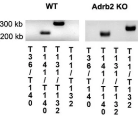
Genotyping was performed by PCR analysis using genomic DNA isolated from tail snips of mice (Sigma-Aldrich). Briefly, genomic DNA is extracted from the mice tail sample that has been incubated in Tissue Preparation Solution and Extraction Solution for 10 minutes at room temperature. The sample is heated to 95uC for 3 minutes and then mixed with a neutralized solution to neutralize inhibitory substances prior to PCR. An aliquot of the DNA extract is then added directly to the optimized PCR mix supplied. PCR was carried out using GoTaqH Hot Start Polymerase kit (Promega) with the following conditions: 98uC 90 s (1 cycle); 98uC 30 s, 64uC 40 s, 72uC 50 s (2 cycles); 95uC 30 s, 64uC 40 s, 72uC 50 s (33 cycles); 72uC 5 min (1 cycle). Primers used for the analysis were as follow:
T11: GGGGCTGCTAAAGCGCATGCTCC (23) pPGK pro-moter of NeoR, anti
T36: GGGAAGACAATAGCAGGCATGCT (23) bGHpA of PGK-NeoR, sense
T131: CGCTATGTTGCTATCACATCGCC (23) mb2 gene,
sense
T132: GATTTGTCTATCTTCTGCAGCTGCC (25) mb2
gene, anti
T140: GGCTCTCTACACCTTGGACTCCG (23) mb1 gene,
anti
T141: CCGCCGCCGTCCCCCGG (17) mb1 gene, sense The mice used for the experiment were sacrificed and ocular tissues collected at 2 months of age. PCR images are presented as Figure 1.
Electroretinogram (ERG)
Prior to sacrifice for morphological and biochemical analyses, animals were subjected to ERG analyses to evaluate the changes in the electrical activity of the retina as we have done previously [5,7]. Briefly, mice were dark-adapted overnight. ERG responses were recorded from both eyes together using platinum wire corneal electrodes, forehead reference electrode, and ground electrode in the tail. Pupils were fully dilated using 1% tropicamide solution (Alcon). Methylcellulose (Celluvise; Allergan, Irvine, CA) drops were applied as well to maintain a good electrical connection and body temperature was maintained at 37uC by a water-based heating pad. All ERG experiments were approved by the University of Tennessee Institutional Animal Care and Use Committee on Protocol#1992. ERG waveforms were recorded with a bandwidth of 0.3–500 Hz and samples at 2 kHz by a digital acquisition system and were analyzed a custom-built program (MatLab). Statistics was done on the mean6SD amplitudes of the a- and b- wave of each treatment group.
Figure 1. Genotyping results.Results of the genotyping to verify that theb2-adrenergic receptor is eliminated in the KO mice. Numbers on the bottom correspond to primers described in the methods to demonstrate effectiveb2-adrenergic receptor knockout.
doi:10.1371/journal.pone.0070555.g001
Figure 2. ERG levels.Mean ERG amplitudes for theb2-adrenergic receptor knockout and wildtype mice. Left panel is the A-wave, middle panel represents the B-wave, and the right panel is the amplitudes for the oscillatory potentials. N = 6 mice in each group.
doi:10.1371/journal.pone.0070555.g002
Neuronal Analyses
Formalin-fixed paraffin sections were stained with hematoxylin and eosin for light microscopy and morphometry of retinal thickness as previously described [6]. Photomicrographs were assessed for retinal thickness and the number of cells in the ganglion cell layer using methods previously described. The thickness of the retina and the cell count were measured using OpenLab software (Improvision, Lexington, MA) [7].
Vascular Analyses
Retinas from 1 eye of wildtype and b2-adrenergic receptor knockout mice were used to count degenerate capillaries. The eyes were enucleated, suspended in 10% buffered formalin for 5 days, and the retina was dissected in 3% crude trypsin solution (Difco Bacto Trypsin 250, Detroit, MI). The retinal vascular tree was dried onto a glass slide and stained with hematoxylin-periodic acid-Shiff. Degenerate capillaries were counted and identified as previously described [7].
Western Blot Analysis
Equal amounts of protein from the tissue extracts were separated on the pre-cast tris-glycine gel (Invitrogen, Carlsbad, CA), blotted onto a nitrocellulose membrane. After blocking in TBST (10 mM Tris-HCl buffer, pH 8.0, 150 mM NaCl, 0.1% Tween 20) and 5% (w/v) BSA, the membrane was treated with appropriate primary antibodies followed by incubation with secondary antibodies labeled with horseradish peroxidase. Anti-gen-antibody complexes were detected by chemilluminescence reagent kit (Thermo Scientific). Primary antibodies used were phosphorylated Akt (Serine 473), Akt, Cytochrome C, Bax, Bcl-xL, SOCS3, GFAP, phosphorylated insulin receptor (tyrosine 1150/1151), insulin receptor (all purchased from Cell Signaling, Danvers, MA), and beta actin (Santa Cruz).
ELISA Analysis
all wells to allow for comparisons based on O.D. For the TNFa ELISA, 50 ug protein was loaded into all wells, with analyses based on a standard curve.
Terminal Deoxynucleotidyl Transferase Mediated dUTP Nick End Labeling Assay (TUNEL)
TUNEL analyses were completed on C57/BL6 mice andb 2-adrenergic receptor knockout mice at 2 months of age. TUNEL was done according to manufacturer’s instructions using the ApopTag-Red kit (Millipore, Bilerica, MA).
Immunohistochemical Staining
b2-adrenergic receptor KO and C57/BL6 wildtype mice at 2 months of age were perfused, under deep Avertin anesthesia, with 4% paraformaldehyde for 15 min. Eyes were then removed, followed by a 30 min immersion in the same fixative. Ten mm cryostat sections contained retinas were collected and underwent double immunofluorescent staining. Briefly, the sections were incubated in 1% bovine serum albumin and 0.3% Triton X-100 (Sigma) in PBS for 1 h at room temperature, followed by incubation with rabbit anti-GFAP and mouse anti-insulin recep-tor, or mouse anti-GFAP and rabbit anti TNFa (all primary antibodies were purchased from Abcam, MA) overnight at 4uC. Following rinses in PBS, sections were incubated with Alexa Fluor 594 or Alexa Fluor 488 secondary antibody for 1 h at room temperature. Sections were counterstained with DAPI (1mM, Invitrogen,Carlsbad, CA) to label all cell nuclei for 3 min at room
temperature. Sections were observed and analysis using a Zeiss710 Confocal microscope.
Statistics
For all analyses, all experiments were done in triplicate. Data is presented as mean6SEM, with statistical analyses using Kruskal-Wallis non-parametric testing, followed by Dunn’s test. For Western blot analyses, data was normalized to beta actin levels.
Results
Previous studies in various mouse models of diabetic retinopathy have established four major functional and morphological markers that help to define the disease. Retinas from b2-adrenergic receptor KO mice displayed three of the four markers as described below.
Marker#1
B-wave and oscillatory potential amplitudes were reduced inb2-adrenergic receptor KO mice compared to wild type at 2 months of age. At all light intensities evaluated, the b2-adrenergic receptor knockout mice had reduced B-wave (Figure 2 middle) and oscillatory potential (Figure 2 right) amplitudes. A-wave responses of KO and wild type were statistically significantly different at high light intensity.
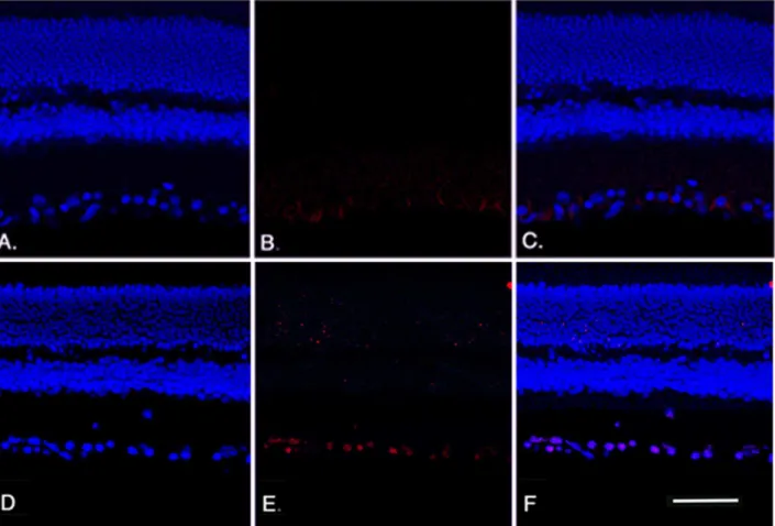
Figure 4. Apoptosis staining analyses.TUNEL results for wildtype mice (A-C) andb2-adrenergic receptor KO mice (D-F) showing DAPI staining (A, D) and TUNEL labeling (B, E) with overlay provided in panels C, F. Scale bar is 50 um.
doi:10.1371/journal.pone.0070555.g004
Marker# 2
b2-adrenergic receptor knockout mice had thinner retinas overall and fewer cells in the ganglion cell layer. We measured the retinal thickness and cell number in the ganglion layer at 2 months of age in theb2-adrenergic receptor knockout mice. The knockout mice had significantly decreased retinal thickness and cell number in the ganglion cell layer compared to wild type (Figure 3).
The loss of cells in the ganglion cell layer was due to apoptosis, reflected in an increase in TUNEL labeling in the ganglion cell layer of theb2-adrenergic receptor knockout mice. We performed TUNEL labeling on retinal sections to measure cell death in the b2-adrenergic receptor knockout mice. Figure 4 (Panel E) shows increased TUNEL-labeling in the ganglion cell layer of b 2-adrenergic receptor knockout mice compared to their wildtype littermates (Panel B).
In addition to TUNEL labeling, we used protein analyses of whole retinal lysates from the wildtype andb2-adrenergic receptor knockout mice for key pro- and anti-apoptotic proteins. We found that all pro-apoptotic proteins evaluated (cleaved caspase 3, bax, cytochrome C) were increased in the b2-adrenergic receptor knockout mice (Figure 5, top panels), while the anti-apoptotic proteins (phosphorylated Akt and Bcl-xL) were significantly reduced (Figure 5, bottom panels). The altered levels of these proteins is likely involved in the decreased ERG signal from the inner retina (B-wave), as well as the increased TUNEL labeling.
Marker#3
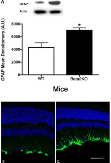
Increased GFAP labeling and protein levels in b 2-adrenergic receptor knockout mice. We have previously published that b2-adrenergic receptors are critical in insulin receptor signaling in retinal Mu¨ller cells [11]. To evaluate whether retinal Mu¨ller cells were activated in theb2-adrenergic receptor knockout mice, we measured protein levels of GFAP and performed immunofluorescent staining for GFAP, which is increased in retinal Mu¨ller cells once activated [13]. We found that GFAP protein levels were significantly increased in theb 2-adrenergic receptor knockout mice compared to wildtype (Figure 6).
Marker#4
Retinas from 2 month old b2-adrenergic receptor KO mice did not show the increase in degenerate capillary numbers normally associated with the diabetic phenotype. We measured the numbers of degenerate capillaries in theb2-adrenergic receptor KO mice at 2 months of age and found a trend towards increased capillary degeneration in the receptor knockout animals, but this did not achieve statistical significance versus that seen in wild type retinas. (Figure 7). We have previously shown increases in degenerate capillaries inb 1-adrenergic receptor knockout mice [10] and animals treated with streptozotocin to induce diabetes [6,7]; however, these changes were not observed until 4–6 months of age or 6 months after treatment. Thus the assessment in b2-adrenergic receptor KO Figure 5. Apoptotic protein levels.Cleaved caspase 3 ELISA and Western blot results for wildtype mice andb2-adrenergic receptor KO mice for pro-apoptotic proteins (top panel-cleaved caspase 3, Bax, Cytochrome C) and anti-apoptotic proteins (bottom panel-phosphorylated Akt, Bcl-xL). A representative Western blot is provided. All Western blot data were normalized to beta actin levels. ELISA data was normalized to protein loaded into well. *P,0.05 vs. wildtype. N = 6 mice in each group.
mice at 2 months may not have allowed sufficient time for degenerate capillaries to form. Experiments are now underway to repeat these experiments using KO mice at 6 months of age.
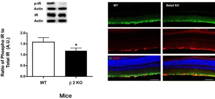
Insulin Receptor Phosphorylation was Reduced inb 2-adrenergic Receptor Knockout Mice
We have previously reported that hyperglycemia significantly reduced insulin receptor phosphorylation on tyrosine 1150/1151, which was restored following treatment with a b-adrenergic receptor agonist, Compound 49b [7]. In retinal Mu¨ller cells, we reported that insulin receptor signaling was altered when cells were grown in high glucose, which led to increased apoptosis that was prevented when Mu¨ller cells were treated with the b 2-adrenergic receptor agonist, salmeterol [11]. To confirm those studies in vivo, we measured insulin receptor phosphorylation in theb2-adrenergic receptor knockout mice. As predicted, receptor phosphorylation was significantly reduced (Figure 8), and the decrease was specifically associated with retinal Mu¨ller cells based on immunoflourescent staining of the receptor that co-localized with GFAP-labeled Muller cells.
TNFawas Increased inb2-adrenergic Receptor KO Mice In retinal endothelial cells, we have shown that TNFa can inhibit insulin receptor signaling through activation of suppressor
of cytokine 3 (SOCS3), coincident with increased retinal endothelial cell apoptosis [14]. We found that loss of b 2-adrenergic receptor signaling triggering significant increases in TNFa(Figure 9) This suggests that loss ofb2-adrenergic receptor signaling changes cellular signaling in a manner similar to that seen in the hyperglycemic state.
Discussion
Loss ofb2-adrenergic Receptor Expression Results in Neuroglial Abnormalities of Common to Diabetic Retinopathy
A major finding from these studies is that b2-adrenergic receptor knockout mice display neuronal and functional markers similar to those observed in diabetic mice, despite the presence of normal levels of glucose. This is consistent with our hypothesis that loss of b2-adrenergic receptor input is a major trigger for the retinal changes observed in diabetes. Since retinalb2-adrenergic receptors are primarily localized to Mu¨ller cells, our findings also highlight the specific role Mu¨ller cells play in this blinding disorder. Similar to other glial cells in CNS, Mu¨ller cells display increased GFAP labeling in response to various types of stress. We find that in our KO model, GFAP expression is increased in Mu¨ller cells, indicative of a direct stress response to the loss of input through theirb2-adrenergic receptors.
TNFa is Increased inb2-adrenergic Receptor KO Mice Since we have previously reported thatb-2-adrenergic receptors can regulate TNFalevels [9,11], we measured retinal TNFalevels in retinal lysates fromb2-adrenergic receptor knockout mice. Our results show a significant increase in overall levels and, based on immunofluorescence studies, the increase is largely localized to Mu¨ller cells. Our results show for the first time TNFalevels are increased in retinal lysates fromb2-adrenergic receptor knockout mice. The mechanisms and specific cell types involved in the TNFapathways are currently under investigation.
Figure 6. Mu¨ller cell staining for GFAP.Western blot results (A) and GFAP labeling in wildtype (B) andb2-adrenergic receptor knockout mice (C). *P,0.05 vs. wildtype. N = 5 for Western blot results. doi:10.1371/journal.pone.0070555.g006
Figure 7. Vascular Analyses. Panel A shows degenerate capillary numbers inb2-adrenergic receptor knockout mice and wildtype mice. N = 5 mice in each group.
doi:10.1371/journal.pone.0070555.g007
b2-adrenergic Receptor Signaling Helps Maintains Insulin Receptor Signaling
Retinal Mu¨ller cells undergo increased apoptosis when cultured in high glucose conditions that are known to lead to insulin resistance [15], Our previous work in vitro has suggested that a functional link may exist between maintenance ofb2-adrenergic receptor function and maintenance of insulin receptor anti-apoptotic pathways. We observed that decreased insulin receptor signaling could subsequently be reversed by restoring b
-2-adrenergic receptor stimulation [11]. We also showed that the opposite is true: loss ofb2-adrenergic receptor stimulation leads to decreased insulin receptor signaling. This agrees with the current
in vivofindings, as insulin receptor phosphorylation is reduced in theb2-adrenergic receptor knockout mice.
Figure 8. Insulin Receptor Levels and localization.Left Panel: Western blot results for phosphorylated insulin receptor (Tyr 1150/1151) inb 2-adrenergic receptor knockout mice vs. wildtype. Western blot data were normalized to beta actin. N = 5 mice in each group. *P,0.05 vs. wildtype. Right Panel: Confocal microscopy with co-labeling for insulin receptor (green), GFAP (red), nuclei (blue). Scale bar is 50 um.
doi:10.1371/journal.pone.0070555.g008
Figure 9. TNFalevels and localization.Top Left Panel: TNFaconcentrations in retinal lysates fromb2-adrenergic receptor knockout and wildtype mice. *P,0.05 vs. wildtype. ELISA data was normalized to protein loaded into well. N = 6 mice in each group. Top Right Panel: Confocal microscopy for TNFa(red), GFAP (green), and nuclei (blue). Scale bar is 50 um.
b2-adrenergic Receptor Signaling Prevents Expression of Inhibitors of Insulin-dependent Pro-apoptotic Pathways
Our results demonstrate that TNFaa negative regulators of the insulin receptor pathway is increased in the absenceb2-adrenergic receptor input. Once produced, these negative regulators can block the insulin-induced activation/phosphorylation of a key apoptotic factor, Akt. Thus, we hypothesize that the diabetes-induced, systemic loss ofb2-adrenergic receptor input to retinal cells, causes further damage to the retina by triggering production of TNFa, which in turn disables the anti-apoptotic actions of insulin receptor pathways. Loss of insulin signaling through insulin resistance, loss ofb2-adrenergic receptor input, and production of the anti-apoptotic inhibitors, TNFa, could lead to retinal cell death associated with retinopathy.
In conclusion, absence of b2-adrenegic receptor input in vivo
results in morphological and function damage to the retina including retinal thinning, cell loss in the retinal ganglion cell layer, and a decrease in A-wave, B-wave and oscillatory potential amplitudes - all markers associated with diabetes in rodents. We do
want to state that we cannot completely rule out that some of the observed differences between the two groups could be due to strain-dependent differences, since we used only the C57/B6 background for the wildtype mice. These findings establish b 2-adrenergic receptor agonists as promising candidates for drug therapy to prevent retinal cell apoptosis, potentially acting on Mu¨ller cell pathways to maintain insulin signaling and homeostasis of the retina. We have recently reported that treatment with a Compound 49b, a novel b-adrenergic receptor agonist, signifi-cantly increases insulin receptor phosphorylation, while decreasing TNFaand retinal cell apoptosis [7]. Future studies will focus on further elucidating the actions of Compound 49b, as well as its potential for treatment of early stage diabetic retinopathy.
Author Contributions
Conceived and designed the experiments: JS QZ. Performed the experiments: YJ QZ JT. Analyzed the data: YJ QZ LL JT TK JS. Wrote the paper: JS TK.
References
1. Burnstock G (1990) Changes in expression of autonomic nerves in aging and disease. JAutonNervSyst 30 Suppl.: S25–S34.
2. Wiley LA, Rupp GR, Steinle JJ (2005) Sympathetic innervation regulates basement membrane thickening and pericyte number in rat retina. Invest Ophthalmol Vis Sci 46: 744–748.
3. Steinle JJ, Kern TS, Thomas SA, McFadyen-Ketchum LS, Smith CP (2009) Increased basement membrane thickness, pericyte ghosts, and loss of retinal thickness and cells in dopamine beta hydroxylase knockout mice. Exp Eye Res 88: 1014–1019.
4. Steinle JJ, Smith PG (2002) Role of adrenergic receptors in vascular remodelling of the rat choroid. Br J Pharmacol 136: 730–734.
5. Jiang Y, Steinle JJ (2010) Systemic Propranolol Reduces B-Wave Amplitude in the ERG and Increases IGF-1 receptor Phosphorylation in Rat Retina. Invest Ophthalmol Vis Sci 51: 2730–2735.
6. Jiang Y, Walker RJ, Kern TS, Steinle JJ (2010) Application of isoproterenol inhibits diabetic-like changes in the rat retina. Exp Eye Res 91: 171–179. 7. Zhang Q, Guy K, Pagadala J, Jiang Y, Walker RJ, et al. (2012) Compound 49b
Prevents Diabetes-Induced Apoptosis through Increased IGFBP-3 Levels. Invest Ophthalmol Vis Sci 53: 3004–3013.
8. Steinle JJ, Booz GW, Meininger CJ, Day JN, Granger HJ (2003) Beta 3-adrenergic receptors regulate retinal endothelial cell migration and proliferation. J Biol Chem 278: 20681–20686.
9. Walker RJ, Steinle JJ (2007) Role of beta-adrenergic receptors in inflammatory marker expression in Muller cells. Invest Ophthalmol Vis Sci 48: 5276–5281. 10. Panjala SR, Jiang Y, Kern TS, Thomas SA, Steinle JJ (2011) Increased tumor
necrosis factor-alpha, cleaved caspase 3 levels and insulin receptor substrate-1 phosphorylation in the beta-adrenergic receptor knockout mouse. Mol Vis 17: 1822–1828.
11. Walker RJ, Anderson NM, Jiang Y, Bahouth S, Steinle JJ (2011) Role of beta-adrenergic receptors regulation of TNF-alpha and insulin signaling in retinal Muller cells. Invest Ophthalmol Vis Sci.
12. Chruscinski AJ, Rohrer DK, Schauble E, Desai KH, Bernstein D, et al. (1999) Targeted disruption of the beta2 adrenergic receptor gene. J Biol Chem 274: 16694–16700.
13. Barber AJ, Antonetti DA, Gardner TW (2000) Altered expression of retinal occludin and glial fibrillary acidic protein in experimental diabetes. The Penn State Retina Research Group. Invest Ophthalmol Vis Sci 41: 3561–3568. 14. Jiang Y, Zhang Q, Soderland C, Steinle JJ (2012) TNFalpha and SOCS3
regulate IRS-1 to increase retinal endothelial cell apoptosis. Cell Signal 24: 1086–1092.
15. Kusner LL, Sarthy VP, Mohr S (2004) Nuclear translocation of glyceraldehyde-3-phosphate dehydrogenase: a role in high glucose-induced apoptosis in retinal Muller cells. Invest Ophthalmol Vis Sci 45: 1553–1561.