PARAQUAT TOLERANCE3 Is an E3 Ligase
That Switches off Activated Oxidative
Response by Targeting Histone-Modifying
PROTEIN METHYLTRANSFERASE4b
Chao Luo1☯, Xiao-Teng Cai1☯, Jin Du1, Tao-Lan Zhao2, Peng-Fei Wang2, Ping-Xia Zhao1, Rui Liu1, Qi Xie2, Xiao-Feng Cao2, Cheng-Bin Xiang1*
1School of Life Sciences, University of Science and Technology of China, Hefei, Anhui Province, China, 2State Key Laboratory of Plant Genomics and National Center for Plant Gene Research, Institute of Genetics and Developmental Biology, Chinese Academy of Science, Beijing, China
☯These authors contributed equally to this work. *[email protected]
Abstract
Oxidative stress is unavoidable for aerobic organisms. When abiotic and biotic stresses are encountered, oxidative damage could occur in cells. To avoid this damage, defense mech-anisms must be timely and efficiently modulated. While the response to oxidative stress has been extensively studied in plants, little is known about how the activated response is
switched off when oxidative stress is diminished. By studying Arabidopsis mutantparaquat
tolerance3, we identified the genetic locusPARAQUAT TOLERANCE3(PQT3) as a major
negative regulator of oxidative stress tolerance.PQT3, encoding an E3 ubiquitin ligase, is
rapidly down-regulated by oxidative stress. PQT3 has E3 ubiquitin ligase activity in ubiquiti-nation assay. Subsequently, we identified PRMT4b as a PQT3-interacting protein. By
his-tone methylation, PRMT4b upregulates the expression ofAPX1andGPX1, encoding two
key enzymes against oxidative stress. On the other hand, PRMT4b is recognized by PQT3 for targeted degradation via 26S proteasome. Therefore, we have identified PQT3 as an E3 ligase that acts as a negative regulator of activated response to oxidative stress and found
that histone modification by PRMT4b atAPX1andGPX1loci plays an important role in
oxi-dative stress tolerance.
Author Summary
Oxidative stress is a major stress in plant cells when biotic and abiotic stresses are imposed. While the response to oxidative stress has been extensively studied, little is known about how the activated response is switched off when oxidative stress is diminished. By studying Arabidopsis mutantparaquat tolerance3, we identified the genetic locusPARAQUAT TOLERANCE3(PQT3) as a major negative regulator of oxidative tolerance.PQT3encodes an E3 ubiquitin ligase and is rapidly down-regulated by oxidative stress. Subsequently, we
a11111
OPEN ACCESS
Citation:Luo C, Cai X-T, Du J, Zhao T-L, Wang P-F, Zhao P-X, et al. (2016) PARAQUAT TOLERANCE3 Is an E3 Ligase That Switches off Activated Oxidative Response by Targeting Histone-Modifying PROTEIN METHYLTRANSFERASE4b. PLoS Genet 12(9): e1006332. doi:10.1371/journal. pgen.1006332
Editor:Christine Helen Foyer, University Of Leeds, UNITED KINGDOM
Received:May 26, 2016
Accepted:August 30, 2016
Published:September 27, 2016
Copyright:©2016 Luo et al. This is an open access article distributed under the terms of theCreative Commons Attribution License, which permits unrestricted use, distribution, and reproduction in any medium, provided the original author and source are credited.
Data Availability Statement:All relevant data are within the paper and its Supporting Information files.
identified PRMT4b as a PQT3-interacting protein. PQT3 was demonstrated to recognize PRMT4b for targeted degradation via 26S proteasome. By histone methylation, PRMT4b may regulate the expression ofAPX1andGPX1, encoding two key enzymes against oxida-tive stress. Therefore, we have identified PQT3 as an E3 ubiquitin ligase that turns off the activated response to oxidative stress. Our study provides new insights into the post-trans-lational regulation of plant oxidative stress response and ROS signaling.
Introduction
Sessile plants cannot avoid harsh living conditions such as drought, salinity, cold and hot tem-perature. These stresses alter the normal cell homeostasis and increase the generation of reac-tive oxygen species (ROS) [1]. ROS could also be generated by paraquat, one widely used herbicide [2]. Accumulation of ROS directly destroys biological membranes and macromole-cules, accelerates cell senescence, induces irreversible damages to cells and even leads to cell death [3]. The level of ROS is increased sharply under stress conditions [1,4–9]. Two protec-tion systems, enzymatic and non-enzymatic, have evolved to scavenge ROS and protect plant cells from oxidative damage. Enzymatic system mainly includes the ascorbate peroxidase (APX), glutathione peroxidase (GPX), catalase (CAT), superoxide dismutase (SOD), and per-oxiredoxin Q (PRXQ) [8,10,11]. The transcript level and activity of antioxidant enzymes cor-relate with paraquat tolerance [12].
The activation of oxidative response involves many layers of regulations [8,13]. Little is known about the regulation by histone methylation of the genes involved in oxidative stress response. Histone methylation plays important roles in the plant development and growth as well as in some stress responses [14–18]. The methylation marks are written on lysines or argi-nines respectively by protein arginine methyltransferases (PRMTs) and histone lysine methyl-transferases (HKMTs). In Arabidopsis, nine PRMTs are found in the genome [14]. It has been reported that PRMTs are involved in salt stress responses, flowering time as well as circadian cycle [19,20]. Two different types of PRMTs catalyze asymmetric di-methylation (ADMA) and symmetric di-methylation (SDMA) on the Arg residues, respectively [17,21]. Theprmt5
mutant has decreased level of histone H4 Arg3-SDMA, leading to enhanced drought tolerance [22]. A pair of PROTEIN ARGININE METHYLTRANSFERASE4 (PRMT4) homologs, AtPRMT4a and AtPRMT4b, is required for the asymmetrical di-methylation of Arg-2, Arg-17, and Arg-26 in histone H3 [14]. Theprmt4aprmt4bdouble mutants are sensitive to salt stress [20]. Protein arginine methylation plays essential roles in diverse biological processes, such as RNA processing and transcriptional regulation [21,23].
Oxidative stress could be perceived by multiple mechanisms, including sensor or cellular receptor. The perception by receptors results in the activation of Ca2+–calmodulin and mito-gen-activated protein kinase (MAPK) cascade signaling transduction pathway. The activation or suppression of different transcription factors regulates a variety of defense pathway subse-quently, such as ROS-scavenging, heat-shock proteins (HSPs), and photosynthesis [8,10,13]. Several paraquat tolerance mutants have been analyzed in Arabidopsis [24]. While much atten-tion has been paid to how plants respond to oxidative stress, we know little about how plants switch off the activated responses when stress is diminished. A common regulatory mechanism is to control the protein level of the stress responsive factors. The most studied mechanism of protein degradation is the ubiquitin/26S proteasome system [25]. The ubiquitin/26S protea-some pathway is involved in different regulatory processes of eukaryotic cells, as it can rapidly eliminate the specific proteins in the cell [25–27]. Ubiquitin, containing 76 amino acids, could be attached to the target protein under the action of three different enzymes [27]. The ubiquitin
Switching off Oxidative Stress Response
PLOS Genetics | DOI:10.1371/journal.pgen.1006332 September 27, 2016 2 / 34
system can identify and modify many intracellular proteins, such as proteins involved in signal transduction, transcription factors, and receptors on cell surface, to participate in the regula-tion of physiological processes [28]. The target proteins with ubiquitin have different fates. For monoubiquitination, one lysine residue of substrate is modified by a single ubiquitin. If several individual lysine residues of target protein are attached with single ubiquitin respectively, the protein modification is named multiubiquitination. Both mono- and multi-ubiquitination could affect protein activity and intracellular localization [29–31]. For polyubiquitination, one ubiquitin is attached to lysine residue of substrate firstly. The C-terminal glycine of next ubi-quitin is linked with lysine residue of the preceding ubiubi-quitin to form polyubiubi-quitin chain sub-sequently [32]. As ubiquitin contains seven lysines, polyubiquitin chains are divided into different types according to different linkages between two adjacent ubiquitins [33]. Proteins with Lys48-Linkage polyubiquitination could be recognized and degraded by the 26S some [34]. It has also been reported that Lys29-Linkage polyubiquitination involved in protea-some-dependent degradation [35]. In addition, Lys63-Linkage polyubiquitination plays roles in endocytosis, repair of DNA damage, protein synthesis and signal transduction [36,37].
In the ubiquitin degradation process, E3 played a crucial role. E3 is responsible for specific recognition of substrate protein and accurate positioning of the binding site between substrate protein and ubiquitin [38]. InArabidopsis thaliana, the genes encoded the subunits of E3 ubi-quitin ligases make up approximately 90 percent of about 1400 genes encoded the components of ubiquitin/26S proteasome pathway. [26,39]. The large and diverse family of plant E3 tin ligases can be divided into HECT domain- and RING/U-box domain-containing E3 ubiqui-tin ligases [25]. The HECT family is relatively small compared with the RING domain-containing family that contains several hundreds of proteins and can be further divided into single subunit RING/U-box E3 ligases and multi subunit RING E3 ligases [26,40]. The large number of E3 ubiquitin ligases in higher plants indicates their important regulatory roles in diverse biological processes [41].
Several RING E3 ligases play positive roles in ABA-mediated drought tolerance [42–45]. In addition, overexpression of RING E3 ligases Rma1 inhibited the trafficking of aquaporin PIP2;1 and promote protein degradation of PIP2;1 to enhance drought tolerance [46]. A few E3 ligases have also been identified to turn off the activated stress responses [47]. HOS1, as a RING E3 ligase, acts as a negative regulator in cold tolerance. HOS1 inhibits the expression of CBFs and downstream cold-responsive genes through the degradation of ICE1 in ubiquitin-proteasome pathway [48–50]. Both PUB22 and PUB23, as E3 ligases contained U-box, play roles as negative regulators in drought tolerance. They could recognize and degrade RPN12a, a subunit of 19S regulatory particle belonged to 26S proteasome, to affect Arabidopsis drought tolerance [51]. The negative function of PUB18 and PUB19 in ABA-mediated drought toler-ance has also been reported [52]. The drought-induced AtERF53 could be degraded by RING E3 Ligase RGLG1 and RGLG2 that acts as negative regulators in drought tolerance [53]. Over-expression of salt-induced RING E3 ligase OsSIRP1 reduced salt tolerance in Arabidopsis [54]. In addition, several substrate receptors of CUL4 E3 ligases, DWAs, regulate ABA responses negatively [55,56]. The function of an enormous number of E3 ligases still remains to be iden-tified. Here we report a novel negative regulator of oxidative stress response by PARAQUAT TOLERANCE3 (PQT3) in Arabidopsis. We identified a paraquat tolerant mutant,paraquat
plants from oxidative stress. When oxidative stress is diminished, PQT3 level increases and acts as E3 ubiquitin ligase to specifically target PRMT4b for degradation. Based on our results, PQT3 is a negative regulator that turns off the activated response of oxidative stress.
Results
Loss of
At4g17410
confers the paraquat tolerance of
pqt3
The mutantpqt3was obtained after screening an activation-tagging library [59]. This library con-sisting of approximately 55, 000 independent lines was screened for mutants with enhanced tol-erance to different stresses [60,61]. To isolate tolerant mutant to oxidative stress, we germinated seeds on MS medium with 2 μM paraquat. Growing green seedlings were selected as putative mutants and were namedparaquat tolerance(pqt) because of their enhanced tolerance to para-quat. Thepqt3, one of such mutants, was further characterized and marked aspqt3-1. The enhanced oxidative tolerance ofpqt3-1mutant was confirmed by germinating seeds on MS medium containing 0 or 2 μM paraquat. Based on the observation of green cotyledons, survival ratio were counted. In presence of paraquat, more than 60%pqt3-1seeds germinated with green cotyledons but only 2% wild type seeds did, while all seeds of both wild type andpqt3-1survived on MS medium without paraquat (Fig 1A and 1B). Genetic analysis showed that the mutation was recessive. All F1 backcross offsprings (pqt3-1x wild type) were paraquat sensitive and F2 self-ing population showed typical 3:1 segregation ratio (sensitive: resistant; 85:27,χ2= 0.0476). The result suggested thatpqt3-1mutant may have a more efficient mechanism of ROS scavenge, which was caused by loss-of-function mutation in a single nuclear genePQT3(At4g17410).
Inpqt3-1mutant, a single T-DNA insertion was located in the fourth intron ofAt4g17410
(S1A Fig). The exact integration site of the T-DNA right border was 803bp downstream of the ATG initiation codon ofAt4g17410. As a result, the expression ofAt4g17410was completely disrupted as confirmed by RT-PCR analysis (S1B and S1D Fig). The expressions of its neigh-boring genes,At4g17390andAt4g17420, were not affected (S1B Fig). TheAt4g17410locus includes 13 exons and 12 introns. By prediction, the open reading frame encodes a 91 kD poly-peptide composed of 827 amino acids. Based on the conserved RING/U-box domain, this pro-tein is predicted as an E3 ubiquitin ligase.
To further determine whether the loss ofAt4g17410resulted in the enhanced oxidative tol-erance ofpqt3-1mutant, we used another allele ofpqt3, the T-DNA insertion mutant
Salk_065409, which was ordered from Arabidopsis Biological Resource Center (ABRC) and its T-DNA insertion was confirmed by RT-PCR (S1A, S1C and S1D Fig). As the first identified
pqt3mutant was named aspqt3-1, the Salk_065409 was marked aspqt3-2. Thepqt3-2mutant showed similar enhanced oxidative tolerance to paraquat and had high survival ratio under dif-ferent concentrations of paraquat treatment aspqt3-1did (Fig 1C). The survival ratio ofpqt3-1
andpqt3-2were 50% and 20%, respectively, under 2 μM paraquat treatment, while none of the wild type seedlings survived under the same condition. In addition, bothpqt3-1andpqt3-2
showed a late-flowering phenotype (Fig 1D).
To confirm further, we generated functional complementation (FC) lines and35Spro:PQT3
overexpression lines (S1E and S1F Fig). FC lines and35Spro:PQT3lines showed similar if not higher paraquat sensitivity to wild type under 2 μM paraquat treatment while thepqt3-1and thepqt3-2mutants displayed enhanced paraquat tolerance (Fig 1E and 1F). These results indi-cate that PQT3 is a negative regulator of oxidative stress tolerance and is responsible for the phenotype ofpqt3mutants.
In addition, we assayed H2O2level in the leaf with DAB staining, in which the chemical
reaction between hydrogen peroxide and DAB lead to the formation of brown precipitate that indicates hydrogen peroxide distribution and oxidative damage. After 6 μM paraquat
Switching off Oxidative Stress Response
treatment for 12 or 24 hours, the result of 3,30- diaminobenzidine (DAB) staining showed that
the brown precipitate in the leaves of the wild type was more than that ofpqt3mutants (Fig 1G–1R). As several stresses could cause oxidative damage to plants, the sensitivity ofpqt3
mutants to other environmental stresses was analyzed subsequently. The result indicated that
pqt3mutants have enhanced tolerance to CdCl2, mannitol, NaCl, and drought stress (S2 Fig).
Spatiotemporal expression pattern and protein localization of
PQT3
To investigate the spatiotemporal pattern ofPQT3expression, we generatedPQT3pro:GUS
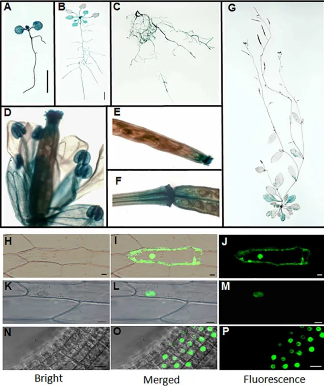
reporter lines. GUS staining results showed thatPQT3was expressed in both shoot and root tissues under normal condition (Fig 2A–2G). GUS expression was detected in the root tissues at all developmental stages we analyzed (Fig 2A–2C). For the 1-week-old seedlings, strong GUS staining was observed in the cotyledons, hypocotyls and root tissues (Fig 2A). For the 3-week-old seedlings, strong GUS staining was also detected in cotyledons, young leaves, and root tissues, but weakly stained in older leaves (Fig 2B). In 7-week-old adult plants, GUS expression was detected in rosette leaves, cauline leaves, the tip and basal junction of siliques, and was significantly higher in the flower petals, stamens and stigma of pistil (Fig 2D–2G).
PQT3 has two predicted nuclear localization signals (NLSs) in the carboxyl terminus (S3 Fig), implicating its nuclear localization. To confirm this, the35Spro:PQT3-GFPconstruct was made and transiently expressed in onion epidermal cells. The35Spro:GFPconstruct was used as control (Fig 2H–2J). PQT3-GFP signal was indeed detected in the nucleus (Fig 2K–2M).
For further confirmation, we transformed thePQT3pro:PQT3-GFPfusion construct into the Arabidopsis and obtained transgenic plants. Fluorescent microscopy results showed that GFP signal was accumulated in the nucleus of root cells (Fig 2N–2P), which is in agreement with the presence of two NLSs of the PQT3.
PQT3
is rapidly down-regulated by stress treatments
The expression ofPQT3was rapidly down-regulated by paraquat treatment and maintained at a low level as long as the paraquat treatment was applied (Fig 3A). This result is supportive for our previous opinion that PQT3 is a negative regulator of plant oxidative tolerance. The suppressed transcript level ofPQT3was restored to the previous level when PQ stress diminished (S4 Fig). By extrapolation, the expression ofPQT3could be down-regulated by other stress conditions. Indeed, our results showed that the expression ofPQT3was down-regulated by H2O2(Fig 3B), mannitol
(Fig 3C), drought (Fig 3D) and CdCl2(Fig 3E) at the indicated time points. Among these stresses,
CdCl2treatment led to the most significant reduction of the expression ofPQT3(Fig 3E). The
expression ofPQT3could also be down-regulated by NaCl stress at 3h. However, unlike the above results, the expression ofPQT3was activated by salt treatment at other time points (Fig 3F).
Fig 1. Phenotype ofpqt3-1andpqt3-2mutants. (A)Confirmation of paraquat (PQ) tolerant phenotype. 7-day-old seedlings of wild type andpqt3mutant were grown on MS medium supplemented with 0 or 2μM paraquat.(B)Survival ratio of wild type andpqt3mutant grown in (A). Values are mean±SD (n = 30 plants,***P<0.001). Asterisks indicate Student’s t-test significant differences.(C)Multiple mutant alleles analysis of paraquat tolerance for theAt4g17410locus. Survival ratio of wild type,pqt3-1andpqt3-2(Salk_065409) grown in the 0, 0.5, 1, and 2μM paraquat medium was counted. Values are mean±SD (n = 30 plants,***P<0.001). Asterisks indicate Student’s t-test significant differences.(D)Late-flowering phenotype ofpqt3-1andpqt3-2compared with wild type. Plants were grown under long day photoperiod (16 h light and 8 h dark).(E)The phenotype of 7-day-oldpqt3-1, wild type, function complementation (FC),pqt3-2, and35Spro: PQT3seedlings under 0μM (the top picture) or 2μM (the bottom picture) paraquat treatment. Bar = 0.5 cm.(F)The survival ratio ofpqt3-1, wild type, FC,pqt3-2, and35Spro:PQT3seeds germinated and grown on MS medium containing 0μM or 2μM paraquat for 7 days was counted. Values are mean±SD (n = 30 plants,***P<0.001). Asterisks indicate Student’s t-test significant differences.(G-R)DAB staining. The leaves of wild type,pqt3-1, andpqt3-2were treated without(H-J)or with 6μM paraquat for 12(L-N)or 24 h(P-R). 10 mM Na2HPO4was used as the negative control to stain the leaves of wild type treated without(G)or with 6μM paraquat for 12(K)or 24 h(O). Bar = 0.5 cm.
doi:10.1371/journal.pgen.1006332.g001
Switching off Oxidative Stress Response
Fig 2. Expression pattern and subcellular localization of PQT3. (A)GUS expression in 1-week-old seedling. Transgenic Arabidopsis plants expressingPQT3pro:GUSwere generated and analyzed for GUS expression. Bar = 0.5 cm.(B)GUS expression in 3-week-old seedling. Bar = 0.5 cm.(C)GUS expression in root tissue of 7-week-old plant.(D)GUS expression in flower tissue.(E and F)GUS expression in silique tip(E)and junction(F).(G)GUS expression in 7-week-old adult Arabidopsis.(H-J)35Spro:GFPwas transiently expressed in onion epidermal cells as control. The GFP can be observed in both plasma membrane and nucleus.(K-M)Nucleus localization of the PQT3-GFP fusion protein in onion epidermal cell was observed. Bar = 20μm.(N-P)The nucleus localization of PQT3-GFP fusion protein in the root tissue of stable transgenic seedlings expressingPQT3pro:PQT3-GFP. Bar = 20μm.
Switching off Oxidative Stress Response
ThePQT3pro:GUSpattern was observed under different stresses subsequently (Fig 3G– 3DD). Compared with transgenic seedlings under normal conditions, GUS staining was weaker in the seedlings under paraquat, H2O2, mannitol, CdCl2, and NaCl treatment. The changed
PQT3pro:GUSpattern was consistent with the altered expression ofPQT3detected by quanti-tative RT-PCR under different stresses.
APX1
and
GPX1
are up-regulated in
pqt3
mutant
Enzymatic protection systems are very important for ROS elimination. The transcript levels of ascorbate peroxidase (APX), glutathione peroxidase (GPX), catalase (CAT), cytosolic Cu/Zn SOD (CSD1), plastidic Cu/Zn SOD (CSD2), FeSOD (FSD), atypical Cys-His rich thioredoxin (ACHT), glutaredoxin C (GRXC), 2-Cys peroxiredoxin B (2CPB), peroxiredoxin Q (PRXQ) and mitochondrial MnSOD (MSD) were analyzed by quantitative RT-PCR inpqt3and wild type. The results show that transcript levels ofAPX1andGPX1were up-regulated inpqt3
under normal conditions compared with that in the wild type (Fig 4A–4J). The elevated tran-script levels ofAPX1andGPX1may contribute to the improved oxidative tolerance.
The enzyme activity of APX and GPX in wild type andpqt3mutant was also detected. The
pqt3mutant had higher enzyme activity of APX and GPX compared with wild type (Fig 4K and 4L).
PQT3 interacts with PRMT4b
To study the molecular mechanisms that underlie the enhanced stress tolerance ofpqt3, we screened cDNA library for potential candidate target proteins of PQT3 using yeast-two-hybrid (Y2H). Several proteins were isolated from the screen. Among these candidate interactors, PRMT4b, a member of arginine methyl transferase family, was frequently presented. To reveal the domain of PQT3 responsible for the interaction with PRMT4b, the PQT3 protein was divided into four parts: N-terminal DWNN, zfCCHC, U-box (RING finger), and C-terminal section containing the NLS1 and NLS2 domains, based on the predicted domains of PQT3 pro-tein (S3A Fig). Full-length PQT3 and four propro-tein sections were used for Y2H assay as baits. Full-length protein and the C-terminus of PQT3 were able to interact with PRMT4b in Y2H assays (Fig 5A). The interaction between PQT3 and PRMT4b was confirmed by colonies that grew on the SD-Leu-Trp-His plate with 50 mM 3-amino-1, 2, 4-triazole (3-AT) and displayed the blue color in X-gal assay (Fig 5A). However, PRMT4a was never isolated in the screening. The Y2H assay was also performed to further study the potential interaction between PQT3 and PRMT4a, sincePRMT4ais a close related gene ofPRMT4b. The result showed that the PQT3 did not interact with PRMT4a (S5 Fig).
The interaction between PQT3 and PRMT4b was further confirmed by protein pull-down assayin vitro. MBP-PQT3-C66 protein containing the NLS domain (S3A Fig) and His-PRMT4b protein were expressed inE.coliand purified subsequently. His-PRMT4b was incu-bated with amylose resin bound with recombinant MBP-PQT3-C66 protein. Pulled-down
Fig 3. The expression ofPQT3is rapidly down-regulated by various oxidative stress conditions. (A)The transcript level of PQT3was down-regulated by paraquat treatment. 1-week-old seedlings were treated with 6μM paraquat for the indicated times before RNA extraction for quantitative RT-PCR analysis. Values are mean±SD (n = 3 experiments,***P<0.001). Asterisks indicate Student’s t-test significant differences.(B-F)The expression ofPQT3was down-regulated by other stress conditions. 1-week-old seedlings were treated by 10 mM H2O2(B), 200 mM mannitol(C), drought(D), 200 mM CdCl2(E), and 150 mM NaCl (F)for the indicated times before RNA extraction for quantitative RT-PCR analysis. Values are mean±SD (n = 3 experiments, *P<0.05,**P<0.01,***P<0.001). Asterisks indicate Student’s t-test significant differences.(G-DD)GUS staining of 7-day-old PQT3pro:GUStransgenic lines without or with 6μM paraquat, 10 mM H2O2, 200 mM mannitol, 200 mM CdCl2, 150 mM NaCl treatment for 3 h. GUS expressions were significantly reduced in primary root tip(G-L), root elongation zone(M-R), root maturation zone(S-X), root zone with LRP(Y-DD)under stress conditions. Bar = 50μm.
Switching off Oxidative Stress Response
protein complex was detected by SDS-PAGE (Fig 5B) and western blotting using His anti-body (Fig 5C). The pull-down result clearly shows that PQT3 interacts with PRMT4bin vitro.
To determine whether the interaction also occurin vivo, we used the bimolecular fluores-cence complementation (BiFC) system. The N-terminus of yellow fluorescent protein (YFP) was fused to full-lengthPQT3cDNA, while full-lengthPRMT4bcDNA was fused to the C-ter-minal region of YFP. The empty plasmids were used as negative controls. Different plasmid combinations were co-infiltrated into epidermal cell ofN.benthamianaleaves. The yellow fluo-rescence was observed in epidermal cell contained both NE-PQT3 (the N-terminus of YFP fused with PQT3) and CE-PRMT4b (the C-terminus of YFP fused with PRMT4b) (Fig 5D). No fluorescence was observed from the negative controls (NE-PQT3/CE, NE/CE-PRMT4b and NE/CE) (Fig 5D). The nuclei were stained by Hoechst and detected by confocal. These results indicate that PQT3 can interact with PRMT4b in the nucleus of plant cell.
PQT3 has E3 ubiquitin ligase activity and can ubiquitinate PRMT4b
Not all the proteins with the predicted RING domain function as an ubiquitin ligase [62]. The E3 activity of PQT3 was determined via self-ubiquitination system. Both full-length(GST-PQT3) and C-terminal deletion (GST-PQT3-N40) proteins showed the E3 ubiquitin ligase activity (Fig 6AandS3B Fig). The ubiquitinated bands of PQT3 were detected by western blotting in the presence of E1 (from wheat), E2 (UBCh5b, from human), and 6×His-tagged ubiquitin (UBQ14, from Arabidopsis). When any of essential reaction components was miss-ing, self-ubiquitination of PQT3 was not detected (Fig 6A).
To determine whether PRMT4b is a substrate recognized by PQT3, we resorted to thein plantaubiquitination assay [63]. Leaf infiltration was conducted viaAgrobacterium tumefa-ciensstrains containing different combination of constructs. The infiltrated parts ofN.
benthamianaleaves were harvested. Total protein was extracted and detected via western blot-ting with anti-HA antibody. A smear of bands, which were larger than the size of HA-PRMT4b and showed the features of ubiquitinated form of the PRMT4b proteins, could be detected by anti-HA antibody in the samples co-infiltrated with PQT3 and HA-PRMT4b (Fig 6B). The cell lysates were immunoprecipitated with anti-HA antibody subsequently. Immunoprecipi-tated samples were detected via western blotting with anti-ubiquitin antibody. In the PQT3-PRMT4b co-infiltration sample, these high molecular size bands could also be detected by anti-ubiquitin antibody (Fig 6B). These results indicated that these high molecular size bands were ubiquitinated forms of PRMT4b. PQT3 protein could ubiquitinate the PRMT4b protein in tobacco. The decline of PRMT4b protein was also found in the samples co-infiltrated with PQT3 and HA-PRMT4b (Fig 6B).
In addition, PRMT4b protein levels in differentPQT3genetic background under MG132 treatments were also supportive of PRMT4b as a substrate of PQT3. As shown in theFig 6C and 6D, the protein level of PRMT4b inpqt3-1was higher than that of the wild type under nor-mal conditions. Under paraquat treatment, it remained lower in wild type than that ofpqt3-1
at the same time point, although protein level of PRMT4b increased gradually in wild type and
Fig 4. The analysis of antioxidant enzymes in wild type andpqt3mutant. (A-J)Quantitative RT-PCR analysis of transcript levels of antioxidant enzyme genes. RNA samples were isolated from 7-day-old wild type andpqt3seedlings for quantitative RT-PCR analysis. The transcript levels of APX(A), GPX(B), ACHT (C), FSD(D), CAT(E), GRXC(F), CSD(G), PRXQ(H), 2CPB(I)and MSD(J)were analyzed. Values are mean±SD (n = 3 experiments,***P<0.001). Asterisks indicate Student’s t-test significant differences.(K and L)Enzyme activity of APX(K)and GPX(L)in wild type andpqt3mutant. Thepqt3mutant has higher enzyme activity of APX and GPX than wild type. Values are mean±SD (n = 3 experiments,***P<0.001). Asterisks indicate Student’s t-test significant differences.
Switching off Oxidative Stress Response
pqt3-1with the prolonged paraquat treatment (Fig 6C and 6D). The transcript level of
PRMT4bwas unlikely to cause the observed difference of protein levels between the mutant and wild type (S6 Fig). Under MG132 treatment, the protein level of PRMT4b was increased in both wild type andpqt3-1mutant. When the seedlings were co-treated with paraquat and MG132 for 12 h, PRMT4b was accumulated in both wild type andpqt3-1mutant, and no sig-nificant difference of PRMT4b protein level was found between the wild type andpqt3-1
mutant (Fig 6C and 6D) because the degradation of the PRMT4b protein through ubiquitina-tion-26S proteasome pathway was inhibited by MG132.
In order to confirm further the PQT3-dependent ubiquitination of PRMT4b, the phenotype of wild type andpqt3-1mutant treated without or with paraquat in presence or absence of protea-some inhibitor MG132 was studied. Survival ratio ofpqt3-1seedlings was higher than that of wild type under paraquat treatment. No significant difference of survival ratio could be observed when the wild type andpqt3-1seedlings were co-treated with paraquat and MG132 (Fig 6E). The wild type gained enhanced paraquat tolerance under MG132 treatment to the level ofpqt3-1mutant.
PRMT4b
is a positive regulator in the oxidative stress tolerance
To reveal whether PRMT4b could play any roles in oxidative tolerance of plants, we obtained the
prmt4bmutant and35Spro:PRMT4blines (S1G, S1H and S1L Fig) and observed the phenotypes of
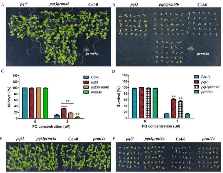
prmt4band35Spro:PRMT4bunder different concentrations of paraquat treatment. Survival ratio of wild type,pqt3-1,pqt3-2,prmt4band35Spro:PRMT4bwere counted (Fig 7A).35Spro:PRMT4b
had similar phenotype aspqt3-1andpqt3-2, while theprmt4bmutant was more sensitive to para-quat treatment than wild type (Fig 7A). Under CdCl2treatment, primary root elongation of
prmt4bmutant was also slower than that of wild type (Fig 7B). Furthermore, the overexpression lines ofPRMT4bwere analyzed under other stress conditions.35Spro:PRMT4bincreased tolerance to CdCl2and NaCl stresses compared with wild type. The35Spro:PRMT4bshowed the opposite
phenotype of the35Spro:PQT3which was more sensitive to CdCl2and NaCl stresses as compared
with wild type (S7 Fig). These results show that PRMT4b is a positive regulator for plant oxidative tolerance, which is also consistent with the function of PQT3. Theprmt4amutant and
prmt4aprmt4bdouble mutants were subsequently examined (S1I–S1K Fig). Theprmt4aprmt4b
double mutants had the similar phenotype toprmt4bunder paraquat treatment, and the knockout ofPRMT4adid not affect the oxidative tolerance (Fig 7C–7E). In addition, the transcript levels of
APX1,GPX1and other antioxidant enzyme genes were down-regulated inprmt4bmutant and up-regulated in35Spro:PRMT4bas compared with that in the wild type (Fig 7FandS8 Fig).
Increased H3R17 methylation on
APX1
and
GPX1
chromatin in
pqt3
The transcript level ofAPX1andGPX1was higher inpqt3than that in wild type under normal conditions (Fig 4A and 4B), which could be regulated by PRMT4b. The modification status of
Fig 5. PQT3 interacts with PRMT4b. (A)Y2H assay. PQT3 and its four protein sections were used as the bait. PRMT4b was used as the prey. Krev1/RalGDS-wt act as strong positive control and Krev1/RalGDS-m1 act as weak positive control. Krev1/RalGDS-m2 was used for negative control. The yeast harboring various constructs was grown on SD-Leu-Trp medium (upper panel). The yeast was transferred to SD-Leu-Trp-His medium with 50 mM 3-AT (middle panel) or used for X-gal staining (lower panel).(B and C)The pull-down assay between PQT3 and PRMT4b. His-PRMT4b was incubated with amylose resin bound with recombinant MBP-PQT3-C66 protein. Pulled-down protein complex was detected by SDS-PAGE(B)and western blot using anti-His antibody (C). MBP protein was used as a negative control.(D)BiFC assay. Different plasmid combinations were expressed in epidermal cells ofN.benthamianaleaves. Yellow fluorescence protein (YFP) was observed in epidermal cell expressing both NE-PQT3 (the N-terminal part of YFP fused with PQT3) and CE-PRMT4b (the C-terminal part of YFP fused with PRMT4b). No fluorescence was observed from the negative controls: NE-PQT3 / CE, NE / CE-PRMT4b, and NE / CE. The nuclei were stained by Hoechst and the fluorescence was detected by confocal. NE indicates pSPYNE vector and CE indicates pSPYCE vector.
Switching off Oxidative Stress Response
H3R17me2a in the chromatin ofAPX1andGPX1was compared between wild type andpqt3
mutant via chromatin immunoprecipitation (ChIP) assays. ChIP assays were performed with wild type andpqt3plants using antibody against H3R17me2a. As shown in theFig 8A and 8B, theAPX1andGPX1chromatin was divided into different regions and the enriched chromo-some fragments were detected by quantitative RT-PCR. The results showed that histone H3R17me2a modification ofAPX1andGPX1chromatin was increased inpqt3mutant (Fig 8C and 8D). The enriched chromosome fragments ofAPX1(C, D and E fragments) andGPX1(C, D and I fragments) were further analyzed inprmt4bmutant andpqt3prmt4bdouble mutants without or with paraquat treatment (Fig 8E–8J). Histone H3R17me2a modification ofAPX1
(C, D and E fragments) andGPX1(C, D and I fragments) chromatin was decreased inprmt4b
mutant andpqt3prmt4bdouble mutants without paraquat treatment as compared with wild type. It remained lower in wild type than that ofpqt3mutant, although H3R17me2a modifica-tion ofAPX1(C, D and E fragments) andGPX1chromatin (C, D and I fragments) in both wild type andpqt3mutant were enhanced under paraquat treatment. H3R17me2a modification of
APX1(C, D and E fragments) andGPX1chromatin (C, D and I fragments) was kept at a low level, although the modification inprmt4bmutant andpqt3prmt4bdouble mutants could also be affected by paraquat treatment. These results suggest that PRMT4b may targetAPX1and
GPX1to enhance the oxidative tolerance by increasing asymmetric dimethylation of H3 at R-17 inAPX1andGPX1chromatin.
pqt3prmt4b
double mutants in oxidative tolerance
To confirm further that PRMT4b is the target of PQT3, thepqt3prmt4bdouble mutants were obtained (S1M and S1N Fig). The survival ratio ofpqt3prmt4bunder paraquat treatment was intermediate betweenpqt3andprmt4b, which demonstrates that the PRMT4b protein is one of the targets of PQT3 and suggests that PQT3 may also target other proteins that contribute to the tolerance to oxidative stress (Fig 9A–9C).
We also obtained thepqt3prmt4adouble mutants (S1O and S1P Fig) and found that the sur-vival ratio ofpqt3prmt4aunder paraquat treatment had no significant difference from that ofpqt3
mutant (Fig 9D–9F), again indicating thatPRMT4ais not involved in oxidative stress response.
Discussion
PQT3 is a member of Arabidopsis RING-finger/U-box E3 ligase family. Secondary structure prediction using InterProScan protein sequence analysis software revealed four conserved
Fig 6. PRMT4b is a substrate recognized by PQT3. (A)In vitroubiquitination assay. GST-PQT3-N40 and GST-PQT3 were expressed inE.coliand purified. Nickel-HRP was used to detect His-tagged ubiquitin. The ubiquitination activity of PQT3 was observed only in the presence of E1 (from wheat), E2 (UBCh5b, from human), and 6×His-tagged ubiquitin (Ub). GST was used as negative control. The numbers on the left indicate the molecular masses of marker proteins.(B)In planta ubiquitination assay. Total protein samples were isolated from the infiltrated parts ofN.benthamianaleaves 1 day after agroinfiltration. The total protein was analyzed via western blot using anti-HA antibody (upper panel). Immunoprecipitated samples were analyzed using western blot with anti-ubiquitin antibody subsequently (middle panel). Ponceau S staining of the Rubisco protein was shown as a loading control (lower panel). The numbers on the left indicate the molecular masses of marker proteins.(C and D)The effect of proteasome inhibitor MG132 on the protein level of PRMT4b in wild type and pqt3-1mutant without or with paraquat treatment in time-course. Wild type andpqt3-1mutant were grown for 14 days, then the seedlings were treated without or with 6μM paraquat for 6 or 12 h in presence or absence of 50μM proteasome inhibitor MG132. The total proteins were extracted. Western blot was performed using anti-PRMT4b antibody(C). Ponceau S staining of the rubisco protein serves as a loading control. Band densities were quantified using Quantity One software (Bio-Rad, USA)(D). Values are mean±SD (n = 3 experiments,***P<0.001). Asterisks indicate Student’s t-test significant differences.(E)The survival ratio of wild type andpqt3mutant germinated and grown on MS medium containing 0μM or 1μM paraquat without or with 15μM MG132 for 7 days were counted. DMSO was used as control. Values are mean±SD (n = 30 plants,*P<0.05). Asterisk indicates Student’s t-test significant difference.
Switching off Oxidative Stress Response
domains including DWNN (domain with no name), zinc finger domain, RING-finger domain, and U-box domain in N-terminus of PQT3 protein (S9A and S9B Fig). DWNN is a novel ubi-quitin-like domain, which is a highly-conserved domain in eukaryotic plants and animals [64]. The DWNN domain of PQT3 contains 76 amine acids. DWNN domain is only found in the N-terminus of the members in the splicing-associated RBBP6 (Retinoblastoma Binding Protein 6) protein family [64]. The RING-finger domain was also found in RBBP6 protein family. The existence of the RING-finger domain suggests that DWNN domain may play its role as ubiqui-tin-like regulatory factors [64]. CCHC-type Zinc finger domain is also known as zinc knuckle, which can be found in a large number of RNA binding proteins [65,66]. As mentioned above, RING-finger domain can combine with the E2 in the cascade reaction of ubiquitination sys-tem,while U-box is a modified RING-finger [67]. In addition, two predicted NLS sequences (471–477 and 696–711 amino acids) were also found in the C-terminus of PQT3, which is con-sistent with nuclear localization of the protein (Fig 2H–2PandS9A and S9B Fig). Phylogenetic tree analysis further revealed the high homology proteins of PQT3 in other species (S9C Fig). DWNN domain and RING-finger/U-box domain in N-terminus of PQT3 were highly con-served in homologous proteins in different plant species (S10 Fig). The PQT3 may play its role as an ubiquitin ligase in different species and its function may be conserved throughout the plant kingdom.
In vitroubiquitination assay shows that PQT3 has the E3 ligase activity (Fig 6A). We noticed that thein vitroactivity was not as high as expected, which could be contributed by suboptimal reaction conditions such as E2 source. This E3 activity was confirmed byin planta
ubiquitination assay, in which PRMT4b protein could be ubiquitinated by expressed PQT3 protein inN.benthamiana(Fig 6B). Consistent with this result,pqt3-1mutant had higher pro-tein level of PRMT4b than the wild type, as the PRMT4b propro-tein was degraded by PQT3 in wild type under normal conditions (Fig 6C and 6D). The transcript level ofPRMT4bcould be induced in wild type under paraquat treatment (S6 Fig), while the transcript level ofPQT3was decreased by paraquat treatment (Fig 3A). In this case, PQT3-mediated PRMT4b degradation could be weakened in wild type. Consequently, the level of PRMT4b protein in wild type was elevated, but lower than that ofpqt3(Fig 6C and 6D). The proteasome inhibitor MG132 could block the degradation of the PRMT4b protein in wild type and enhance paraquat tolerance of wild type (Fig 6C–6E). Taken together, PQT3, as an E3 ubiquitin ligase, play its role in oxida-tive tolerance through the ubiquitination-degradation of PRMT4b.
A series of environmental stress could lead to oxidative damage to plants [10,13,68]. Both biotic stress and abiotic stress result in the production of ROS which in excess cause oxidative stress. Besides paraquat tolerance,pqt3mutants have enhanced tolerance to various environ-ment stresses (Fig 1andS2 Fig). Further studies may reveal new mechanisms of PQT3 in mul-tiple stress tolerance. Analysis of PQT3-interacting proteins may be a good start point to further understand the function ofPQT3. In the screen for PQT3-interacting proteins with Y2H, we isolated 66 positive colonies based on the expression of reporter genes included His3
Fig 7. PRMT4b is involved in oxidative tolerance of Arabidopsis. (A)Paraquat tolerance assay. Survival ratio of wild type,pqt3-1mutant, pqt3-2mutant (Salk_065409),prmt4bmutant and35Spro:PRMT4bgrown on 0, 2, 3, or 5μM paraquat medium were counted. The assay was repeated for three times. Values are mean±SD (n = 50 plants,*P<0.05,**P<0.01,***P<0.001). Asterisks indicate Student’s t-test significant differences.(B)Primary root elongation of wild type andprmt4bmutant seedlings grown on MS without or with 150μM CdCl2was measured. Values are mean±SD (n = 30 plants,**P<0.01). Asterisks indicate Student’s t-test significant differences.(C-E)The phenotype of wild type,prmt4a,prmt4b, andprmt4aprmt4bgrown on MS without(C)or with 1μM paraquat(D). The survival ratio of wild type,prmt4a, prmt4b, andprmt4aprmt4bgerminated and grown on MS medium containing 0μM or 1μM paraquat for 5 days were counted(E). Values are mean±SD (n = 36 plants,*P<0.05,**P<0.01). Asterisks indicate Student’s t-test significant differences.(F)The transcript levels ofAPX1 andGPX1in wild type,prmt4bmutant and35Spro:PRMT4b. RNA samples were isolated from 7-day-old seedlings for quantitative RT-PCR analysis. Values are mean±SD (n = 3 experiments,**P<0.01,***P<0.001). Asterisks indicate Student’s t-test significant differences.
Switching off Oxidative Stress Response
Fig 8. Arg-17 methylation in specific regions ofAPX1andGPX1chromatin is increased inpqt3mutant. (A and B) The illustration ofAPX1andGPX1chromatin. A to F represent different regions ofAPX1chromatin(A). A to I represent different regions ofGPX1chromatin(B). Promoters (black lines), exons (white boxes), and introns (black lines) ofAPX1 andGPX1were shown.(C and D)ChIP-PCR assay. Quantitative PCR was performed to verify each chromatin region of APX1andGPX1using specific primers. Fragments C, D and E inAPX1chromatin were enriched by anti-H3R17 antibodies inpqt3mutant(C). Fragments C, D and I inGPX1chromatin were enriched by anti-H3R17 antibodies inpqt3 mutant(D).UBQ5was used as an internal control. Values are mean±SD (n = 3 experiments,**P<0.01,***P<0.001). Asterisks indicate Student’s t-test significant differences.(E-G)The enrichment of C fragment(E), D fragment(F)and E fragment(G)inAPX1chromatin was analyzed in wild type,pqt3mutant,prmt4bmutant andpqt3prmt4bdouble mutants without or with paraquat treatment.UBQ5was used as an internal control. Values are mean±SD (n = 3 experiments, **P<0.01,***P<0.001). Asterisks indicate Student’s t-test significant differences.(H-J)The enrichment of C fragment (H), D fragment(I)and I fragment(J)inGPX1chromatin was analyzed in wild type,pqt3mutant,prmt4bmutant and pqt3prmt4bdouble mutants without or with paraquat treatment.UBQ5was used as an internal control. Values are mean±SD (n = 3 experiments,**P<0.01,***P<0.001). Asterisks indicate Student’s t-test significant differences.
doi:10.1371/journal.pgen.1006332.g008
Fig 9. The phenotypes ofpqt3prmt4bandpqt3prmt4adouble mutants under paraquat treatment. (A and B)The phenotypes ofpqt3, pqt3prmt4b, wild type andprmt4bseedlings grown on MS without(A)or with 2μM paraquat(B).(C)The survival ratio ofpqt3,pqt3prmt4b, wild type andprmt4bunder 2μM paraquat treatment for 7 days were counted. Values are mean±SD (n = 30 plants,**P<0.01,***P<0.001). Asterisks indicate Student’s t-test significant differences.(D)The survival ratio ofpqt3,pqt3prmt4a, wild type andprmt4aunder 1μM paraquat treatment for 7 days were counted. Values are mean±SD (n = 30 plants,**P<0.01).(E and F)The phenotypes ofpqt3,pqt3prmt4a, wild type andprmt4aseedlings grown on MS without(E)or with 1μM paraquat(F).
and LacZ. Several proteins were identified from these positive colonies repeatedly. The most frequent interacting partner is 20S core protease subunit of 26S proteasome. The result sug-gested that PQT3 act as an E3 ubiquitin ligase. Among these proteins, PRMT4b, as previously mentioned, may be responsible for the increased degree of Arg-17 methylation which may fur-ther regulateAPX1andGPX1genes to enhance oxidative tolerance of plants (Figs4A, 4Band 8). It has been reported that arginine methylation was involved in signal transduction, tran-scriptional control, DNA repair, RNA processing, and nuclear transport [17,19,23,69–72]. The functions of PRMTs have been extensively analyzed [15,19,20]. Here, theprmt4bmutant was found to be more sensitive to paraquat and CdCl2treatment than wild type (Fig 7A–7E).
Thus, PRMT4b plays a role in oxidative stress response of plants. It is known that PRMT4-me-diated methylation at 17 of histone H3 is linked to transcription activation [73]. The Arg-17 of histone H3 is the major site of PRMT4-mediated methylation, although it was reported that other protein site could also be methylated by PRMT4 [74–76]. We suggest that the increased Arg methylation degree of specific regions inAPX1andGPX1chromatin was caused by PRMT4b (Fig 8).
The increased transcript level ofAPX1andGPX1may be caused by PRMT4b-mediated his-tone Arg methylation (Figs4A, 4Band8). In presence of ascorbate as electron donor, the cyto-solic enzyme, APX1, catalyze the degradation of H2O2[77]. The response ofAPX1to oxidative
stress has been studies in Arabidopsis. The crucial role of APX1 in multiple stress response was reported [78]. It could be activated by multiple stresses to protect plants against oxidative stress [79–81]. The tobacco overexpressingAPX1could be more tolerant against UV-C-caused oxi-dative damage [82]. GPXs also have important functions in oxioxi-dative signaling, which can pro-tect plants from harmful effects of excessive oxidation [83]. It has also been reported that enhanced peroxide scavenging and decreased oxidative damage was found in transgenic tobacco seedlings overexpressing tobacco glutathione S-transferase that showed glutathione peroxidase (GPX) activity [84].
Thepqt3mutants also have late-flowering phenotype (Fig 1D). It has been reported that
prmt4aprmt4bdouble mutants display late-flowering phenotype [15]. The Y2H result showed that the PQT3 can not interact with PRMT4a (S5 Fig). As compared with wild type, theprmt4a
mutant also has no significant difference in the oxidative tolerance (Fig 7C–7E). The pheno-type ofpqt3prmt4adouble mutants demonstrate that PRMT4a protein was not involved in the response regulation of oxidative stress by PQT3 (Fig 9D–9F). The late-flowering phenotype of
pqt3may be regulated by other mechanisms, rather thanPRMT4aandPRMT4b. The flower-ing-related transcription factor AGAMOUS (AG) was found to be a potential target interacted with PQT3 in Y2H library screening. AG is involved in carpel development, leaf development, identification of floral organs, and stamen development [85]. Targeted removal of AG by PQT3 may be related to the late-flowering phenotype ofpqt3. In addition, one member of a large G protein family and the proteins with unknown function were also found to be potential targets interacted with PQT3 in Y2H library screening. By interacting with different partners, PQT3 may mediate multiple functions in diverse biological processes.
Thepqt3has enhanced tolerance to multiple stresses. PQT3 is also down-regulated by vari-ous oxidative stresses. PQT3 act as negative regulator in multiple stress responses. The interact-ing partner will be further analyzed to reveal the molecular function ofPQT3. As research continues, other combinations, also targeted by PQT3, may be revealed, where PQT3 act as a positive regulator. We could improve crop tolerance to multiple stresses throughPQT3 muta-tion. More candidate genes could be further determined from the potential targets interacted with PQT3. These genes may also have high application value after the revelation of molecular mechanisms.
Switching off Oxidative Stress Response
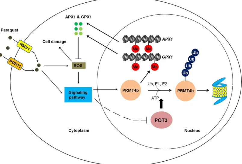
In conclusion, paraquat may enter the cell through plasma membrane-localized paraquat trans-porters PDR11 and RMV1 [59,86]. The intracellular paraquat may further activate different downstream signaling pathways to regulate the expression ofPQT3andPRMT4b. In addition, paraquat stress increases the generation of ROS. ROS is also an important signal molecule that mediate the responses to environmental stress [87]. ROS-activated signaling pathways may also be responsible for the regulation ofPQT3andPRMT4b. The oxidative stress activates the expression ofPRMT4b, represses the expression ofPQT3, and weakens PQT3-mediated the ubiquitinated degradation of PRMT4b, synergistically resulting in increased accumulation of PRMT4b. Conse-quently, the increased level of PRMT4b protein may lead to higher degree of histone methylation on theAPX1andGPX1chromatin. As a result, the transcription ofAPX1andGPX1is activated, leading to more APX1 and GPX1 enhancing oxidative tolerance of plants. When the stress disap-pears, transcription repression ofPQT3by oxidative stress is removed. The function of PQT3, as a negative regulator of oxidative stress response, is restored. The PRMT4b is then degraded by PQT3 in ubiquitination pathway. The activated response to oxidative stress is switched off. We propose a working model for PQT3 as a negative regulator of oxidative stress response (Fig 10).
Materials and Methods
Mutant screen from the activation-tagging library
Approximately 55,000 individual lines were screened for paraquat tolerant mutant, which came from the activation-tagging library constructed with Arabidopsis Columbia ecotype and pSKI015 vector [59].
Plant material and growth conditions
All Arabidopsis transgenic lines and mutants were based on the genetic background of Columbia ecotype used as wild type in the study. Salk_065409 (pqt3-2), Salk_097442C (prmt4b) and Salk_033423 (prmt4a) were obtained from ABRC (Arabidopsis Biological Resource Center). The seeds were sterilized in 10% bleach for 10 min. Then the seeds were washed for 5 times at least with sterile water. For vernalization, the seeds were kept in the dark with water at 4°Cfor 3 days to ensure the synchronous germination. Sterile seeds were germinated on MS medium. The seedlings were grown at 22°C under 16-h-light /8-h-dark cycle with light intensity 100 μE m-2s-1.
Transformation of Arabidopsis
The constructs were electroporated into competent cell ofAgrobacterium tumefaciensC58C1. The floral-dip method was used to transfer these constructs into Arabidopsis as described [88,89].
PCR analysis
The different tissues of plants were used for RNA extraction with Trizol method. Then RNA reverse reaction was carried out by TransScript Kit (TransGen Biotech). Specific primers were designed for RT-PCR analysis and the PCR products were detected by agarose gel electropho-resis. Applied Biosystem Step One real-time PCR system was used for the quantitative RT-PCR detection with specific primers listed in theS1 Tableand Premix Ex Taq II SYBR (TaKaRa).
UBQ5was used as the internal control.
previously described to confirm the results of genomic PCR. The35Spro:PQT3and35Spro:
PRMT4bplasmids were transformed into Col-0 to obtain the overexpression lines ofPQT3
andPRMT4b. For the FC line, the35Spro:PQT3was transformed intopqt3mutant, and the line with the same expression level ofPQT3as wild type was chosen and used as FC line.
35Spro:PQT3,35Spro:PRMT4band FC line were identified by glufosinate screening and quan-tified by RT-PCR or quantitative RT-PCR.
Stress tolerance assay
The seeds were germinated on MS medium containing different concentrations of paraquat, mannitol, CdCl2and NaCl, respectively. The phenotype was observed and survival ratio was
scored at the indicated time points.
For drought tolerance assay, the wild type,pqt3-1mutant andpqt3-2mutant seeds were ger-minated in one pot at same density. The 15-day-old seedlings were used for drought tolerance
Fig 10. A working model for PQT3 acting as a negative regulator of oxidative stress response.Many environmental stresses cause oxidative stress in plants. Under oxidative stress, stress signaling up-regulatesPRMT4bexpression and down-regulatesPQT3expression, leading to higher PRMT4b activity that will activateAPX1andGPX1and enhance antioxidation capacity of APX1 and GPX1. When oxidative stress is diminished,PRMT4bexpression is decreased andPQT3expression is increased. As a result, PQT3 activity is increased, leading to faster removal of PRMT4b via 26S proteasome. Together with decreasedPRMT4bexpression, PRMT4b activity will rapidly drop, leading to decreased expression ofAPX1andGPX1. The activated response of oxidative stress is then switched off.
doi:10.1371/journal.pgen.1006332.g010
Switching off Oxidative Stress Response
assay. Watering was withheld for another 15 days before re-watering. The photos were taken before re-watering and after re-watering for 1 day and 7 days. The survival ratio was scored after re-watering for 1 day and 7 days.
DAB staining
DAB staining was performed as described [91]. DAB staining solution (pH 6.0) was prepared by adding 0.05% (v/v) Tween-20 and 10 mM Na2HPO4to the DAB solution (1 mg/ml DAB,
pH3.0). For each treatment condition, at least 3 leaves per plant were obtained from 3 indepen-dent plants for each line (Col-0,pqt3-1andpqt3-2). Arabidopsis leaves from different lines were treated using MS liquid medium without or with 6 μM paraquat for 12 hr or 24 hr. These leaves were stained in 6-well culture plates with DAB staining solution subsequently and 10 mM Na2HPO4(pH 6.0) was used as the negative control. The 6-well plates were covered with
aluminum foil and placed on a shaker for 4–5 h. Follow the incubation, the bleaching solution (glycerol: acetic acid: ethanol = 1:1:3) was used in the discoloration after DAB staining solution was removed. The 6-well plates were placed into boiling water bath at 95°C for 15–20 min. Dis-coloration process was repeated using fresh bleaching solution. The brown precipitate caused by the reaction between DAB and H2O2could be observed on the leaves. Photos were taken
using a camera. Special attention should be paid to light avoidance through the whole opera-tion. The experiment was repeated for three times.
The detection of GUS activity
The promoter ofPQT3was cloned into pCB308R [92,93]. The transgenic lines containing
PQT3pro:GUSwere isolated by glufosinate screening. The T2 population was used for GUS staining. Histochemical staining for GUS activity in Arabidopsis was carried out as described previously [94] and GUS staining solution was prepared as described before [59]. The experi-mental materials were incubated in staining solution at 37°C. Then Arabidopsis tissues were destained and stored in 70% ethanol. The GUS activities of individual parts were observed via a light microscope Axio skop2 plus (ZEISS, Germany) with a video camera.
Subcellular localization assay
The full length CDS ofPQT3was cloned into the binary vector pCB2008E to construct the PQT3-GFP fusion vector [93]. After the detection of gene sequencing for inserted sequence, the recombinant plasmid was transducted into the epidermal cells of onion along with particle gun bombardment for transient expression assay. PQT3-GFP was also transferred into Col-0 via floral-dip method to create transgenic plant for the analysis of PQT3-GFP fusion protein localization. ZEISS fluorescence microscope (Axio skop2 plus) with video camera was used to observe the green fluorescence in both the epidermal cells of onion and the root cells of Arabi-dopsis transgenic lines.
Enzyme activity assay
For APX and GPX enzyme activity assay, Arabidopsis seedlings were ground in liquid nitrogen and resuspended in precooling Enzyme extraction buffer (50 mM phosphate buffer (Na2
H-PO4-NaH2PO4), pH = 7.0; 2% (w/v) polyvinylpolypyrrolidone (PVPP); 0.05% (v/v)
modifications [95]. For APX activity, 50 μl enzyme and 2950 μl reaction mixture (50 mM Tris-HCl, pH7.0; 0.1 mM EDTA; 0.1 mM H2O2and 0.5 mM ascorbic acid) was mixed. The
decreased OD290was recorded per 10 s. The enzyme amount oxidized 1 mM AsA in one
min-ute set as one activity unit (U) of APX. The APX activity was defined as U • g-1protein. GPX activity was measured indirectly through the detection of glutathione reductase (GR) activity using GPX Activity Measurement Kit (Beyotime Biotech, China). GR activity was detected as described with modifications [96]. The OD340was recorded per 30 s. The enzyme amount
con-sumed 1 mM NADPH in one minute set as one activity unit (U) of GPX. The GPX activity was also defined as U • g-1protein.
Western blot
For western blot analysis, proteins were electroblotted from 12% acrylamide gel to nitrocellu-lose membrane (Immobilon-P, MILLIPORE Corporation, USA) after the separation of SDS-PAGE. Antibodies used in western blot were as follows: anti-HA antibody (M20003, Mouse mAb, Abmart, Shanghai, China), 1:1,000 for western blot; anti-PRMT4b antibody, 1:500 for western blot; anti-Ubiquitin antibody (ab7254, abcam, USA), 1:1,000 for western blot; anti-His antibody (M30111, Mouse mAb, Abmart, Shanghai, China), 1:1,000 for western blot and goat anti-mouse lgG-HRP (Santa Cruz Biotechnology, USA), 1:5,000 for western blot. Image Quant LAS 4000 (GE, USA), as the CCD camera system, was used for the result exami-nation with Super Signal West Femto Trial Kit (Thermo, USA). Band densities were quantified via Quantity One software (Bio-Rad, USA).
In vitro
E3 ubiquitin ligase activity assay
Thein vitroE3 ubiquitin ligase activity assay was carried out as described previously [43]. GST-PQT3 fusion protein was obtained fromE.coiland purified subsequently. His-tagged ubi-quitin of Arabidopsis (UBQ14) was also expressed using bacterial expression system and puri-fied. In addition, the wheat (Triticum aestivum) E1 (GI: 136632) and human E2 (UBCh5b) were also used in the reaction. Reactions were performed for 1.5 h at 30°C. For the immuno-blot, His-tagged ubiquitin of Arabidopsis was detected using Nickel-HRP (Kirkegaard & Perry Laboratories, nickel–nitrilotriacetic acid agarose conjugated to horseradish peroxidase).
Y2H screening and confirmation
The bait plasmid was transformed into Mav203 strain of yeast, which was constructed with pDEST32 vector and the full-lengthPQT3cDNA. Yeast two-hybrid screening was performed using two-hybrid cDNA library of Arabidopsis. The cDNA library containing fragments of fusion proteins composed of prey proteins and GAL4—AD was used to transform Mav203 cells harboring bait plasmid in Y2H assay. Positive clones were screened using SD/-Leu-Trp-His and X-gal assay. Then it was identified by nucleotide sequencing with corresponding primers. Two-hybrid screening was carried out based on the protocol described in Two-Hybrid System Manual (Invitrogen, USA). The result of screening was further confirmed by the two-hybrid assay. The full-length CDS ofPQT3and the four segments ofPQT3were inserted into pDEST32 vector to construct the bait plasmid, while the prey plasmid was constructed with pDEST22 vector and the full-length CDS ofPRMT4b. For two-hybrid assay, the primers could be found inS1 Table.
Pull-down assay
MBP-PQT3-C66 and His-AtPRMT4b fusion protein were expressed using prokaryotic expres-sion system and purified. MBP-PQT3-C66 fuexpres-sion protein was incubated with MBP beads
Switching off Oxidative Stress Response
(amylose resin) at 4°C for 2 h, and the MBP tag was used as a negative control. The beads were cleaned with washing buffer for 4 times. Then the beads were incubated with His-AtPRMT4b at 4°C for 2 h respectively. The beads were cleaned with washing buffer for 4 times. Western blot was used to detect the SDS-PAGE separation results of pulled-down mixtures in nitrocel-lulose membrane with anti-His antibody.
Agroinfiltration procedure
Agrobacterium tumefaciensstrain C58C1 was used in the experiments. Agroinfiltration proce-dure was carried out as described previously [63]. At first, these strains were grown on LB medium with Gentamicin and Kanamycin. Single colony was transferred into 5 ml LB liquid medium containing same resistance and grown for 48 h in a 28°C shaker. The bacteria solution was inoculated into new LB liquid medium containing 40 μM acetosyringone (1:100 ratio, v/v) and 10 mM 2-(N-morpholine)-ethanesulfonic acid (MES; pH 5.6). Bacteria were developed in a 28°C shaker until OD600reached 3.0 approximately. The bacteria were collected gently by
means of 10 min centrifugation (3,200 g/min), and the resuspension of pellets was performed with 10 mM MgCl2until OD600reached 1.5 approximately. The bacteria solution was kept at
room temperature with a final concentration of 200 μM acetosyringone for at least 3 h without shaking. The different plastid combinations were transformed into epidermal cells ofN.
benthamianaleaves by disposable syringe.
BiFC analysis
NE-PQT3 (the N-terminus of YFP fused with PQT3) and CE-PRMT4b (the C-terminus of YFP fused with PRMT4b) were constructed. These constructs were transferred to Agrobacter-ium strains C58C1 respectively. As mentioned above, the different plastid combinations were transformed into epidermal cells ofN.benthamianaleaves by agroinfiltration. YFP was observed 1–2 days after leaf infiltration using confocal. The nuclei were stained by Hoechst subsequently and fluorescence detection by confocal was performed. For BiFC assay, the prim-ers could be found inS1 Table.
Protein extraction
Native extraction buffer 1 [10 mM EDTA; 50 mM TRIS-MES, pH 8.0; 1 mM MgCl2; 5 mM
DTT; 0.5 M sucrose; protease inhibitor cocktail for plant cell and tissue extracts (Sigma, USA)] was chosen for protein extraction buffer. Other steps of protein extraction were carried out as described previously [63].
In planta
ubiquitination assay
Image Quant LAS 4000 (GE, USA), as the CCD camera system, was used for the result examination.
Chromatin immunoprecipitation-PCR assay
The wild type,pqt3mutant,prmt4bmutant andpqt3prmt4bdouble mutants were used for ChIP assay without or with 6 μM paraquat treatment for 24h.UBQ5was chose for internal control. ChIP was performed as previously described [97,98]. The regions with Arg-17 methyl-ation were precipitated via anti-H3R17me2a antibodies (anti-Histone H3 asymmetric dimethyl R17 antibody-ChIP grade, ab8284, Abcam, USA) from input DNA. The corresponding primers were designed for quantitative RT-PCR to detect the enrichments of different DNA fragments inAPX1andGPX1chromatins [99,100]. The primers used in ChIP assay were showed inS1 Table.
Supporting Information
S1 Fig. Identification of mutants, homozygous Salk lines and transgenic lines. (A)The loca-tion of the T-DNA inserloca-tions inpqt3-1mutant andpqt3-2mutant. (Salk_065409). The loca-tions of T-DNA insertion were shown as inverted black triangles. The structure of the
At4g17410locus was shown for exons as red boxes, introns as black lines and UTR as black box.(B)Detection of transcript levels forPQT3and its neighboring genes using RT-PCR. The transcript levels ofAt4g17390,At4g17410(PQT3), andAt4g17420were compared between wild type andpqt3-1mutant.Tubulin8(TUB8) was used as a loading control. The RT-PCR assay was repeated for three times, and a typical result was shown.(C)Genomic PCR analysis of homozygous Salk_065409. Genomic DNA isolated from leaves of Salk_065409 line and wild type was used as template for PCR.(D)RT-PCR analysis ofpqt3-1mutant and homozygous T-DNA insertion mutant of Salk_ 065409 (pqt3-2). RNA was extracted from 2-week-old wild type,pqt3-1mutant and Salk_065409 (pqt3-2). The transcript level ofPQT3was analyzed by RT-PCR. No signal was detected inpqt3-1mutant and the homozygous Salk_065409 (pqt3-2).
TUB8was used as a loading control.(E)RT-PCR analysis ofPQT3transcript levels using RNA samples isolated from wild type,35Spro:PQT3, and FC line.(F)Identification of35Spro:PQT3
using quantitative RT-PCR. RNA was extracted from 4-week-old wild type and35Spro:PQT3
lines. Values are mean ± SD (n = 3 experiments,P<0.05). Asterisk indicate Student’s t-test significant difference.(G)Genomic PCR analysis of homozygous Salk_097442C (prmt4b). Genomic DNA isolated from leaves of Salk_097442C line and wild type was used as template for PCR.(H)quantitative RT-PCR analysis ofprmt4bmutant (Salk_097442C). RNA was extracted from 4-week-old wild type andprmt4bmutant. No signal was detected inprmt4b
mutant.(I)Genomic PCR analysis of homozygous Salk_033423 (prmt4a). Genomic DNA iso-lated from leaves of Salk_033423 line and wild type was used as template for PCR.(J and K) Genomic PCR analysis ofprmt4aprmt4bdouble mutants. Genomic DNA isolated from leaves ofprmt4aprmt4bdouble mutants and wild type was used as template for PCR.The knockout of
PRMT4a(J)andPRMT4b(K)was identified respectively.(L)Identification of35Spro:PRMT4b
using quantitative RT-PCR. RNA was extracted from 4-week-old wild type and35Spro: PRMT4blines. Values are mean ± SD (n = 3 experiments,P<0.01,P<0.001). Asterisks indicate Student’s t-test significant differences.(M and N)Identification ofpqt3prmt4adouble mutants. The knockout ofPRMT4ainpqt3prmt4adouble mutants was analyzed by Genomic PCR(M). The transcript level ofPQT3was detected by RT-PCR(N). No signal was detected in
pqt3prmt4adouble mutants.TUB8was used as a loading control.(O and P)Identification of
pqt3prmt4bdouble mutants. The knockout ofPRMT4binpqt3prmt4bdouble mutants was analyzed by Genomic PCR(O). The transcript level ofPQT3was detected by RT-PCR(P). No
Switching off Oxidative Stress Response
signal was detected inpqt3prmt4bdouble mutants.TUB8was used as a loading control. (DOCX)
S2 Fig. Phenotype ofpqt3mutants under other environmental stresses lead to oxidative damage. (A)The phenotype of wild type,pqt3-1andpqt3-2mutant grown on MS medium for 12 days. Bar = 1 cm.(B)Primary root elongation of wild type,pqt3-1andpqt3-2mutant grown on MS medium for 12 days was measured. Values are mean ± SD (n = 30 plants).(C)The phenotype of wild type,pqt3-1andpqt3-2mutant grown on MS medium with 150 μM CdCl2for 12 days.
Bar = 1 cm.(D)Primary root elongation of wild type,pqt3-1andpqt3-2mutant grown on MS with 150 μM CdCl2for 12 days was measured. Values are mean ± SD (n = 30 plants,P<0.01).
Asterisks indicate Student’s t-test significant differences.(E)The phenotype of wild type,pqt3-1
andpqt3-2mutant grown on MS medium containing 250 mM mannitol for 12 days. Bar = 1 cm. (F)Primary root elongation of wild type,pqt3-1andpqt3-2mutant grown on MS containing 250 mM mannitol for 12 days was measured. Values are mean ± SD (n = 30 plants,P<0.01,
P<0.001). Asterisks indicate Student’s t-test significant differences.(G)The phenotype of
wild type,pqt3-1andpqt3-2mutant grown on MS medium with 120 mM NaCl for 12 days. Bar = 1 cm.(H)Primary root elongation of wild type,pqt3-1andpqt3-2mutant grown on MS with 120 mM NaCl for 12 days was measured. Values are mean ± SD (n = 30 plants,P<0.05,
P<0.01). Asterisks indicate Student’s t-test significant differences.(I)The schematic diagram
of the locations of wild type,pqt3-1andpqt3-2plants grown in one pot for drought tolerance assay.(J to L)Drought stress assay of thepqt3mutants and wild type grown in the same pot. The wild type,pqt3-1, andpqt3-2plants were grown in the same pot for 15 days before drought stress was imposed. These plants were grown under drought stress for 15 days. The photos were taken before re-watering(J)and after re-watering for 1 day(K)and 7 days(L).(M)Re-water survival ratio of wild type,pqt3-1andpqt3-2mutant after drought stress was counted. Values are
mean ± SD (n = 18 plants,P<0.001). Asterisks indicate Student’s t-test significant differences. (DOCX)
S3 Fig. Sections of PQT3 protein used in Y2H, pull-down andin vitroubiquitination assay. (A)The division of PQT3 protein for Y2H assay was presented by the red lines. The full-length protein sequence of PQT3 was divided into four segments (DWNN, zfCCHC, U-box/RING finger and C-terminus contained the NLS1 and NLS2 domain) and each segment was used as bait. In pull-down assay, the protein section of PQT3 (PQT3-C66) located in the blue box was used.(B)The protein used in Self-ubiquitin assay. Full-length protein sequence of PQT3 and PQT3-N40 (the section of PQT3 protein located in the red box) were selected for the assay. PQT3-N40 (1–360 aa) contains all the conserved domains of an E3 ubiquitin ligase. (DOCX)
S4 Fig. The transcript level ofPQT3after the elimination of PQ stress.1-week-old wild type seedlings were treated by 6 μM paraquat for 3h. PQ stress diminished for 3h subsequently before RNA was extracted for quantitative RT-PCR analysis. Values are mean ± SD (n = 3 experiments,P<0.001). Asterisks indicate Student’s t-test significant differences. (DOCX)
S5 Fig. Y2H assay for PQT3 and PRMT4a.PQT3 and its four protein sections were used as the bait. PRMT4a was used as the prey. Krev1/RalGDS-wt act as strong positive control and Krev1/RalGDS-m1 act as week positive control. Krev1/RalGDS-m2 was used for negative con-trol. The yeast harboring various constructs was grown on SD-Leu-Trp medium (upper panel). The yeast was transferred to SD-Leu-Trp-His medium with 50 mM 3-AT (middle panel) or used for X-gal staining (lower panel).