Spirostanol glucosides from the leaves of
Cestrum laevigatum
L.
Paulo Riceli Vasconcelos Ribeiro
a, Ana Jérsia Araújo
b, Letícia Veras Costa-Lotufo
c, Raimundo Braz-Filho
d,e,
Hélio Vitoriano Nobre Junior
f, Cecília Rocha da Silva
f, João Batista de Andrade Neto
f,
Edilberto Rocha Silveira
a, Mary Anne Sousa Lima
a,⇑aDepartamento de Química Orgânica e Inorgânica, Centro de Ciências, Universidade Federal do Ceará, CP 12.200, CEP 60.021-940 Fortaleza, CE, Brazil bDepartamento de Farmacologia e Fisiologia, Faculdade de Medicina, Universidade Federal do Ceará, CP 12.200, 60430-270 Fortaleza-CE, Brazil cDepartamento de Farmacologia, Instituto de Ciências Biomédicas, Universidade de São Paulo, CEP 05508-900 São Paulo, SP, Brazil
dLaboratório de Ciências Químicas, Universidade Estadual do Norte Fluminense Darcy Ribeiro, 28013 602 Campos dos Goytacazes, RJ, Brazil eDepartamento de Química, Universidade Federal Rural do Rio de Janeiro, CP 74541, 23890-000 Seropédica, RJ, Brazil
fDepartamento de Farmácia, Universidade Federal do Ceará, CP 12.200, 60430-170 Fortaleza, CE, Brazil
a r t i c l e
i n f o
Article history:
Received 22 June 2015
Received in revised form 21 October 2015 Accepted 10 December 2015
Available online 17 December 2015
Keywords:
Solanaceae
Cestrum laevigatum
Spirostanol glycosides Antifungal
Cytotoxic
a b s t r a c t
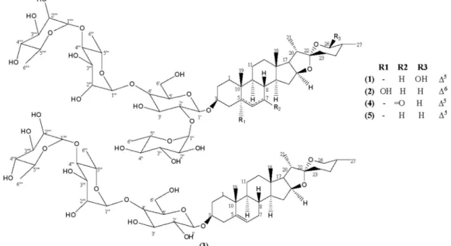
Two new steroidal saponins, (25R)-spirost-5-ene-3b,26b-diol 3-O-a-L-rhamnopyranosyl-(1?4)-a-L -rhamnopyranosyl-(1?4)-[(1?2)-a-L-rhamnopyranosyl]-b-D-glucopyranoside (1) and (25R )-spirost-6-ene-3b,5b-diol 3-O-a-L-rhamnopyranosyl-(1?4)-a-L-rhamnopyranosyl-(1?4)-[(1?2)-a-L -rhamno-pyranosyl]-b-D-glucopyranoside (2), along with the known diosgenin 3-O-a-L-rhamnopyranosyl-(1?
4)-a-L-rhamnopyranosyl-(1?4)-b-D-glucopyranoside (3), chonglouoside SL-5 (4) and Paris saponin Pb (5) were isolated from the leaves ofCestrum laevigatum. The structures of the compounds were deter-mined using spectroscopic analyses including HRESI-MS, 1D and 2D NMR data, followed by comparison with data from the literature. Among them, two are particularly unique, compound1is the first6D -spir-ostanol saponin and compound2has an unusual C-26 hydroxyl in the5D-spirostanol skeleton. Antifungal testing showed a potent activity to formosanin C againstCandida albicans andCandida parapsilosis. Evaluation of the cytotoxic activity indicated that compound1has a moderate activity against HL-60 and SF-295 cell lines, while compound2were active only against HL-60.
Ó2015 Elsevier Inc. All rights reserved.
1. Introduction
Cestrumis the second largest genus of the Solanaceae family, with 150 species distributed in tropical and subtropical America, 50 of which occurring in Brazil[1–2]. As particular to some Solana-ceae genera, several species have been reputed as poisonous plants and the search for active compounds revealed a prolific source of bioactive steroids saponins containing spirostanol or furostanol glycoside skeletons[2–7].
Cestrum laevigatum L. is an evergreen shrub native to South
America and has been introduced to South Africa. It is widespread into natural grasslands, forests, riparian habitats, and coastal dunes, where is popularly known as ‘‘coerana”, ‘‘lady of the night” and ‘‘corana”. The dried leaves are used in traditional medicine as treatment for malaria and fever[8], and smoked by the Mapuche Indian of southern Chile as a substitute for cannabis[9]. Although commonly used for ornamental purposes,C. laevigatumis
consid-ered the most lethal plant to mammals, among the group of com-mon invasive species that cause liver damage. For this reason, is one of toxic plants of greater importance in Brazilian livestock for its wide distribution and economic losses, and its growth is severely controlled or eradicated into pastureland[10,11].
In the course of our search for bioactive natural compounds we report the isolation and structural characterization of two new sapogenins (25R)-spirost-5-ene-3b,26b-diol 3-O-
a
-L-rhamnopyra-nosyl-(1?4)-a
-L-rhamnopyranosyl-(1?4)-[(1?2)-a
-L-rhamnopyranosyl]-b-D-glucopyranoside (1) and (25R)-spirost-6-ene-3b,5b -diol 3-O-
a
-L-rhamnopyranosyl-(1?4)-a
-L-rhamnopyranosyl-(1?4)-[(1?2)-
a
-L-rhamnopyranosyl]-b-D-glucopyranoside (2)from the leaves ofC. laevigatum, in addition to the known diosgenin O-
a
-L-rhamnopyranosyl-(1?4)-a
-L-rhamnopyranosyl-(1?4)-b-D-glucopyranoside (3) [12], chonglouoside SL-5 (4) [13] and for-mosanin C (5)[14]. The screening for antimicrobial activities against
Candida parapsilosis (ATCCÒ
22019TM), Candida albicans (ATCCÒ 10231TM),Candida krusei(ATCCÒ
14243TM),Pseudomonas aeruginosa (ATCCÒ
9027TM),Staphylococcus aureus(ATCCÒ
6538TM) andBacillus
subtilis(ATCCÒ
6633TM), and evaluation of cytotoxicity using human
http://dx.doi.org/10.1016/j.steroids.2015.12.006
0039-128X/Ó2015 Elsevier Inc. All rights reserved. ⇑ Corresponding author.
E-mail address:[email protected](M.A.S. Lima).
Contents lists available atScienceDirect
Steroids
promyelocytic leukemia (HL-60), ovarian carcinoma (OVCAR-8), colorectal adenocarcinoma (HCT-116) and glioma (SF-295) cell lines were performed (seeFig. 1).
2. Experimental
2.1. General experimental procedures
Melting points were obtained on a MetllerToledo-FP82HT and were uncorrected. IR spectra were recorded as KBr pallets on a Perkin-Elmer FT-IR Spectrum 1000 using KBr. The NMR spectra were performed on Bruker Avance DRX 300 or on Avance DRX500 MHz equipped with an inverse detection probe head and
z-gradient accessory. The1H and13C chemical shifts are expressed
in thedscale and were referenced to TMS through the residual sol-vent. High-resolution mass spectra were recorded on a Waters Acquity UPLC system coupled with a Quadrupole/Time-of-Flight system (UPLC/Qtof MSE spectrometer) in the positive mode. The TOF conditions were as follow: source temperature 120°C; desolvation temperature 350°C; desolvation gas flow of 350 L/h; capillary voltage 2 kV; collision Energy Ramp 20 eV. The mode was acquired from 110 to 1200 Da. Optical rotations were obtained on a Perkin-Elmer Q-2000 polarimeter, at 589 nm and 25°C. Column chromatography was performed over Sephadex LH-20 (Pharmacia) and SPE C18 cartridge (Phenomenex), while TLC was performed on precoated silica gel aluminum sheets (Merck). The compounds were visualized by UV detection and by spraying with vanillin/perchloric acid/EtOH solution, followed by heating.
2.2. Plant material
C. laevigatumL. was collected at the Guaramiranga Mountain, Pacoti, Ceará State, Northeast of Brazil. Voucher specimens (#38643) were deposited at the Herbário Prisco Bezerra (EAC) and identified by MSc. Edson de Paula Nunes, Departamento de Biologia, Universidade Federal do Ceará, Ceará, Brazil.
2.3. Extraction and isolation
Leaves ofC. laevigatum(1.14 kg) were pulverized and extracted with EtOH (39.0 L) at room temperature. The solvent was removed under reduced pressure to yield a dark green residue (131.4 g).
Part of EtOH extract (91.0 g) was dissolved in a mixture of MeOH:H2O (1:1 v/v) and submitted to liquid–liquid partition
chro-matography with hexane, CHCl3, EtOAc andn-BuOH to give four
fractions: hexane (0.11 g), CH2Cl2 (27.1 g), EtOAc (11.3 g) andn
-BuOH (32.0 g).
An aliquot of the n-BuOH fraction (2.0 g) was rechro-matographed on Sephadex LH-20 (10.0 g) (column 2.5 cm10 cm) to afford forty-five fractions (3.0 mL) that were pooled together into five resulting sub-fractions after TLC analysis. Sub-fraction F-3 (1.1 g) was further chromatographed on a SPE C18 (5.0 g) cartridge using MeOH/H2O 1:1 (30.0 mL), MeOH/H2O 7:3
(3.00 mL), MeOH/H2O 8:2 (40.0 mL), MeOH/H2O 9:1 (30.0 mL)
and MeOH (50.0 mL), yielding five fractions. Sub-fraction F-5 (0.360 g) was submitted to semi-preparative RP-18 HPLC analysis, using MeOH/H2O (87:13) as eluent, to afford1(10.6 mg).
An Aliquot of the EtOAc fraction (2.1 g) was rechromatographed on Sephadex LH-20 (10.0 g) (column 2.5 cm10 cm) to give forty sub-fractions (3.0 mL), which were combined into four resulting sub-fractions according to TLC analysis. Sub-fraction F-3 (0.70 g) which was then chromatographed on a SPE C18 cartridge by elu-tion with MeOH/H2O 1:1 (30.0 mL), MeOH/H2O 7:3 (30.0 mL),
MeOH/H2O 8:2 (40.0 mL), MeOH/H2O 9:1 (30.0 mL) and MeOH
(40.0 mL), yielding five fractions. Fraction F-3 (0.26 g) was submit-ted to semi-preparative RP-18 HPLC chromatography, using an iso-cratic mixture MeOH/H2O 8:1 to afford2(10.0 mg),3(8.0 mg),4
(10.5 mg) and5(19.5 mg).
2.3.1. (25R)-spirost-5-ene-3b,26b-diol 3-O-
a
-L-rhamnopyranosyl-(1?4)-
a
-L-rhamnopyranosyl-(1?4)-[(1?2)-a
-L-rhamnopyranosyl]-b-D-glucopyranoside (1)
White amorphous powder, mp 240–242°C; [
a
]D20-172.8 (c0.10, MeOH); Rf 0.22 (CH2Cl2/MeOH 25%); 1H and13C NMR data: seeTable 1; IR (KBr) 3355; 2928; 1640; 1452; 1036 cm1. HRESIMS m/z: 1031.5468 [M+H]+(calcd for C
51H83O21[M+H], 1031.5427).
2.3.2. (25R)-spirost-6-ene-3b,5b-diol 3-O-
a
-L-rhamnopyranosyl-(1?4)-
a
-L-rhamnopyranosyl-(1?4)-[(1?2)-a
-L-rhamnopyranosyl]-b-D-glucopyranoside (2)
White amorphous powder, mp 178–179°C; [
a
]D20-132.7 (c0.16, MeOH); Rf 0.20 (CH2Cl2/MeOH 25%);1H and 13C NMR data: seeTable 1; IR (KBr) 3362; 2928; 1653; 1453; 1037. HRESIMS m/z: 1031.5427 [M+H]+(calcd for C
51H83O21[M+H], 1031.5427).
2.3.3. (25R)-spirost-5-ene-3b-ol 3-O-
a
-L-rhamnopyranosyl-(1?4)-a
-L-rhamnopyranosyl-(1?4)-b-D-glucopyranoside (3)[
a
]D20-55.5°(c0.09; MeOH) {[a
]D24-62.0 (c0.09; MeOH)[15]}; Rf0.50 (CH2Cl2/MeOH 25%).
2.3.4. (25R)-7-oxo-espirost-5-ene-3b-ol 3-O-
a
-L-rhamnopyranosyl-(1?4)-
a
-L-rhamnopyranosyl-(1?4)-[(1?2)-a
-L-rhamnopyranosyl]-b-D-glucopyranoside (4)
[
a
]D20-115.8°(c0.11; MeOH) {[a
]D15-32.8°(c0.12; MeOH)[13]};Rf 0.23 (CH2Cl2/MeOH 25%).
2.3.5. (25R)-espirost-5-ene-3b-ol 3-O-
a
-L-rhamnopyranosyl-(1?4)-a
-L-rhamnopyranosyl-(1?4)-[(1?2)-a
-L-rhamnopyranosyl]-b-D-glucopyranoside (5)
[
a
]D20-116.7°(c0.20; MeOH) {[a
]D21-118.6°(c0.20; MeOH)[15]};Rf 0.23 (CH2Cl2/MeOH 25%).
2.4. Acid hydrolysis and sugar analysis
Compounds1and2(5.0 mg) were dissolved in 2 M HCl (diox-ane/H2O, 1:1, 2 mL) and stirred at 90°C for 2 h. After cooling, the
reaction mixture was neutralized with solution of 1 M NaOH, extracted with CH2Cl2(35 mL), and the aqueous layer was evap-orated to give a mixture of monosaccharides. The residue was dis-solved in hexamethyldisilazane/trimethylchlorosilane/pyridine (3:1:9), and stirred at 70°C for 60 min. The supernatants (3
l
L) were analyzed by GC Agilent model GC-789/MSD-5977A (quadru-pole), under the following conditions: CP-ChiraSil-L-Val column, 0.25 mm25 m; temperatures for detector and injector 150 and 200°C respectively; temperature gradient system for the oven, 100°C for 1 min and then raised to 180°C; rate 5°C/min kept for 5 min. The configurations of sugars were determined by compar-ison of the retentions times of the corresponding derivatives with those of standards treated simultaneously with same silylating reagents (L-rhamnose 11.15 and D-glucose 16.57). Peaks of the hydrolysates of1and2were detected at 11.20 (L-rhamnose) and 16.57 (D-glucose). Co-injection of each hydrolysate with standard D-glucose andL-rhamnose gave single peaks.2.5. Cytotoxic activity
The tested tumor cell lines (colorectal adenocarcinoma HCT-116, ovarian carcinoma OVCAR-8, human promyelocytic leukemia HL-60 and glioma SF-295) were kindly donated by the National Cancer Institute (Bethesda, MD, USA). Cells were maintained in RPMI 1640 medium supplemented with 10% fetal bovine serum, 2 mM glutamine, 100 U mL1penicillin, and 100
l
g mL1strepto-mycin at 37°C with 5% CO2. The cytotoxicity of the isolated com-pounds was tested the against tumor cell lines using the 3-(4,5-dimethyl-2-thiazolyl-2,5-diphenyl-2H-tetrazolium bromide) (MTT) (Sigma Aldrich Co., St. Louis, MO/USA) reduction assay [16]. Cells were plated in 96-well plates (105 cells per well for
adherent cells or 3105 cells per well for suspended cells in 100
l
L of medium) and compounds (0.05–25l
g mL1) weredis-solved in DMSO, added to each well using the HTS – high-through-put screening-biomek 3000-Beckman Coulter (Inc. Fullerton, California, USA), and incubated for 72 h. Doxorubicin was used as Table 1
NMR spectral data of compounds1and2(1H – 500 MHz;13C – 125 MHz; CD 3OD).
Position 1 2
dC dH dC dH
1 38.1 1.82 (m) 29.6 1.68 (m)
1.02 (m) 1.40 (m)
2 30.6 1.90 (m) 32.3 1.79 (m)
1.60 (m) 1.57 (m)
3 79.4 3.54 (m) 75.2 4.17 (m)
4 39.6 2.44 dd (12.5, 3.0) 39.3 2.50 (m) 2.35 t (12.5) 2.36 t (12.7)
5 142.1 – 84.2 –
6 122.8 5.38 d (4.4) 134.4 5.72 d (10.2) 7 33.3 2.00 (m) 131.7 5.62 dd (10.2, 2.1)
1.58 (m)
8 33.0 1.28 (m) 39.8 2.17 (m)
9 51.2 0.90 (m) 45.4 1.80 (m)
10 38.2 – 39.6 –
11 22.2 1.55 (m) 21.7 1.54 (m) 1.37 (m) 12 41.1 1.78 (m) 41.8 1.75 (m)
1.20 (m) 1.19 (m)
13 41.6 – 42.7 –
14 58.0 1.18 (m) 54.7 1.40 (m) 15 1.70 (m) 32.4 2.10 (m)
1.58 (m) 1.40 (m)
16 82.5 4.58 (m) 82.3 4.40 (m) 17 63.9 1.82 (m) 63.5 1.80 (m) 18 16.9 0.80 (s) 17.0 0.82 (s) 19 20.0 1.04 (s) 16.0 0.98 (s) 20 43.1 1.92 (m) 42.8 1.92 (m) 21 15.0 1.01 d (6.5) 14.9 0.96 d (6.9)
22 113.8 – 110.6 –
23 32.9 2.00 (m) 33.2 2.50 (m)
1.50 (m) 1.50 (m)
24 29.2 1.60 (m) 29.8 1.90 (m)
1.50 (m) 1.60 (m)
25 38.2 1.32 (m) 31.3 1.60 (m) 26 97.2 4.52 d (8.0) 67.9 3.45 (m) 3.38 (m) 27 17.3 0.93 d (8.0) 17.6 0.79 d (6.4)
b-D-glycopyranosyl(1?3)
10 100.6 4.49 d (8.0) 100.8 4.50 d (7.8) 20 79.5 3.40 (m) 79.4 3.40 (m) 30 78.1 3.56 (m) 78.0 3.58 (m) 40 79.6 3.52 (m) 79.9 3.54 (m) 50 76.8 3.30 (m) 76.6 3.32 (m) 60 62.1 3.80 (m) 62.0 3.81 (m)
3.65 (m) 3.68 (m)
a-L-rhamnopyranosyl(1?2)
100 102.5 5.18 d (1.5) 102.3 5.20 (s) 200 73.0 3.78 (m) 72.0 3.94 (m) 300 72.5 3.65 (m) 72.3 3.61 dd (9.5, 3.2) 400 74.0 3.38 (m) 73.8 3.40 (m) 500 69.2 4.05 (m) 69.7 4.12 (m) 600 18.7 1.29 d (6.5) 17.9 1.25 d (6.0)
a-L-rhamnopyranosyl(1?4)
1000 102.8 4.83 d (1.5) 102.6 4.83 d (1.3) 2000 72.5 3.93 (m) 72.8 3.79 (m) 3000 73.0 3.76 (m) 72.9 3.75 (9.1, 3.1) 4000 81.0 3.57 (m) 80.8 3.55 (m) 5000 70.0 4.14 (m) 69.2 4.03 (m) 6000 18.0 1.24 d (5.5) 18.6 1.29 d (6.2)
a-L-rhamnopyranosyl(1?4)
1000 0 103.3 5.18 d (1.5) 103.0 5.19 (s) 2000 0 72.5 3.93 (m) 72.0 3.94 (m) 3000 0 72.3 3.60 (m) 72.3 3.68 (m) 4000 0 74.1 3.38 (m) 73.9 3.40 (m) 5000 0 70.6 3.70 (m) 70.4 3.70 (m) 6000 0 18.1 1.25 d (6.5) 18.0 1.25 d (6.0)
the positive control. Control groups received the same amount of DMSO. After 69 h of incubation, the supernatant was replaced by fresh medium containing MTT (0.5 mg mL1). Three hours later,
the MTT formazan product was dissolved in 150
l
L of DMSO, and the absorbance was measured at 595 nm (DTX 880 Multimode Detector, Beckman Coulter, Inc. Fullerton, CA, USA).2.6. Antimicrobial activity
2.6.1. In vitro antibacterial activity
Tests were performed according to the M02-A11 (CLSI, 2012) protocol with modifications[17]. Wells with 6 mm diameter were made in the agar overlay of the Petri dish[18]. To those wells, a volume of 20
l
L (1000l
g mL1) of the obtained compounds wasapplied. The plates were incubated for 20 h at 35°C. The solvents and diluents used in the compounds dissolution were used as negative control.
2.6.2. In vitro antifungal activity
The broth microdilution (BMD) antifungal susceptibility test was performed according to M27-A3 protocol using RPMI broth (pH 7.0) buffered with 0.165 M MOPS [3-(N -morpholino)propane-sulfonic acid] (Sigma–Aldrich, St Louis, MO, USA)[19]. Compounds were dissolved in dimethyl sulfoxide (DMSO; Sigma–Aldrich) and tested at concentrations ranging from 1.95 to 1000
l
g mL1. Theyeasts and compounds were incubated in 96-well culture plates at 35°C for 24 h and the results were examined visually. The minimum inhibitory concentration (MIC) of each compound was determined as the concentration that inhibited 50% of fungal growth.
3. Results and discussion
Compound 1 was isolated as a white solid. Its molecular formula was defined as C51H82O21 by the protonated molecular
ion peak at m/z 1031.5468 [M+H] (calcd 1031.5427) in the HRESIMS spectrum. The IR spectrum displayed an absorption band relative to hydroxyl at 3355 cm1.
The1H NMR spectrum revealed the presence of two singlet
methyls atdH0.80 (s, H-18), 1.04 (s, H-19) and five doublets at dH0.93 (J= 8.0 Hz, H-27), 1.01 (J= 6.5 Hz, H-21), 1.24 (J= 5.5 Hz,
H-6000), 1.29 (d,J= 6.5 Hz, H-600) and 1.25 (J= 6.5 Hz, H-6000 0), besides a vinyl hydrogen attached of a trissubstituted double bond atdH
5.38 (H-6, J= 4.4 Hz), and several signals in the range at dH
3.30–5.18, indicating1to be a steroidal glycoside.
The13C NMR spectrum displayed forty-four signals, some of
which were oxygenated and superimposed and twenty-seven others attributable to the aglycone moiety. The presence of charac-teristic signals related to one spiro acetal carbon atdC113.8 (C-22),
besides two olefinics atdC142.1 (C-5) and 122.8 (C-6), after
com-parison to the literature data for steroidal sapogenins containing a D5-spirostanol skeleton, showed a good match with those related to gitogenin[20]. The only difference was the replacement of the signal of the oxymethylene C-26 in gitogenin atdC 67.1, for one
additional dioxymethine carbon at dC 97.2 in 1, suggesting
hydroxylation of C-26 on the ring F.
All proton and carbon signals were fully assigned heteronuclear single-quantum coherence (HSQC) and HMBC spectra (Table 1). The four sugar units were characterized by the HSQC correlations of the signals of the acetal carbon at dC 100.6 (C-10) with the
anomeric proton at dH4.49 (d,J= 8.0 Hz, H-10), the carbon atdC
102.8 (C-1000) with the hydrogen atd
H 4.83 (d,J= 1.5 Hz, H-1000),
while the two others atdC102.5 (C-100) and 103.3 (C-1000 0) showed
correlation with the signal atdH5.18 (d,J= 1.5 Hz). This suggestion
was corroborated by sequential loss of the four hexoses moieties
showed by the fragment ion peaks atm/zat 885.4839, 739.4393, 557.3475 and 413.3046 in the HRESIMS spectrum. The identity of the monosaccharides as oneD-glucose and threeL-rhamnose units were carried out by NMR analyses and comparison with the liter-ature data[20], whose absolute configuration of glucose was deter-mined asDand those of the rhamnoses asL, on the basis of the acid hydrolysis and HPLC analysis as described. Ab-anomeric configu-ration of the glucopyranosyl moiety was supported by the relatively large vicinal coupling constant value of 8.0 Hz for the anomeric proton atdH4.49 (H-10), while the
a
-anomericconfigura-tion of the three rhamnopyranosyl units atdH4.83 (H-1000) and 5.18
(H-100 and H-10000), was deduced by the value ofJ= 1.5 Hz.
The HMBC spectrum permitted to establish the sequence of all sugar units by long-range correlations between the proton signal at dH5.18 (H-10000) of the first rhamnopyranose unit with the carbon at dC81.0 (C-4000) of the second, whose anomeric hydrogen atdH4.83
(H-1000) showed correlation with the carbon atd
C79.6 (C-40) of the
glycopyranose. The anomeric hydrogen atdH5.18 (H-100) of third
rhamnopyranose also showed correlation with the glycopyranose moiety through the carbon atdC79.5 (C-20). Finally, the anomeric
hydrogen of the glycopyranose unit atdH4.49 (H-10) showed
corre-lation with the C-3 carbon of the aglicone portion atdC79.3 (C-3).
These findings confirmed the attachment sequence of the sugar chain at C-3 to be 3-O-
a
-L-rhamnopyranosyl-(1?4)-a
-L-rhamnopyranosyl-(1?4)-[(1?2)-a
-L-rhamnopyranosyl]-b-D -glucopyranoside. The locations of the 26-hydroxyl and 27-methyl group equatorially positioned were confirmed by long range corre-lations between the methyl group atdH 0.93 (CH3-27) with theacetal carbon atdC 97.2 (C-26), and based on the large value of
the coupling constant of 8.0 Hz observed for the dioxymethine hydrogen atdH4.52 (H-26). Thus, the structure of1was elucidated
as the new (25R)-spirost-5-ene-3b,26b-diol 3-O-
a
-L-rhamnopyra-nosyl-(1?4)-a
-L-rhamnopyranosyl-(1?4)-[(1?2)-a
-L-rhamnopy-ranosyl]-b-D-glucopyranoside.
Compound2was isolated as a yellow resin. The support for a spirostanol steroid skeleton came from the comparative analysis of its MS and NMR data with those observed for compound1. It had also a molecular formula C51H83O21on the basis of HRESIMS
ions peak atm/z1031.5427 (calcd 1031.5427).
The1H NMR spectral data of2were also quite similar to those
of1, except the by the absence of the dioxymethyne hydrogen atdH
4.52 (H-26) observed in1, and the presence of an additional vicinal vinyl hydrogens as a pair of doublets atdH5.62 (dd,J= 10.2, 2.1 Hz,
H-7) and 5.72 (d,J= 10.2 Hz, H-6) in2, instead of the single signal in1 at dH 5.38 (H-6), indicating that the trissubstituted double
bond in1was rearranged to acisconfiguration in2.
In the13C NMR spectrum of2was identified an additional
oxy-genated quaternary carbon atdC 84.2 (C-5), and similar signals
related to the four sugar anomeric carbons at dC 100.8 (C-10),
102.3 (C-100), 102.6 (C-1000) and 103.0 (C-1000 0) showing HSQC corre-lations with corresponding hydrogens signals at dH 4.50 (d, J= 7.8 Hz, H-1’), 5.20 (d,J= 1.5 Hz, H-100), 4.83 (d,J= 1.3 Hz, H-1000) and 5.19 (d,J= 1.5 Hz, H-1000 0), respectively, in addition to deoxy-genated monohydrodeoxy-genated carbon atdC110.6 (C-22). Moreover,
thecisdisubstituted double bond was confirmed by the two signals of unsaturated monohydrogenated carbons atdC131.7 (C-7) and
134.4 (C-6), showing correlations with the hydrogens atdH5.62
(H-7) and 5.72 (H-6), respectively.
The assignments of the signals related to the sugar carbons, as well as their sequence were established by long-range connectivi-ties in the HMBC spectrum (Table 1). Moreover, the location of the hydroxylated carbon at C-5 and theD6-double bond was deter-mined by the three bond correlations of the hydrogens of the methyl group at dH 0.98 (CH3-19) and olefin proton at dH 5.62
(H-7) with the oxygenated carbon atdC84.2 (C-5), besides
unsaturated carbon atdC134.4 (C-6). The shielding effects of the 13C NMR signals observed for the carbons d
C 29.6 (C-1), 75.2
(C-3) and 45.4 (C-9) as compared with those to compound 1, suggested that these carbons were subjected to a
c
-gauche shield-ing effect of the C-5 hydroxyl group, that consequently was axially oriented. In addition, the methyl group equatorially-oriented was determined by the diagnostic assignments observed for the car-bons at C-23, C-24, C-25 and C-26 in comparison with those reported by Agrawal et al.[19].As observed in 1, the glucopyranosyl moiety presented a b-anomeric configuration based on the large vicinal coupling constant value of 7.8 Hz of the anomeric proton atdH4.50 (H-10),
while the three rhamnopyranosyl units have an
a
-anomeric configuration by the value ofJ= 1.5 Hz, for the hydrogens atdH4.83 (H-1000), 5.19 (H-10000) and 5.20 (H-100). The absolute configura-tion of the glucose unit was also determined asDand that of rham-nose asL, through the acid hydrolysis and the HPLC analysis. In addition, the diagnostic signals at m/z 1013.5504, 867.4950, 721.4130, 575.3622 and 413.3011 in the HRESIMS, showed the subsequent loss of H2O and the four sugar moieties and confirmed
the proposed structure.
Thus, the structure of2was elucidated as the (25R )-spirost-6-ene-3b,5b-diol 3-O-
a
-L-rhamnopyranosyl-(1?4)-a
-L-rhamnopy-ranosyl-(1?4)-[(1?2)-
a
-L-rhamnopyranosyl]-b-D-glucopyranoside.
The known compounds were identified as (25R )-spirost-5-ene-3b-ol 3-O-
a
-L-rhamnopyranosyl-(1?4)-a
-L-rhamnopyra-nosyl-(1?4)-b-D-glucopyranoside (3)[12], (25R
)-7-oxo-espirost-5-ene-3b-ol 3-O-
a
-L-rhamnopyranosyl-(1?4)-a
-L-rhamnopyra-nosyl-(1?4)-[(1?2)-
a
-L-rhamnopyranosyl]-b-D-glucopyranoside(Chonglouoside SL-5) (4) [13] and (25R)-espirost-5-ene-3b-ol 3-O-
a
-L-rhamnopyranosyl-(1?4)-a
-L-rhamnopyranosyl-(1?4)-[(1?2)-
a
-L-rhamnopyranosyl]-b-D-glucopyranoside (5)[14]afterextensive NMR spectral data analysis and comparison of its spectroscopic data with those reported in the literature.
The isolation of steroidal saponins is already reported for sev-eral species of different genera of Solanaceae as spirostane, furos-tane and spirofurosfuros-tane sapogenins skeletons[21-24]. Although a large number of compounds belonging to the5D-spirostane series
have been reported, the structure of 6D-spirostane skeleton as
observed for compound2, is a feature never reported for this class of compounds. In addition, the uncommon hydroxylation at C-26 on the six membered pyran ring observed for1is noteworthy.
The structure–cytotoxicity relationship of steroidal saponins has been discussed and it has been established that aglycone played an important role in biological activity, while the type, length, linkage as well as the substituents of the glycosyl chain influenced the cell recognition and regulation of biological activity [25]. A literature survey has revealed that the cytotoxic activity of formosanin C (5) has been well documented and it has recently emerged as potential antitumor agent[26–28].
Compounds1–5were evaluated against four human cancer cell lines colorectal adenocarcinoma (HCT-116), ovarian carcinoma (OVCAR-8), human promyelocytic leukemia (HL-60) and glioma (SF-295) (Table 2). As expected, our results showed compound5
as the most active showing effective action against all tested cell lines with IC50ranging to 0.6 to 2.4
l
g mL1. Compound1exhib-ited a moderate effect against HL-60 and SF-295, compound 2
exhibited a selective activity against HL-60 with a IC50 value of
7.3
l
g mL1, compound 4showed moderated activity to all cell lines, while compound3was inactive. The antimicrobial activities of the isolated compounds againstC. parapsilosis(ATCCÒ22019TM),
C. albicans(ATCCÒ
10231TM),C. krusei(ATCCÒ
14243TM),P. aeruginosa (ATCCÒ
9027TM), S. aureus (ATCCÒ
6538TM) and B. subtilis (ATCCÒ 6633TM) were evaluated. The results showed that only formosanin C (5) presented antifungal activity against C. parapsilosisand C. albicanswith values of IC50values of 3.2 and 1.9
l
g mL1,respec-tively, while compounds1–4were inactive.
These findings can suggest that the (1?2)-
a
-L-rhamnopyra-nosyl linkage of the inner glucosyl moiety can be important for cytotoxic activity, since the compound3was inactive in all cancer cell lines and microorganisms. The modifications on the diosgenyl aglycone structure by the introduction of a hydroxyl group at C-26 on compound1, one carbonyl group at C-6 on4, or changes of the double bond in the6D-spirostene skeleton showed only weak
cyto-toxic results, and no antimicrobial activities were observed.
4. Conclusion
The isolation of spirastone glycosides in C. laevigatum is in accordance with the previous reports about the Cestrum genus and the Solanaceae family. However, the occurrence of a6D
-spiros-tane skeleton is an unprecedented feature in the literature. The cytotoxic activity against HCT-116, OVCAR-8, HL-60 and SF-295 cancer cell lines showed formosanin C (5) as the most active against all cell lines, while compounds1and2showed moderated activity against HL-60. Formosanin C (5) also showed selective antimicrobial effects against C. parapsilosis andC. albicans while compounds 1–4 were inactive. The current results suggest that theCestrumgenus is an important source of structurally interest-ing and biologically active steroidal saponins.
Acknowledgements
The authors are grateful to CNPq/CAPES/PRONEX/FUNCAP for the fellowships and financial support. We also thank to CENAUR-EMN of Universidade Federal do Ceará and Embrapa Agroindustria Tropical-CE for NMR and high-resolution mass spectra, respectively.
Table 2
Cytotoxicity of compounds against four human cancer cell lines.a
Compounds HL-60 OVCAR-8 HCT-116 SF-295
1 6.5 (5.2–8.1) 10.3 (5.4–19.9) 10.1 (4.5–23.0) 7.7 (4.2–14.1)
2 7.3 (6.7–7.9) 15.3 (11.9–19.6) 11.4 (9.6–13.5) 12.9 (10.8–15.4)
3 >25 >25 >25 >25
4 8.2 (7.4–9.1) 10.8 (9.4–12.4) 8.6 (7.6–9.9) 6.9 (5.6–8.4)
5 0.6 (0.4–0.7) 2.4 (1.9–2.9) 1.01 (0.74–1.37) 1.3 (1.0–1.6)
Doxorubicinb 0.02 (0.01–0.02) 0.3 (0.2–0.3) 0.1 (0.1–0.2) 0.2 (0.2–0.3)
a Results are expressed as IC
50values inlM and means ± standard deviation of three independent replicates. b Positive control.
Appendix A. Supplementary data
Supplementary data associated with this article can be found, in the online version, athttp://dx.doi.org/10.1016/j.steroids.2015.12. 006.
References
[1]A.T. Hunziker, South American Solanaceae, a synoptic survey, in: J.G. Hawkes, R.N. Lester, A.D. Skelding (Eds.), The Biology and Taxonomy of the Solanaceae, Academic Press, New York, 1979, pp. 49–86.
[2]M. Nee, An overview ofCestrum, in: R.G. Van der Berg, G.W.N. Barendse, Van der Weeden, C. Mariani (Eds.), Solanaceae V: Advances in Taxonomy and Utilization, Nijmegen University Press, Nijmegen, 2001, pp. 109–136. [3]M. Haraguchi, Y. Mimaki, M. Motidome, H. Morita, K. Takeya, H. Itokawa, A.
Yokosuka, Y. Sashida, Steroidal saponins from the leaves of Cestrum sendtenerianum, Phytochemistry 55 (2000) 715–720.
[4]F.T. Baqai, A. Ali, V.U. Ahmad, Two new spirostanol glycosides fromCestrum parqui, Helv. Chim. Acta 84 (2001) 3350–3356.
[5]Y. Mimaki, K. Watanabe, H. Sakagami, Y. Sashida, Steroidal glycosides from the leaves ofCestrum nocturnum, J. Nat. Prod. 65 (2002) 1863–1868.
[6]M.A. Fouad, K.M. Mohamed, M.S. Kamel, K. Matsunami, H. Otsuka, Cesdiurins I–III, steroidal saponins fromCestrum diurnumL., J. Nat. Med. 62 (2008) 168– 173.
[7]M. Galarraga, M. Elier, A. Anne-Claire, T. Miyamoto, C. Tanaka, L. Pouysegu, S. Quideau, L.B. Rojas, M. Lacaille-Dubois, Steroidal saponins from the fruits of
Cestrum ruizteranianum, Nat. Prod. Commun. 6 (2011) 1825–1826.
[8]M.D.C. Madureira, A.P. Martins, M. Gomes, J. Paiva, A.P. Cunha, V. Rosário, Antimalarial activity of medicinal plants used in traditional medicine in S. Tomé and Príncipe islands, J. Ethnopharmacol. 81 (2002) 23–29.
[9]M. Pennacchio, L. Jefferson, K.H. Medard, E. Welch, Uses and Abuses of Plant-derived Smoke: Its Ethnobotany as Hallucinogen, Perfume, Incense, and Medicine, Oxford University Press, United Kingdom, 2010. p. 264.
[10] C.H. Tokarnia, M.F. Brito, J.D. Barbosa, P.V. Peixoto, J. Dobereiner, Plantas Tóxicas do Brasil, Rio de Janeiro: Ed. Helianthus; 2012, p. 566.
[11]J.D. Barbosa, C.M.C. Oliveira, C. Pinheiro, C.T.A. Lopes, D. Marquiore, M.F. Brito, E.M. Yamasaki, C.H. Torkania, Intoxicação por Cestrum laevigatum (Solanaceae) em bubalinos, Pesq. Vet. Bras. 30 (2010) 1049–1052.
[12]C.H. Sun, W. Ni, Z.H. Liu, L. Yang, Y.A. Si, Y. Hua, C.X. Chen, L. He, J.H. Zhao, H.Y. Liu, Steroidal saponins with induced platelet aggregation activity from the aerial parts ofParis verticillata, Steroids 92 (2014) 90–95.
[13]X. Qin, D. Sun, W. Ni, C. Chen, Y. Hua, L. He, H. Liu, Steroidal saponins with antimicrobial activity from stems and leaves of Paris polyphylla var.
yunnanensis, Steroids 77 (2012) 1242–1248.
[14]H. Yu, X.W. Han, X. Liu, B. Yu, Y.Z. Hui, X. Bao, NMR studies on synthesized diosgenyl saponin analogs, Magn. Res. Chem. 38 (2000) 704–706.
[15]K.Y. Jung, J.C. Do, K.H. Son, The Structures of two diosgenin glycosides isolated from the subterranean parts ofAllium fistuiosum, J. Korean Soc. Food Sci. Nutr. 22 (1993) 313–316.
[16]T.J. Mosmann, Rapid colorimetric assay for cellular growth and survival: application to proliferation and cytotoxicity assays, J. Immunol. Methods 65 (1983) 55–63.
[17] Performance Standards for Antimicrobial Disk Susceptibility Tests. Approved Standard M02-A11, Clinical and Laboratory Standards Institute, Wayne, 2012. [18] Reference method for broth dilution antifungal susceptibility testing of yeasts. Approved standard M27-A3. Wayne: Clinical and Laboratory Standards Institute; 2008.
[19]J.B.A. Neto, C.R. da Silva, F.B.S.A. Nascimento, L.S. Sampaio, A.R. da Silva, Screening of antimicrobial metabolite yeast isolates derived biome Ceará against pathogenic bacteria, including MRSA: antibacterial activity and mode of action evaluated by flow cytometry, Int. J. Curr. Microbiol. Appl. Sci. 4 (2015) 459–472.
[20]P.K. Agrawal, D.C. Jain, R.K. Gufta, R.S. Thakur, Carbon-13 NMR spectroscopy of steroidal sapogenins and steroidal saponins, Phytochemistry 24 (1985) 2479– 2496.
[21]S. Whankhede, M. Saxena, B. Yadav, Determination of triterpenoids (Quillaja saponaria Molina) from roots of Whitania somnifera (l.) dunal by high performance liquid chromatography, Asian J. Chem. 23 (2011) 693–696. [22]C.L. Lee, T.S. Hwang, J.C. Yang, H.T. Cheng, W.T. He, C.T. Yen, C.L. Kuo, C.J. Chen,
Anti-inflammatory spirostanol and furostanol saponins from Solanum macaonense, J. Nat. Prod. 77 (2014) 1770–1783.
[23]M. Iorissi, V. Lanzoti, G. Ranalli, Marino S. De, F. Zollo, Antimicrobial furostanol saponins from the seeds ofCapsicum annuumL. var.acuminatum, J. Agric. Food Chem. 50 (2002) 4310–4316.
[24]I.A. Nwabunike, A.C. Ezike, N.J. Nwodo, S.U. Udegbunam, C.O. Okoli, Bioactivity-guided studies on the antiinflammatory activity of extract of aerial parts ofSchwenckia americanaL. (Solanaceae), J. Med. Plant Res. 8 (2014) 794–801.
[25]S.L. Man, Y.L. Wang, Y.Y. Li, W.Y. Gao, X.X. Huang, C.Y. Ma, Phytochemistry, pharmacology, toxicology, and structure-cytotoxicity relationship ofParidis rhizomesaponin, Chin. Herbal Med. 5 (2013) 33–46.
[26]A. Yokosuka, Y. Mimaki, Steroidal glycosides from the underground parts of
Trillium erectumand their cytotoxic activity, Phytochemistry 69 (2008) 2724– 2730.
[27]X. Xiao, J. Zou, T.M. Bui-Nguyen, P. Bai, L. Gao, J. Liu, S. Liu, J. Xiao, X. Chen, X. Zhang, H. Wang, Paris saponin II ofRhizoma paridis– a novel inducer of apoptosis in human ovarian cancer cells, BioSci. Trends 6 (2012) 201–211. [28]S. Man, W. Gao, Y. Zhang, C. Ma, L. Yang, W. Li, Paridis saponins inhibiting