Fibrosis: Predominance of TNF-a, IL-4 and IL-10
Aline S. Bossa1,2, Vera M. C. Salemi3, Susan P. Ribeiro2, Daniela S. Rosa5,
Ludmila Rodrigues Pinto Ferreira1,2, Suzete C. Ferreira3, Anna Shoko Nishiya4, Charles Mady3, Jorge Kalil1,2, Edecio Cunha-Neto1,2*
1Laboratory of Immunology, Heart Institute (InCor), University of Sa˜o Paulo School of Medicine, Sa˜o Paulo, Brazil,2Division of Clinical Immunology and Allergy, University of Sa˜o Paulo School of Medicine, Sa˜o Paulo, Brazil,3Cardiomyopathy Unit, Heart Institute (InCor), University of Sa˜o Paulo School of Medicine, Sa˜o Paulo, Brazil,4 Pro´-Sangue Foundation, Sa˜o Paulo, Brazil; Institute for Investigation in Immunology (iii), INCT, Sa˜o Paulo, Brazil,5Division of Immunology, Department of Microbiology, Immunology and Parasitology-Federal University of Sa˜o Paulo-UNIFESP, Sa˜o Paulo, Brazil
Abstract
Background: The participation of immune/inflammatory mechanisms in the pathogenesis of tropical endomyocardial fibrosis (EMF) has been suggested by the finding of early blood and myocardial eosinophilia. However, the inflammatory activation status of late-stage EMF patients is still unknown.
Methodology/Principal findings:We evaluated pro- and anti-inflammatory cytokine levels in plasma samples from late stage EMF patients. Cytokine levels of Tumor Necrosis Factor (TNF)-a, Interferon (IFN)-c, Interleukin (IL)-2, IL-4, IL-6, and IL-10 were assayed in plasma samples from 27 EMF patients and compared with those of healthy control subjects. All EMF patients displayed detectable plasma levels of at least one of the cytokines tested. We found that TNF-a, IL-6, IL-4, and IL-10 were each detected in at least 74% of tested sera, and plasma levels of IL-10, IL-4, and TNF-awere significantly higher than those of controls. Plasma levels of such cytokines positively correlated with each other.
Conclusions/Significance:The mixed pro- and anti-inflammatory/Th2circulating cytokine profile in EMF is consistent with the presence of a persistent inflammatory stimulus. On the other hand, the detection of increased levels of TNF-amay be secondary to the cardiovascular involvement observed in these patients, whereas IL-4 and IL-10 may have been upregulated as a homeostatic mechanism to buffer both production and deleterious cardiovascular effects of pro-inflammatory cytokines. Further studies might establish whether these findings play a role in disease pathogenesis.
Citation:Bossa AS, Salemi VMC, Ribeiro SP, Rosa DS, Ferreira LRP, et al. (2014) Plasma Cytokine Profile in Tropical Endomyocardial Fibrosis: Predominance of TNF-a, IL-4 and IL-10. PLoS ONE 9(10): e108984. doi:10.1371/journal.pone.0108984
Editor:Naglaa H. Shoukry, University of Montreal Hospital Research Center (CRCHUM), Canada ReceivedMarch 27, 2014;AcceptedSeptember 2, 2014;PublishedOctober 10, 2014
Copyright:ß2014 Bossa et al. This is an open-access article distributed under the terms of the Creative Commons Attribution License, which permits
unrestricted use, distribution, and reproduction in any medium, provided the original author and source are credited.
Funding:This research was supported by the Brazilian Council for Scientific and Technological Development - CNPq and the Sa˜o Paulo State Research Funding Agency - FAPESP. ASB is recipient of a Sa˜o Paulo State Research Funding Agency - FAPESP fellowship. ECN and JK are recipients of Brazilian Council for Scientific and Technological Development - CNPq productivity awards. The funders had no role in study design, data collection and analysis, decision to publish, or preparation of the manuscript.
Competing Interests:The authors have declared that no competing interests exist. * Email: [email protected]
Introduction
Tropical endomyocardial fibrosis (EMF) is a restrictive cardio-myopathy characterized by fibrous tissue deposition of the endomyocardium of one or both ventricles, associated with diastolic heart failure (HF), secondary valvular dysfunction, and atrial arrhythmias, such as atrial fibrillation (AF). The etiopatho-genesis of EMF is still obscure [1]. Several factors involving immune mechanisms have been suggested to play a pathogenetic role, including infections, chronic helminthic infection-related hypereosinophilia, allergy, auto-immunity, and malnutrition. One of the major pathogenetic theories states that EMF could be considered a late effect of helminthic infection-induced eosinophil degranulation in the heart,due to its similarities with the eosinophilic endocarditis (EE) of Loeffler’s syndrome [2–4]. At the late stage of the disease, the presence of a focal perivascular chronic inflammatory infiltrate deep within the endomyocardium, predominantly composed by lymphocytes and macrophages, with
very rare eosinophils is consistent with a role of persistent immune-mediated inflammation [5]. Cytokines are key mediators of immunity, modulating the nature of the immune and inflamma-tory responses. Proinflammainflamma-tory cytokines such as TNF-aand
IL-6 have been found to be increased both in peripheral blood and heart tissue, in several cardiovascular (CV) diseases including HF [6–8] and have prognostic significance [9,10].
late-stage EMF, it is yet unknown whether such patients display systemic inflammatory activation.In order to assess whether there is systemic inflammation in the late stages of EMF, we assessed the circulating levels of pro- and anti-inflammatory/Th2 cytokines in EMF patients and controls.
Methods
The protocol was approved by the Institutional Review Board of the University of Sa˜o Paulo School of Medicine (Protocol number 0569/10), and written informed consent was obtained from all the subjects.
Patient selection
We evaluated cytokine plasma levels of 27 EMF outpatients (24 female, 34.6615.5 years) followed at the Cardiomyopathy Unit of the Heart Institute (InCor), University of Sa˜o Paulo Medical School between 2004 and 2012, and 38 healthy blood donors (26 female, 33.9612.0 years) from Pro´-Sangue Foundation, as control subjects. Patients had undergone bidimensional Doppler
echocar-diography and gadolinium-enhanced magnetic resonance imag-ing. The major inclusion criteria included clinical signs suggestive of diastolic HF, apical obliteration of one or both ventricles [16,17] and late gadolinium enhancement magnetic resonance imaging showing the typical pattern of fibrous tissue deposition [18]. There were no exclusion criteria.Nine (30%) patients hadbiventricular, seven (26%) right ventricular, and eleven (44%) left ventricular (LV) involvement. Patients came from lower socioeconomic strata, with poor housing and evidence of protein malnutrition during childhood. Twenty-one patients (78%) underwent surgical resec-tion of endomyocardial fibrous tissue; gross anatomy and histopathology confirmed the diagnosis.
Sample collection and cytokine quantification
EDTA-anticoagulated peripheral blood samples were collected from the patients and from healthy controls. Plasma samples were stored at280uC until cytokine assays were performed, using the Th1/Th2 II human bead array (BD Biosciences), according to the manufacturer’s recommendations. Samples were analyzed with
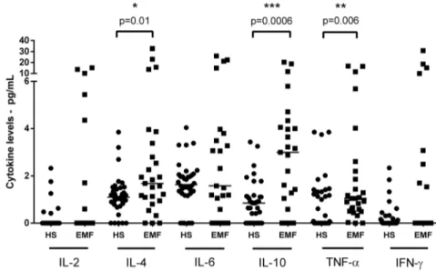
Figure 1. Cytokine plasma levels of endomyocardial fibrosis patients and healthy subjects. Dot plot represents cytokine levels (interleukin 2, 4, 6, 10, TNF-aand IFN-c) from endomyocardial fibrosis patients (EMF) and healthy subjects (HS), evaluated by CBA. Horizontal lines indicate the median values. *Differences whereP#0.05 are indicated. (
N
Healthy Subjects;&EMF patients). doi:10.1371/journal.pone.0108984.g001Table 1.Demographic and echocardiographic data from EMF patients and healthy subjects.
Variable EMF HS
Gender (Male/Female) 3/24 12/26
Age (years - Male/Female) 34.6615.5 33.9612
Bilateral/RV/LV EMF (%) 49.5613.3 53.9612,9
Mitral regurgitation (%) * 30/26/44 NA
Tricuspid regurgitation (%)* 55.5%/26%/7.4% NA
Diastolic dysfunction grade (%)** 37%/18.5%/7.4% NA
Systolic dysfunction (%) 60,90% NA
AF (%) 21.4 NA
NA – Not applicable; AF – Atrial Fibrillation; (Percentage of EMF patients with the conditionregistered in the medical records); Systolic dysfunction: Ejection Fraction, 55%; *Valvar regurgitation level: mild, moderate, and severe, respectively; Diastolic dysfunction: grades mild, moderate, and severe ** It was not possible to evaluate diastolic function in 4 patients, due to the presence of pacemaker or bioprosthetic heart valve.
doi:10.1371/journal.pone.0108984.t001
the FACSCanto flow cytometer (BD Biosciences), and the FCAP Array software (BD Biosciences) was used for data analysis.
Statistical Methods
Cytokine levels were compared among EMF patients and subject controls by using the Mann-WhitneyUtest. Correlations
of data were analyzed by the non-parametric Spearman test. All analysis was performed with Graph Pad Prism version 5 software (GraphPad Software, La Jolla, California, USA). APvalue,0.05
was considered statistically significant.
Results
EMF and healthy subjects were age and sexmatched (Table 1). Plasma samples from all 27 EMF patients examined in this study showed detectable levels of at least one of the assayed cytokines. Increased levels of interleukin (IL)-4 (4.5167.79 pg/mL), IL-10 (4.1165.27 pg/mL), and tumor necrosis factor alpha (TNF-a)
(2.7764.64 pg/mL) were detected in a high proportion of EMF patients (88.8%, 92.6%, and 77.7%, respectively) compared with healthy control samples (1.2260.87 pg/mL - P= 0.001, 0.996
0.89 pg/mL-P= 0.0001 and 0.9460.24 pg/mL -P= 0.006,
res-pectively - Mann Whitney U Test) - (Figure 1 and 2). There was no significant difference in IL-6, IFN-cand IL-2 levels compared
with control samples. Interestingly, circulating IL-4 was present in 20/21 samples in which IL-10 was detectable. In addition, a positive correlation was observed between all cytokine levels in EMF patients, with the exception of TNF-aand IFN-c(P= 0.08;
Figure 2).
Discussion
Our study shows that EMF patient shave elevated plasma levels of pro- and anti-inflammatory cytokines, especially TNF-a, IL-4, and IL-10, which were significantly higher than those in healthy subjects. The correlation between levels of pro- and anti-inflammatory/Th2 cytokines may suggest that either the stimuli that induce both kinds of cytokines are the same, or that IL-10 and IL-4 may have a regulatory role and control the levels and deleterious effects of proinflammatory cytokines.
Multiple cardiovascular disorders are associated with increased levels of circulating pro-inflammatory cytokines, especially TNF-a
and IL-6, which are related to a common cytokine profile found in acute and chronic HF patients independently of etiology [19,20]. Mann proposed that acute increased levels of IL-6, TNF-aand IL-1 could be an adaptive response for cardiac injury, with cardioprotective effects, whereas the chronic elevation of these mediators could be a maladaptive response and promote cardiac decompensation [21]. However, the underlying cause for the presence of the inflammatory response in different CV diseases is still under investigation [22]. It has been suggested that the predominant mechanism of upregulation of TNF-aproduction is
secondary to advanced heart failure itself, such as low cardiac output and intestinal bacterial translocation [23].
In addition, a limited number of reports on HF have found increased plasma levels of the other cytokines detected in our study (IL-10, IL-4, and to a lesser extent IFN-cand IL-2) [13,24–26]. It
is thus possible that the triggers of cytokine production in EMF are similar to those found in HF with other causes, such as Figure 2. Correlation among cytokine plasma levels of endomyocardial fibrosis patients.Dot plots represent correlation between cytokine levels (interleukin 2, 4, 6, 10, TNF-aand IFN-c) from endomyocardial fibrosis patients, evaluated by CBA.
hypertensive or idiopathic dilated cardiomyopathy. On the other hand, our findings that late-stage EMF patients display increased IL-4 and IL-10 levels are also consistent with the observed early eosinophilia and helminthic infections in EMF, once this type of infection is usually associated with increased levels of these cytokines [27–30]. Whichever the stimulus for cytokine production may be, results suggest a possible relevance of a persisting Th2 (IL-4 and IL-10) cytokine-driven immune mechanism in the patho-genesis of EMF. Significantly, IL-10 is an anti-inflammatory cytokine that may reduce TNF-a production, which may be
clinically significant in the pathogenesis of HF [31]. In our study, the almost universal co-detection of inflammatory and anti-inflammatory cytokines, as well as the correlation between their levels is consistent with such an antagonistic effect. On the other hand, along with its known anti-inflammatory effects, long-term overexpression of IL-10 has been associated with lung fibrosis [32]. Although blood eosinophilia (BE) has been reported in EMF cases [29], less than 40% of our patients displayed BE ($
500 eosinophils/mm3) during follow-up in the chronic phase of this disease. Our data are consistent with those reported by Patel and associates (1977) [33], who observed that the absolute BE in African EMF patients was similar to that of healthy control subjects. In a recent study from a similar cohort of patients in our Hospital, Iglezias et al. [5] found no eosinophils in the heart lesions of EMF patients. Together, these results suggest that neither blood nor endocardial eosinophilia are essential components of late-stage EMF. Our data are consistent with the hypothesis that early helminthic infestation could cause a waning eosinophilia that might be involved in initial heart damage in EMF pathogenesis, and along with a long-lasting Th2 response whose pathogenic or protective potential is yet unclear. However, we cannot exclude that the anti-inflamatory cytokine levels are raised as a
homeo-static mechanism to buffer both production and effects of pro-inflammatory cytokines. Although persistent IL-10 production may lead to lung fibrosis [33], it is unknown whether the cytokine could accelerate fibrous tissue deposition in the endomyocardium of EMF patients. One limitation of this study is the lack of information about helminthic infection status in our patient group. In summary, we have shown for the first time that late-stage EMF patients display detectable plasma levels of a mixed pro- and anti-inflammatory/Th2 cytokine profile, predominantly composed of TNF-a, IL-10 and IL-4 levels. The finding of such a mixed
cytokine profile may either reflect the multiple cardiovascular disorders also experienced by EMF patients, or indicate a common persistent stimulus for production of both pro- and anti-inflammatory/Th2 cytokines. On the other hand, anti-inflamma-tory/Th2 cytokines IL-4 and IL-10 may either be upregulated by previous helminthic infection, or as a homeostatic mechanism to buffer both production and effects of pro-inflammatory cytokines. This antagonism is consistent with the almost universal co-detection of inflammatory and anti-inflammatory cytokines in EMF plasma samples, as well as the positive correlation between their plasma levels. Further studies may allow the identification of the stimulus for chronic cytokine production, and to establish whether cytokines play a role in pathogenesis or have a prognostic value for rates of disease progression or post-surgical follow-up.
Author Contributions
Conceived and designed the experiments: ASB VMCS ECN CM. Performed the experiments: ASB LRPF SPR ASN DSR SCF. Analyzed the data: ASB SPR ECN JK. Contributed reagents/materials/analysis tools: SPR LRPF ASN DSR ECN JK SCF. Wrote the paper: ASB VMCS ECN.
References
1. Davies JN (1948) Pathology of Central African Natives; Mulago Hospital post mortem studies. East Afr Med J 25: 454–467.
2. Roberts WC, Liegler DG, Carbone PP (1969) Endomyocardial disease and eosinophilia. A clinical and pathologic spectrum. Am J Med 46: 28–42. 3. Satoh M, Minami Y, Takahashi Y, Nakamura M (2008) Immune modulation:
role of the inflammatory cytokine cascade in the failing human heart. Curr Heart Fail Rep 5: 69–74.
4. Brockington IF, Olsen EG (1972) Eosinophilia and endomyocardial fibrosis. Postgrad Med J 48: 740–741.
5. Iglezias SD, Benvenuti LA, Calabrese F, Salemi VM, Silva AM, et al. (2008) Endomyocardial fibrosis: pathological and molecular findings of surgically resected ventricular endomyocardium. Virchows Arch 453: 233–241. 6. Torre-Amione G, Kapadia S, Benedict C, Oral H, Young JB, et al. (1996)
Proinflammatory cytokine levels in patients with depressed left ventricular ejection fraction: a report from the Studies of Left ventricular Dysfunction (SOLVD). J Am Coll Cardiol 27: 1201–1206.
7. Mocelin AO, Issa VS, Bacal F, Guimara˜es GV, Cunha E, et al. (2005) The influence of aetiology on inflammatory and neurohumoral activation in patients with severe heart failure: a prospective study comparing Chagas’ heart disease and idiopathic dilated cardiomyopathy. Eur J Heart Fail 7: 869–73. 8. Ferreira RC, Ianni BM, Abel LC, Buck P, Mady C, et al. (2003) Increased
plasma levels of tumor necrosis factor-alpha in asymptomatic/"indeterminate" and Chagas disease cardiomyopathy patients. Mem Inst Oswaldo Cruz 98: 407–11.
9. Rauchhaus M, Doehner W, Francis DP, Davos C, Kemp M, et al. (2000) Plasma cytokine parameters and mortality in patients with chronic heart failure. Circulation 102: 3060–3067.
10. Oru’s J, Roig E, Perez-Villa F, Pare´ C, Azqueta M, et al. (2000) Prognostic value of serum cytokines in patients with congestive heart failure. J Heart LungTransplant 19: 419–425.
11. Haudek SB, Taffet GE, Schneider MD, Mann DL (2007) TNF provokes cardiomyocyte apoptosis and cardiac remodeling through activation of multiple cell death pathways. J Clin Invest 117: 2692–2701.
12. MacDonald AS, Araujo MI, Pearce EJ (2002) Immunology of parasitic helminth infections. Infect Immun 70: 427–33.
13. Sato Y, Takatsu Y, Kataoka K, Yamada T, Taniguchi R, et al. (1999) Serial circulating concentrations of C-reactive protein, interleukin (IL)-4, and IL-6 in patients with acute left heart decompensation. Clin Cardiol 22: 811–3.
14. Amir O, Rogowski O, David M, Lahat N, Wolff R, et al. (2010) Circulating interleukin-10: association with higher mortality in systolic heart failure patients with elevated tumor necrosis factor-alpha. Isr Med Assoc J 12: 158–162. 15. Sliwa K, Mocumbi AO (2010) Forgotten cardiovascular diseases in Africa. Clin
Res Cardiol 99: 65–74.
16. Salemi VM, Leite JJ, Picard MH, Oliveira LM, Reis SF, et al. (2009) Echocardiographic predictors of functional capacity in endomyocardial fibrosis patients. Eur J Echocardiogr 10: 400–405.
17. Salemi VM, Picard MH, Mady C (2004) Assessment of diastolic function in endomyocardial fibrosis: value of flow propagation velocity. Artif Organs 28: 343–346.
18. Salemi VM, Rochitte CE, Shiozaki AA, Andrade JM, Parga JR, et al. (2011) Late gadolinium enhancement magnetic resonance imaging in the diagnosis and prognosis of endomyocardial fibrosis patients. Circ Cardiovasc Imaging 4: 304– 311.
19. Anker SD, von Haehling S (2004) Inflammatory mediators in chronic heart failure: an overview. Heart 90: 464–470.
20. Aukrust P, Gullestad L, Ueland T, Dama˚s JK, Yndestad A (2005) Inflammatory and anti-inflammatory cytokines in chronic heart failure: potential therapeutic implications. Ann Med 37: 74–85.
21. Mann DL (2003) Stress-activated cytokines and the heart: from adaptation to maladaptation. Annu Rev Physiol 65: 81–101.
22. Mehra VC, Ramgolam VS, Bender JR (2005) Cytokines and cardiovascular disease. J Leukoc Biol 78: 805–818.
23. Muller-Werdan U, Engelmann H, Werdan K (1998) Cardiodepression by tumor necrosis factor-alpha. Eur Cytokine Netw 9: 689–691.
24. Sato Y, Takatsu Y, Kataoka K, Yamada T, Taniguchi R, et al. (1999) Serial circulating concentrations of C-reactive protein, interleukin (IL)-4, and IL-6 in patients with acute left heart decompensation. Clin Cardiol 22: 811–813. 25. Gage JR, Fonarow G, Hamilton M, Widawski M, Martı´nez-Maza O, et al.
(2004) Beta blocker and angiotensin-converting enzyme inhibitor therapy is associated with decreased Th1/Th2 cytokine ratios and inflammatory cytokine production in patients with chronic heart failure. Neuroimmunomodulation.11: 173–180.
26. Guinjoan SM, Vigo DE, Castro MN, Tateosian N, Chuluyan E, et al. (2009) Mood, Th-1/Th-2 cytokine profile, and autonomic activity in older adults with acute/decompensated heart failure: preliminary observations. World J Biol Psychiatry 10: 913–918.
27. Yu L, Sun X, Yang F, Yang J, Shen J, et al. (2012) Inflammatory cytokines
IFN-c, IL-4, IL-13 and TNF-a alterations in schistosomiasis: a meta-analysis. Parasitol Res 110: 1547–52.
28. Ive FA, Willis AJ, Ikeme AC, Brockington IF (1967) Endomyocardial fibrosis and filariasis. Q J Med 36: 495–516.
29. Andy JJ, Ogunowo PO, Akpan NA, Odigwe CO, Ekanem IA, et al. (1988) Helminth associated hypereosinophilia and tropical endomyocardial fibrosis (EMF) in Nigeria. Acta Trop 69: 127–140.
30. Schopf LR, Hoffmann KF, Cheever AW, Urban JF, Wynn TA (2002) IL-10 is critical for host resistance and survival during gastrointestinal helminth infection. J Immunol. 168: 2383–2392.
31. Yamaoka M, Yamaguchi S, Okuyama M, Tomoike H (1999) Anti-inflammatory cytokine profile in human heart failure: behavior of interleukin-10 in association with tumor necrosis factor-alpha. Jpn Circ J 63: 951–956.
32. Sun L, Louie MC, Vannella KM, Wilke CA, LeVine AM, et al. (2011) New concepts of IL-10-induced lung fibrosis: fibrocyte recruitment and M2 activation in a CCL2/CCR2 axis. Am J Physiol Lung Cell Mol Physiol. 300(3):L341–53. 33. Patel AK, D’Arbela PG, Somers K (1977) Endomyocardial fibrosis and