Inte rle ukin-5 and inte rle ukin-1 0
are m ajo r cyto kine s in ce re bro spinal
fluid fro m patie nts with active
ne uro cystice rco sis
1Laboratório de Imunologia and 2Disciplina de Neurologia,
Faculdade de Medicina do Triângulo Mineiro, Uberaba, MG, Brasil V. Rodrigues Jr.1,
F.A. de-Mello1,
E.P. Magalhães1,
S.B.F. Ribeiro2
and J.O . Marquez2
Abstract
Neurocysticercosis (NCC) is a common neurological disorder espe-cially in developing countries, caused by infection of the brain with encysted larvae of the tapeworm Taenia solium. Seizures are a
com-mon finding associated with this disease. The objective of the present study was to evaluate the correlation between the levels of various cytokines present in the cerebrospinal fluid (CSF) of patients with NCC and the severity of the disease. The levels of the cytokines IL-1ß, TNF-a, IL-5, IL-10 and IFN-g were determined in the CSF of 22 patients with active NCC, 13 patients with inactive NCC and 15 control subjects. CSF from patients with active NCC presented signif-icantly higher IL-5 levels compared to control subjects. IL-5 and IL-10 levels in CSF from NCC patients with inflammatory CSF were signif-icantly higher than those detected in non-inflammatory CSF. These results show a predominant Th2 lymphocyte activation in human NCC and also indicate the possible use of cytokines in the CSF as a marker for the differential diagnosis between inactive disease and the active form of NCC.
Co rre spo nde nce
V. Rodrigues Jr. Laboratório de Imunologia Faculdade de Medicina do Triângulo Mineiro Rua Frei Paulino, 30 38025-180 Uberaba, MG Brasil
Fax: + 55-34-312-6640
E-mail: vrodrigues@ mednet.com.br
Research supported by FAPEMIG and CNPq. F.A. de-Mello and E.P. Magalhães were recipients of CNPq fellowships (PIBIC program).
Received September 9, 1999 Accepted June 12, 2000
Ke y wo rds
·Cysticercosis ·Cytokines ·Cerebrospinal fluid
Intro ductio n
Neurocysticercosis (NCC) is a common neurological disorder in several developed and developing countries. It is endemic in Latin America, Asia and Central and South Africa and is relatively frequent in Portugal, Spain and Eastern European countries (1-3). In other developed countries NCC can be frequent among immigrants. The disease is caused by the encysted larvae of the
tape-worm Taenia solium lodged in the central
nervous system (CNS). In the same commu-nities, teniasis is a public health problem with a frequency of family antecedents reach-ing 34.2% in some regions (4).
the most frequent clinical features. This pleo-morphic disease is a result of the presence of the parasite itself (cysticerci), the inflamma-tory process that surrounds the larvae, and the residual fibrosis and calcification (5,6).
The cysticercus contains a large number of antigens that can elicit a host immune-inflammatory reaction. The immune-inflammatory cellular infiltrate, if present, may be discrete with the presence of lymphocytes and eosin-ophils in the vesicular stage, or may be a more intense lymphocyte infiltrate with gi-ant multinuclear foamy macrophages in the necrotic stage, with parasites in different stages of desintegration (7).
Most of the previous studies involving cerebrospinal fluid (CSF) were limited to an analysis of the presence of specific antibod-ies and nonspecific mediators of the inflam-matory reaction such as parasite-specific an-tibodies, increased protein levels and eosi-nophilia (8). Eosieosi-nophilia provides indirect evidence for the active form of NCC. Re-cently, several reports on cysticercosis in-volving studies of the cellular infiltrate in humans and in animal models have indicated that a Th1 immune response predominates over a Th2 response (9,10), while studies of the CSF have revealed a predominant Th2 response (11,12).
Cytokines are pleotropic hormones that regulate many aspects of the immune re-sponse and of inflammatory reactions. Inter-leukin-5 (IL-5) is produced mainly by Th2 lymphocytes and plays a role in eosinophil maturation, migration and activation and also induces immunoglobulin A switch in B lym-phocytes (13). Interleukin-10 is produced by Th2 lymphocytes, B cells, and macrophages and has strong anti-inflammatory properties (14). Interferon-g (IFN-g) is produced by Th1 lymphocytes and natural killer (NK) cells and is involved in macrophage activa-tion and in immunoglobulin class switch (15). Other cytokines such as IL-1ß and tu-mor necrosis factor alpha (TNF-a) are pro-duced mainly by macrophages and by glial
cells in the CNS, and are involved in the acute phase of inflammatory reactions.
In the present study, we determined the presence of IL-5, IL-10, IFN-g, TNF-a and IL-1ß in CSF from patients with active and inactive NCC and compared their levels with those found in the CSF of unaffected con-trols. The results indicate that high levels of IL-5 are significantly associated with active NCC and that IL-10 reaches higher levels in inflammatory CSF.
Patie nts and Me tho ds
Patie nts
CSF samples collected from 22 patients with active NCC, 13 with inactive NCC and 15 controls (patients with chronic headache, normal topography and standard parameters of CSF analysis within normal values) were analyzed. All patients and controls were sub-mitted to neurological investigation includ-ing clinical examination, brain tomography and standard CSF analysis. CSF analyses consisted of serological tests for anti-cys-ticercus and anti-treponema antibodies, pro-tein concentration, sodium, potassium and chloride measurements, and standard blood cell counts. Patients with active and inactive NCC were classified on the basis of a brain tomography scan (16). All of them exhibited anti-cysticercus antibodies in CSF and were negative for anti-treponema antibodies. This study was approved by the Ethics Commit-tee of the Faculdade de Medicina do Triân-gulo Mineiro.
Cyto kine titratio n
Nonspecific binding was prevented by incu-bating the plates with 3% BSA (Sigma Chem-ical Co., St. Louis, MO, USA) in PBS. Plates were incubated overnight with 100 µl of 1:2 dilution of CFS in PBS, 2% BSA and stan-dard cytokines (Pharmingen and R&D, Minneapolis, MN, USA). Plates were then washed 4 times with PBS and 0.05% Tween and incubated with rabbit anti-IL-1ß antibody (Genzyme), rabbit anti-TNF-a an-tibody (Genzyme), biotinylated anti-IL-5 mAb (Pharmingen), biotinylated anti-IL-10 mAb (Pharmingen) or biotinylated anti-IFN-g mAb (Mabtec) for 4 h. Plates were then washed and incubated for 2 h with alkaline phosphatase-conjugated goat anti-rabbit IgG (Immunotech, Marseille, France) or alkaline phosphatase conjugated to streptavidin. Fi-nally, plates were washed 4 times and enzy-matic activity was developed by incubating the plates with p-nitrophenyl phosphate (Sigma). Absorbance was read at 405 nm in a microplate reader apparatus (Biorad, Her-cules, CA, USA). The sensitivity of the tests was 20 pg/ml.
Re sults
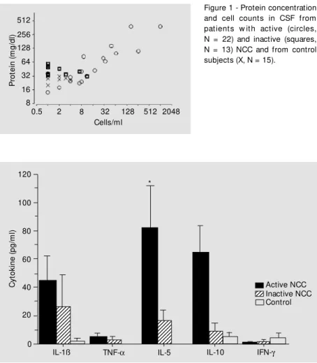
Patients with NCC were classified ac-cording to a computer tomography scan and the presence of anti-cysticercus antibodies (14). As shown in Figure 1, CSF from pa-tients with inactive NCC did not present relevant alterations in cellularity or protein concentration. Nevertheless, 8 patients with active NCC presented elevation of cells and proteins in CSF, with eosinophils ranging from 4 to 9%.
Levels of IL-1ß, TNF-a, IL-5, IL-10 and IFN-g were measured in CSF by ELISA. CSF from the majority of control patients did not present measurable levels of the tested cytokines. The levels of TNF-a and IFN-g were below the limit of detection in the study group. CSF from patients with active NCC showed an elevation of IL-1, IL-5, and IL-10 levels. IL-5 levels were significantly higher
(t-test) than those found in the CSF from
patients with inactive NCC and from control subjects (Figure 2). These findings were par-ticularly clear when patients with alterations in terms of both cellular and biochemical criteria were compared with the other group. In the CSF from patients with active NCC with protein concentration above 50 mg% and more than 20 cells/ml, IL-5 levels ranged from 57 to 792 pg/ml and the levels of IL-10 levels ranged from 38 to 398 pg/ml. In con-trast, the levels of IL-5 and IL-10 were sig-nificantly lower in CSF from patients with active NCC with a non-inflammatory reac-tion (Figure 3).
P
ro
te
in
(
m
g
/d
l)
512
256
128
64
32
16
8
0.5 2 8 32 128 512 2048 Cells/ml
C
y
to
k
in
e
(
p
g
/m
l)
120
12345 12345 1234
1234 1234 1234 12345
12345 12345 12345 12345 12345 12345 12345 123456
123456 12345
12345 12345 12345 12345 12345 12345 12345 12345 12345 12345
100
80
60
40
20
0
IL-1ß TNF-a IL-5 IL-10 IFN-g
123 123 123
Active NCC Inactive NCC Control *
Figure 1 - Protein concentration and cell counts in CSF from pat ient s w it h act ive (circles, N = 22) and inactive (squares, N = 13) NCC and from control subjects (X, N = 15).
D iscussio n
The clinical manifestations of human cysticercosis depend on various factors in-cluding number of cysts, their location, and host reaction. When the cysticerci are lodged in the CNS, the disease is known as neuro-cysticercosis, and the involvement of the CNS represents the most dangerous clinical form of this parasitosis. NCC is usually asymptomatic for several years and during this period, a discrete circumscribed inflam-matory reaction is observed (17). Conversely, symptomatic disease is associated with a prominent inflammatory response, consist-ing of lymphocytes, eosinophils, granulo-cytes and plasma cells (18).
The mechanisms by which cysticercal lesions develop in the CNS depend on a combination of host immune-inflammatory response, mainly mediated by cytokines pro-duced by resident and infiltrating cells acti-vated by cysticercal antigens. The lesions containing viable cysticerci have a small inflammatory infiltrate surrounding the worm, which is usually composed of
mono-nuclear cells and eosinophils (7). These cyst antigens stimulate the production of anti-bodies which are inefficient in killing ma-ture metacestodes. However, the anti-cys-ticercus antibodies can be detected in CSF of patients with active and inactive NCC and are the basis for the immune diagnosis of this disease. The inflammatory reaction of NCC was demonstrated in CSF (19) and confirmed by some studies on experimental models and on human tissues. These studies showed an immune response predominantly consisting of Th1 lymphocytes (9,10) and the later development of a Th2 response associated with parasite destruction (11).
In the present study, we observed that CSF from patients with NCC contains el-evated concentrations of 1ß, 5, and IL-10. The levels of these cytokines in the CSF from the majority of uninfected controls was below ELISA sensitivity. However, statisti-cal analysis demonstrated that IL-5 levels were significantly higher in patients with active NCC than in patients with inactive NCC and in control subjects. Moreover, IL-10 and IL-5 levels were higher in active NCC patient with elevated concentrations of pro-teins and increased cellularity.
Our results are significant because they can help us to understand some of the cellu-lar alterations present in the CSF of NCC patients, i.e., a marked eosinophilia. This alteration may be the result of the intense production of IL-5 during the immune re-sponse to the cysticercal antigens, because one of the main functions of IL-5 is to induce and activate eosinophils to mediate an anti-body-dependent helminthotoxic effect (20). We suggest that the eosinophilia present in the CSF of patients with active NCC is, at least in part, mediated by the IL-5 produced by the host in response to the infectious process. Similarly, the presence of IL-10 and the absence of IFN-g in CSF from patients with NCC confirm a Th2 response elicited by helminthic infection. Moreover, IL-10, as an anti-inflammatory cytokine, may be
in-C
y
to
k
in
e
(
p
g
/m
l)
250
200
150
125
75
25
0
IL-1ß TNF-a IL-5 IL-10 IFN-g
123456 123456 123456 123456 123456
1234567 1234567 1234567 1234567 1234567 1234567 1234567 123456
123456 123456
123456 123456 123456 123456 123456 123456
123 123
Inflammatory Non-inflammatory *
*
Figure 3 - Cytokine levels in CSF from 8 patients w ith active NCC w ith inflammatory CSF (filled bars) and from 14 NCC patients w ith non-inflammatory (striped bars) CSF based on protein concentrations and cell counts. Statistical analysis w as performed on log-trans-formed cytokine levels. * P<0.05 compared to NCC patients w ith non-inflammatory CSF (Student t-test).
225
175
100
volved in the control of inflammatory reac-tions in patients with elevated cell concen-tration in CSF.
Finally, our results show a predominant activation of Th2 lymphocytes in human NCC, with the presence of increased levels of IL-1ß, IL-5, and IL-10 in the absence of IFN-g and TNF-a. These results show that the determination of cytokines in CSF may be an important asset by providing an
addi-tional parameter for the differential diagno-sis between inactive NCC and active dis-ease.
Ackno wle dgm e nts
We would like to thank Dr. Antonio Cam-pos-Neto for a critical review of the manu-script.
Re fe re nce s
1. Ahuja GK, Roy S, Karla G & Vermin V (1978). Cerebral cysticercosis. Journal of the Neurological Sciences, 35: 365-374. 2. Del Bruto OH & Sotelo J (1988).
Neuro-cysticercosis: an update. Review of Infec-tious Diseases, 10: 1075-1087.
3. White Jr AC (1997). Cysticercosis: a ma-jor cause of neurological disease w orld-w ide. Clinical Infectious Diseases, 24: 10-15.
4. Silva-Vergara M L, Prata A, Netto HV, Vieira C de O, Castro JH, M icheletti LG, Otano AS & Franquini Junior J (1998). Risk fac-tors associated w ith taeniasis-cysticerco-sis in Lagamar, M inas Gerais State, Brazil.
Revista da Sociedade Brasileira de M edi-cina Tropical, 31: 65-71.
5. M onteiro L, Coelho T & Stocker A (1992). Neurocysticercosis - A review of 231 cases. Infection, 20: 61-65.
6. Rosenfeld EA, Byrd SE & Shulman ST (1996). Neurocysticercosis among chil-dren in Chicago. Clinical Infectious Dis-eases, 23: 262-268.
7. Pittella JEH (1997). Neurocysticercosis.
Brain Pathology, 7: 681-693.
8. M iller BL, Staugaitis SM , Tourtellotte WW, Shapshak P, Goldberg M , Heiner D & Weil M (1985). Intra-blood-brain barrier IgG synthesis in cerebral cysticercosis.
Archives of Neurology, 42: 782-784. 9. Restrepo BI, Llaguno P, Sandoval M A,
Enciso JA & Teale JM (1998). Analysis of immune lesions in neurocysticercosis pa-tients: central nervous system response to helminth appears Th1-like instead of Th2. Journal of Neuroimmunology, 89: 64-72.
10. Cardona, AE, Restrepo BI, Jaramillo JM & Teale JM (1999). Development of an ani-mal model for neurocysticercosis: Im-mune response in the central nervous sys-tem is characterized by a predominance of gd T cells. Journal of Immunology, 162: 995-1002.
11. Robinson P, Atmar RL, Lew is DE & White Jr AC (1997). Granuloma cytokines in mu-rine cysticercosis. Infection and Immuni-ty, 65: 2925-2931.
12. Evans CA, Garcia HH, Hartnell A, Gilman RH, Jose PJ, M artinez M , Remick DG, Williams TJ & Friedland JS (1998). El-evated concentrations of eotaxin and in-terleukin-5 in human neurocysticercosis.
Infection and Immunity, 66: 4522-4525. 13. Takatsu K, Takaki S & Hitoshi Y (1994).
Interleukin-5 and its receptor system: im-plications in the immune system and in-flammation. Advances in Immunology, 57: 145-190.
14. M oore KW, O’Garra A, de Vaal M alefyt R, Vieira P & M osmann TR (1993). Interleu-kin-10. Annual Review of Immunology, 11: 165-190.
15. Finkelman FD, Holmes J, Katona IM , Ur-ban Jr JF, Beckm ann M P, Park LS, Schooley KA, Coffman RL, M osmann TR & Paul WE (1990). Lymphokine control of
in vivo immunoglobulin isotype selection.
Annual Review of Immunology, 8: 303-333.
16. Sotelo JM D, Guerrero V & Rubio F (1985). Neurocysticercosis: a new classification based on active and inactive forms: A study of 753 cases. Archives of Internal M edicine, 145: 442-445.
17. Ridaura Sanz C (1987). Host response in childhood neurocysticercosis. Childs Ner-vous System, 3: 206-207.
18. Gutierrez Y (1990). Cysticercosis, coenu-rosis, and sparganosis. In: Gutierrez Y (Editor). Diagnostic Pathology of Parasitic Infection w ith Clinical Correlation. Lea & Febiger, Philadelphia, 432-459.
19. Ostrosky-Zeichner L, M endonza EG, Rios C & Sotelo J (1996). Humoral and cellular immune response w ithin the subarach-noid space of patients w ith neurocysticer-cosis. Archives of M edical Research, 27: 513-517.