JULIANA MARTINS AGUIAR
Genotipagem de Giardia duodenalis: detecção de infecções mistas e recombinações gênicas
em amostras de origem humana
JULIANA MARTINS AGUIAR
Genotipagem de Giardia duodenalis: detecção de infecções mistas e recombinações
gênicas em amostras de origem humana
Tese apresentada ao Programa de Pós-Graduação em Epidemiologia Experimental Aplicada às Zoonoses da Faculdade de Medicina Veterinária e Zootecnia da Universidade de São Paulo para obtenção do título de Doutor em Ciências
Departamento:
Medicina Veterinária Preventiva e Saúde Animal Área de Concentração:
Epidemiologia Experimental Aplicada às Zoonoses Orientador:
Prof. Dr. Rodrigo Martins Soares
Autorizo a reprodução parcial ou total desta obra, para fins acadêmicos, desde que citada a fonte.
DADOS INTERNACIONAIS DE CATALOGAÇÃO-NA-PUBLICAÇÃO
(Biblioteca Virginie Buff D’Ápice da Faculdade de Medicina Veterinária e Zootecnia da Universidade de São Paulo)
T.3150 Aguiar, Juliana Martins
FMVZ Genotipagem de Giardia duodenalis: detecção de infecções mistas e recombinações gênicas em amostras de origem humana / Juliana Martins Aguiar. -- 2015.
40 f. : il.
Tese (Doutorado) - Universidade de São Paulo. Faculdade de Medicina Veterinária e Zootecnia. Departamento de Medicina Veterinária Preventiva e Saúde Animal, São Paulo,2015.
Programa de Pós-Graduação: Epidemiologia Experimental Aplicada às Zoonoses.
Área de concentração: Epidemiologia Experimental Aplicada às Zoonoses.
Orientador: Prof. Dr. Rodrigo Martins Soares.
FOLHA DE AVALIAÇÃO
Autor: MARTINS, Juliana Aguiar
Título: Genotipagem de Giardia duodenalis: detecção de infecções mistas e recombinações
gênicas em amostras de origem humana
Tese apresentada ao Programa de Pós-Graduação em Epidemiologia Experimental Aplicada às Zoonoses da Faculdade de Medicina Veterinária e Zootecnia da Universidade de São Paulo para obtenção do título de Doutor em Ciências
Data: ____/____/____
BANCA EXAMINADORA
Prof. Dr. ____________________________________________________________________ Assinatura: ___________________________ Julgamento: ___________________________
Prof. Dr. ____________________________________________________________________ Assinatura: ___________________________ Julgamento: ___________________________
Prof. Dr. ____________________________________________________________________ Assinatura: ___________________________ Julgamento: ___________________________
Prof. Dr. ____________________________________________________________________ Assinatura: ___________________________ Julgamento: ___________________________
Dedicatória
À minha mãe, Cléo, por ter dedicado sua vida para mim, mulher guerreira que mesmo
sem ter tido a oportunidade de estudar sempre batalhou para me conceder a melhor educação, muitas vezes abdicando de seus próprios sonhos, sem ela nada disso seria possível. É meu espelho, exemplo de mãe e avó.
Ao meu querido esposo, Wagner, que caminha comigo há muito tempo, sempre me
encorajando a enfrentar os obstáculos da vida. Por ter me proporcionado a maior alegria do mundo, de ser mãe da pequena Luana, a quem eu também dedico este trabalho, pois tudo que
Agradecimentos
A Deus por me dar sabedoria e força para lidar com todos os momentos da vida, por não me desamparar e por cada porta que se abre.
À Fundação de Amparo à Pesquisa do Estado de São Paulo (FAPESP) pela concessão da bolsa de doutorado (Processo 2011/13472-2).
À Profa. Solange por permitir meu ingresso no VPS/FMVZ/USP, em 2007, quando me aceitou como estagiária no laboratório de Doenças Parasitárias.
Ao Prof. Rodrigo por ser meu orientador desde o mestrado, pelos ensinamentos e paciência.
A Drª Hilda Fátima de Jesus Pena pelos conhecimentos compartilhados, profissional que eu admiro muito.
Ao Valdir Azevedo pela amizade, confiança, ensinamentos e por fornecer as amostras utilizadas neste projeto.
A toda minha família pelo amor, apoio e orações concebidas.
Às amigas Sheila Oliveira e Giselle Ayres, que me acompanham desde o mestrado, que tanto me ajudaram no desenvolvimento deste projeto, por confiarem no meu trabalho e principalmente pela sincera amizade.
Às amigas Nara Thiers e Laila Andréia, as amigas do coração, que fizeram total diferença na minha vida, pela convivência e por deixarem os meus dias mais alegres e divertidos.
Aos professores Leonardo José Richtzenhain, Paulo Eduardo Brandão e Fabio Gregori pelos ensinamentos, disponibilidade e por me receberem de braços abertos no Laboratório de Biologia Molecular.
Ao colega Renato Caravieri pela amizade, prontidão e por estar sempre me incentivando.
Aos funcionários da secretaria do VPS, Danival, Virgínia e Cristina, por toda ajuda prestada.
“Por isso não desanimamos. Embora exteriormente estejamos a desgastar-nos, interiormente estamos sendo renovados dia após dia, pois os nossos sofrimentos leves e momentâneos estão produzindo para nós uma glória eterna que pesa mais do que todos eles. Assim, fixamos os olhos, não naquilo que se vê, mas no que não se vê, pois o que se vê é transitório, mas o que não se vê é eterno”
RESUMO
MARTINS, J. A. Genotipagem de Giardia duodenalis: detecção de infecções mistas e
recombinações gênicas em amostras de origem humana. [Genotyping of Giardia duodenalis: detection of mixed infection and genetic recombination in samples of human origin]. 2015. 40 f. Tese (Doutorado em Ciências) – Faculdade de Medicina Veterinária e Zootecnia, Universidade de São Paulo, São Paulo, 2015.
Giardia duodenalis é um protozoário de distribuição mundial responsável por causar
infecções entéricas em uma grande variedade de mamíferos, incluindo os humanos. Mesmo apresentando pouca variação em sua morfologia, os isolados podem ser diferenciados, de acordo com análises de proteína e polimorfismo de DNA, em pelo menos oito agrupamentos genéticos distintos, denominados assemblages (A-H). Apenas os assemblages A e B têm sido
reportados em humanos e outros mamíferos. Isolados de assemblage A podem, ainda, ser
divididos em quatro sub-assemblages (AI, AII, AIII e AIV). Sequencias heterogêneas têm
sido frequentemente identificadas em estudos de caracterização molecular envolvendo amostras contendo múltiplos cistos do parasita. Buscando estudar a ocorrência dos eventos de heterozigose de sequencia alélica (ASH) e recombinação gênica, o presente trabalho teve como objetivo isolar cistos de G. duodenalis empregando-se a técnica de micromanipulação e
caracterizá-los molecularmente através da análise multilócus envolvendo os genes gdh, tpi,
orfC4 e bg. Dez cistos foram individualizados e utilizados na pesquisa. Todos foram
igualmente identificados por todos os genes, nove cistos caracterizados como assemblage AII
e um cisto caracterizado como assemblage B. Os cromatogramas oriundos do cisto
identificado como assemblages B apresentaram diversos sítios heterogêneos nos genes gdh,
bg e orfC4, sendo que, nesses dois últimos, observaram-se sobreposições dos alelos AII e B
no produto sequenciado (heterozigose inter assemblage). Os produtos de PCR foram clonados
e as sequencias obtidas revelaram a ocorrência dos dois alelos neste único cisto. Os sítios polimórficos encontrados nas sequencias do gene gdh indicaram heterozigose intra
assemblage B. Embora ASH já tenha sido relatada em cistos individualizados de G.
duodenalis, estes são os primeiros resultados indicando a presença dos dois alelos,
simultaneamente, em um único indivíduo. Esses resultados demonstram fortes evidências que ocorre troca genética entre indivíduos geneticamente distintos de G. duodenalis.
ABSTRACT
MARTINS, J. A. Genotyping of Giardia duodenalis: detection of mixed infection and genetic recombination in samples of human origin. [Genotipagem de Giardia duodenalis:
detecção de infecções mistas e recombinações gênicas em amostras de origem humana]. 2015. 40 f. Tese (Doutorado em Ciências) – Faculdade de Medicina Veterinária e Zootecnia, Universidade de São Paulo, São Paulo, 2015.
Giardia duodenalis is a worldwide distribution enteric protozoan responsible for causing
infections in a wide variety of mammals, including humans. Even showing little change in their morphology, isolates can be distinguished, according to the analysis of proteins and DNA polymorphisms in at least eight distinct genetic groups, known assemblages (A - H).
Only assemblages A and B have been reported in humans and other mammals. Isolates of
assemblage A also can be divided into four sub-assemblages (AI, AII, AIII and AIV).
Heterogeneous sequences have been frequently identified in studies involving molecular characterization of samples containing multiple cysts of the parasite. Seeking to study the occurrence of allelic sequence heterozygosity (ASH) and genetic recombination events, the present study aimed to isolate G. duodenalis cysts employing the micromanipulation
technique and characterize them molecularly through multilocus analysis involving gdh, tpi,
orfC4 and bg genes. Ten cysts were individualized and used in the research. All cysts were
equally identified for all genes; nine cysts were characterized as assemblage AII and one
characterized as assemblage B. The chromatograms derived from the cyst identified as
assemblage B presented many heterogeneous sites in gdh, bg and orfC4 genes, and in these
last two, there were overlaps of alleles AII and B in the sequenced product (heterozygous inter assemblage). PCR products were cloned and the sequences obtained revealed the occurrence of two alleles at this single cyst. The polymorphic sites found in the sequences of the gdh gene indicated intra heterozygosity assemblage B. Although ASH has already been
reported in G. duodenalis individualized cysts, these are the first results indicating the
presence of two alleles simultaneously in a single individual. These results demonstrate strong evidence that genetic exchange occurs between individuals genetically distinct of G.
duodenalis.
SUMÁRIO
I. Multilocus amplification of genomic DNA from single cysts of Giardia duodenalis
separated using micromanipulation technique………13
1 INTRODUCTION………14
2 MATERIAL AND METHODS………...14
3 RESULTS AND DISCUSSION………...17
4 CONCLUSION……….……….…...19
.REFERÊNCIAS………..20
II. Evidence of heterozigosity and recombinant alleles in single cysts of Giardia duodenalis……….22
1 INTRODUCTION………23
2 MATERIAL AND METHODS………..……..24
3 RESULTS………..27
3.1Sequence analysis of gdh………...……….27
3.2Sequence analysis of tpi………..29
3.3Sequence analysis of bg………..29
3.4Sequence analysis of orfc4………..33
4 DISCUSSION………..………..34
REFERENCES………..37
13
I. Multilocus amplification of genomic DNA from single cysts of Giardia duodenalis separated using micromanipulation technique.
Juliana Martins Aguiar1; Sheila Oliveira da Silva1; Valdir Antônio dos Santos2; Giselle Ayres Razera Rossa1; Sueli Akemi Taniwaki1; Fabio Gregori1; Lara Borges Keid3, Rodrigo Martins Soares.1
1Departamento de Medicina Veterinária Preventiva e Saúde Animal, Faculdade de Medicina Veterinária e Zootecnia, Universidade de São Paulo, São Paulo, SP, Brasil.
2Departamento de Farmácia e Laboratório Clínico do Hospital Universitário, Universidade de São Paulo, São Paulo, SP, Brasil.
3Faculdade de Zootecnia e Engenharia de Alimentos, Departamento de Medicina Veterinária, Faculdade de Zootecnia e Engenharia de Alimentos, Universidade de São Paulo, São Paulo, SP, Brasil.
ABSTRACTS
Giardia duodenalis is divided into at least eight groups, named assemblages A to H.
Assemblages A and B are the only ones able to infect humans and other mammals. The species status for these assemblies is a moot point, but has not gained general acceptance because sexual activity in Giardia is not completely understood. Heterozygosity in Giardia
duodenalis can be detected through simultaneous identification of multiple loci in single cysts
or trophozoites. In this paper, we describe a technique that enables simultaneous detection of fragments from four genes from single cysts of Giardia duodenalis recovered from stool
samples. Each cyst from a fecal sample of human origin was separated, the DNA was extracted and amplified by means of multiplex PCR directed to four genes and the multiplex PCR product was further re-amplified using four single PCR (one for each gene). The following loci were detected: beta giardin (bg), GLORF-C4 (orfC4), triose phosphate isomerase (tpi) and glutamate dehydrogenase (gdh). This procedure should make it possible to investigate multiple genes from a single cyst of Giardiaduodenalis assemblage A or B.
14
1. INTRODUCTION
Giardia duodenalis (synonyms: G. intestinalis and G. lamblia) is an enteric parasite
that affects a large variety of domestic and wild animals and also humans (Ryan, Caccio, 2013). Giardia duodenalis has been described as a complex species in which the isolates
present few morphological variations but can be differentiated into at least eight genetic groupings called assemblages (A to H) (Caccio, Ryan, 2008). Assemblages A and B are the only ones capable of infecting not only humans but also other mammals, and are therefore considered potentially zoonotic (Lalle et al., 2005; Sprong et al., 2009). The other assemblages seem to be confined to specific hosts. Several genetic markers have been used to differentiate the assemblages of Giardia duodenalis (Ryan, Caccio, 2013), and the
commonest of these comprise investigations of molecular diversity: small subunit ribosomal RNA (SSU rRNA); beta-giardin (bg); glutamate dehydrogenase (gdh); elongation factor 1-α
(el1α); triose phosphate isomerase (tpi); GLORF-C4 (orfC4); spacer region of ribosomal RNA; ferredoxin (fd); histone H2B (H2B); histone H4 (H4); mlh1 (mlh); and ribosomal
protein L7a (rp) (Monis et al., 1999; Caccio et al, 2002; Lasek-Nesselquist et al., 2009;
Almeida et al, 2010).
In the present study, we describe a protocol that enables molecular identification of
Giardia duodenalis cysts individually. Each cyst in a fecal sample is separated out and its
DNA is extracted and then amplified by means of multiplex PCR directed towards four genes. Following this, the product from the multiplex PCR is re-amplified using four different PCRs (one for each gene), in nested PCR format. This technique makes it possible to study the presence of gene sequences originating from a single cyst and thus to ascertain whether this cyst has different copies from a single allele, with the possibility of elucidating questions relating to allele sequence heterozygosity and gene recombination.
2. MATERIAL AND METHODS
Two feces samples of human origin that were positive for G. duodenalis, from
15
Each fecal sample was subjected to the technique of centrifugation-floatation in sucrose (modified Sheather technique) (Sheather, 1923) described elsewhere (Souza et al. 2007).
The cysts were recovered on a sterile Petri dish, after washing the slide and cover slip that had been used in observing the cysts, using 1.5 mL of TE buffer (10 mM Tris-HCL pH 8.0; 1 mM EDTA pH 8.0). The product thus obtained from washing was transferred from the dish to a microtube of capacity 1.5 mL and this was then centrifuged at 12,000 g for 10 minutes. The supernatant was discarded, 1 mL of the same buffer was added and centrifugation was performed again under the same conditions as before. The supernatant was again discarded and the sediment containing the G. duodenalis cysts was immediately
subjected to a micromanipulation technique.
This micromanipulation was performed with the aid of an inverted Nikon Eclipse TS100 microscope, sterile Petri dishes and a device constructed in the laboratory for this study, composed of the following materials: microhematocrit capillary tube with the tips tapered by means of heat treatment (molded in the same way as a Pasteur pipette), filter for syringe with pore size of 0.22 µm, tubing for serum and tip of 20 µl with filter. Aspiration was done with the help of a P20 micropippete (P20 Pipetman®, Gilson Inc, WI, USA).
Approximately 20 µL of each sediment containing the parasite cysts recovered as described above were diluted in a drop of 20 µL of TE placed on a sterile Petri dish. This procedure was repeated successively in serial dilutions while maintaining the same proportions (volume/volume), until a drop of 20 µL was obtained at a concentration that would result in microscope fields with very low cyst density (one cyst for every five microscope fields examined). The observations were made using an inverted microscope at a magnification of 200 X. Each cyst was aspirated and transferred to a microtube for PCR with a capacity of 200 µL that already contained 5 µL of TE. Each cyst was aspirated with a volume of approximately 1 µL of TE. Thus, each 200 µL tube that was used for cyst recovery contained a final volume of 6 µL of TE.
DNA extraction was performed in the same microtubes in which the Giardia
duodenalis cysts had been isolated. A further 5 µL of TE were added to the 6 µL of TE
16
Four pairs of primers were used simultaneously for amplification of fragments of the genes gdh, tpi, orfC4 and bg (Table 1). The primers were designed on sites that are common
to sequences of Giardia duodenalis assemblages A and B that are available in GenBank.
Primers to tpi are at the same binding sites as primers AL3544 and AL3545 described
elsewhere (Sulaiman et al. 2003). Primers to orfC4 are based on consensus sequences to
AF293413, XM001704865, AJ291756, and M90390.
Table 1. Primers used for amplifying Giardia duodenalis cysts that have been individualized
using the micromanipulation method.
Name1 Length2 Sequence3
TPI16F22 520 CCCTTCATCGGIGGTAACTTCA
TPI515R21 ACGCCCGTGCCRATRGACCACA
ORFIV231F20 433 GCTCATCWTCGTCCTCTAGC
ORFIV644R20 GCATACGAYGCRACGACCTT
GDH418F21 550 GTCATGCGCTTCTGCCAGTCC
GDH947R21 CGCARGGCATGATGCAATCCA
BG268F22 383 ATCAAGGAGGAGATCGACACCA
BG633R18 ATCTCCGAGGCGACGTTC
1Names of primers, 2expected lengths of base pairs in the fragments amplified and 3primer sequences. The positions identified with the letters W, R and Y constitute degenerated positions, following the IUPAC code. The letter I corresponds to inosine. BG: beta giardin. ORFIV: GLORF-C4. TPI: triose phosphate isomerase. GDH: glutamate dehydrogenase.
The following reagent mix was added to each microtube from which the cysts had been extracted (containing 11 µL of TE and digested cyst): 29.2 µL of ultrapure water; 1 µL of dNTP (10 mM of each nucleotide); 5 µL of 10X PCR buffer (Platinum® Taq Polymerase, Invitrogen); 1 µL of each primer (sense and antisense) (10 pmol/µL); 1.5 µL of MgCl2 (50 mM); and 0.3 µL Taq DNA Polymerase (5 U/µL) (Platinum® Taq Polymerase, Invitrogen).
17
manufacturer’s recommendations. This was done in order to be able to eliminate residues of unincorporated nucleotides, primers and other molecules from single-strand nucleic acids.
Each nested PCR was performed in a different microtube. The primers used in the nested PCRs were same as those used in the multiplex PCR. The following reagent mix was added to each microtube: 23.2 µL of ultrapure water; 1 µL of dNTP (10 mM of each nucleotide); 5 µL of 10X PCR buffer (Platinum® Taq Polymerase, Invitrogen); 1 µL of each primer (sense and antisense) (10 pmol/µL); 1.5 µL of MgCl2 (50 mM) and 0.3 µL of Taq DNA polymerase (5 U/µL) (Platinum® Taq Polymerase, Invitrogen).
The thermocycling conditions for all the reactions were: initial denaturing at 95 °C for 2 minutes, followed by 35 cycles of denaturing at 95 °C for 30 seconds, hybridization at a temperature of 60 °C for 20 seconds and extension at 72 °C for 20 seconds; and a final extension at 72 °C for seven minutes. The cycle was performed in Veriti™ Thermal Cycler equipment (Applied Biosystems).
The amplified products were viewed by means of the technique of electrophoresis on 1.5% agarose gel stained using a 0.5 µg/mL solution of ethidium bromide as described (Sambrook and Russel, 2001).
3. RESULTS AND DISCUSSION
Micromanipulation of Giardia duodenalis for molecular identification is not a novel
procedure. Ankarklev et al. (2012) demonstrated that heterozygosity occurs in Giardia
duodenalis of human origin, through using micromanipulation with multilocus analysis to
individually identify cysts and trophozoites of the parasite. The procedure described by those authors was essentially similar to what was described in the present study, but it differed in the method used for DNA extraction and also in the number of loci analyzed. In both cases, the extraction and multiplex PCR were performed in the same tube that was used for collecting the cyst that had been isolated. However, in the study by Ankarklev et al. (2012), only two loci were analyzed (loci bg and tpi) and the nested PCR was performed using a multiplex reaction.
18
present in the multiplex PCR mix that would be used as the target in the subsequent PCR, so as to avoid occurrences of multiple spurious bands that might make it difficult to identify the bands of interest.
Ankarklev et al. (2012) used two methods for cyst extraction: firstly based only on freezing and defrosting the cyst and secondly using the DNAreleasy kit. They considered that the protocol using the kit was better than the other protocol because after the final products had been sequenced, double-peak sequences were only recorded in samples extracted by means of the kit, thus probably indicating better reaction efficiency, since more than one allele seemed to have been amplified. Likewise in our study, with the aim of optimizing the DNA extraction process, we evaluated two protocols for extracting individualized cysts: one in which 1 µL of proteinase K (20 mg/ml) and 1 µL of TritonX-100 (10%) were added to the individualized cyst and the other without addition of detergent. The protocol using proteinase K along was superior to the protocol with addition of TritonX-100. Two groups of six cysts each were tested with each of the protocols. In the group treated with proteinase K, all the cysts presented amplification of all four genes. In the other group, only two cysts presented amplification of all four genes and the other four cysts presented amplification of the genes tpi
and orfc4 (data not shown).
The complete procedure using DNA extraction with proteinase K was repeated twice in order to test the repeatability of this method. In the first test, eight of the ten cysts presented amplification of all of the genes and in the second, 10 of the 14 cysts showed amplification of all of the genes.
The need to use the reagent ExoSAP-IT was noted after we observed that suppression of this stage could impair the diagnosis because this would lead to occurrences of many spurious bands, thereby causing difficulty in the processes of DNA purification and isolation of the bands of interest. These occurrences are certainly due to excess single-strand DNA, which accumulates in the multiplex PCR mix and ends up impairing the next reaction, i.e. the assemblage-specific nested PCRs.
In the present study, it was also necessary to adapt the quantities of reagents and temperatures to be used in the PCR cycles. With the aim of minimizing nonspecific amplifications, preference was given to a cycle with short times for hybridization and chain lengthening (20 seconds for both of these) and a hybridization temperature of 60 °C.
19
procedures should be performed on cyst samples that have not been previously frozen and defrosted, since this would theoretically facilitate occurrences of free DNA in the sample. Differentiation between free DNA and DNA originating from individualized cysts would not be possible.
Certification that only one cyst had been isolated was done through examination of the volume aspirated with the cyst once again. After recovery, the cyst was again placed in a Petri dish. The volume pipetted was inspected under a microscope and the material was aspirated again and transferred to a microtube. This procedure may have impaired the sensitivity of the test because of the possible loss of the cyst, although this is a way of ensuring that only one cyst has been manipulated. The negative control for the procedure consisted of aspiration of a volume of TE without any cyst. Verification of absence of any cyst in the aspirated sample was done in a manner that was identical to the procedure that would have been used if a cyst had been aspirated. In addition, this procedure was done to test whether there was residual DNA in the preparation.
4. CONCLUSION
In conclusion, the protocol developed in this study is a promising method that enables simultaneous identification of four gene fragments and can be extended to conventional investigations in samples from multiple cysts. Micromanipulation to isolate single cysts of
Giardia duodenalis is a viable procedure: the present results corroborate those described
previously (Ankarklev et al. 2012) and enable investigation of heterozygosity in this protozoan species. Furthermore, this protocol can be extended for application in cases of experimental infection when inocula containing a single cyst need to be obtained.
ACKNOWLEDGEMENTS
20
REFERENCES
Almeida, A., Pozio, E., Cacciò, S.M., 2010. Genotyping of Giardia duodenalis cysts by new real-time PCR assays for detection of mixed infections in human samples. Applied Environmental Microbiology. 76(6), 1895-901.
Ankarklev, J., Svärd, S.G., Lebbad, M. 2012. Allelic sequence heterozygosity in single Giardia parasites. BMC Microbiology. 3, 12:65.
Cacciò, S.M., De Giacomo, M., Pozio, E., 2002. Sequence analysis of the beta-giardin gene and development of a polymerase chain reaction-restriction fragment length polymorphism assay to genotype Giardia duodenalis cysts from human faecal samples. International Journal for Parasitology. 32(8), 1023-30.
Cacciò, S.M., Ryan, U., 2008. Molecular epidemiology of giardiasis. Molecular and Biochemichal Parasitology. 160, 75–80.
Lalle, M., Pozio, E., Capelli, G., Bruschi, F., Crotti, D., Cacciò, S.M., 2005. Genetic heterogeneity at the beta-giardin locus among human and animal isolates of Giardia duodenalis and identification of potentially zoonotic subgenotypes. International Journal for Parasitology. 35(2), 207-13.
Lasek-Nesselquist, E., Welch, D.M., Thompson, R.C., Steuart, R.F., Sogin, M.L., 2009. Genetic exchange within and between assemblages of Giardia duodenalis. Journal of Eukaryotic Microbiology. 56(6), 504-18.
Monis, P.T., Andrews, R.H., Mayrhofer, G., Ey, P.L., 1999. Molecular systematics of the parasitic protozoan Giardia intestinalis. Molecular and Biology Evolution. 16(9), 1135-44. Ryan, U., Cacciò, S.M., 2013. Zoonotic potential of Giardia. International Journal for Parasitology. 43(12-13), 943-56.
Sambrook, J., Russel, D. W., 2001. Molecular cloning: a laboratory manual, 3rd edition. Cold Spring Harbor Laboratory Press. Cold Spring Harbor, NY.
Sheather, A.L., 1923. The detection of intestinal protozoa and mange parasites by a flotation
technique. Journal for Comparative Pathology. 36, 266-275.
21
dogs, cats and cattle from the state of São Paulo, Brazil, by sequence analysis of fragments of glutamate dehydrogenase (gdh) coding gene. Veterinary Parasitology. Nov 10;149(3-4):258-64.
Sprong, H., Cacciò, S.M., van der Giessen, J.W., 2009. ZOOPNET network and partners. Identification of zoonotic genotypes of Giardia duodenalis. PLoS Neglected Tropical Diseases. Dec 1;3(12):e558.
22
II. "Evidence of heterozigosity and recombinant alleles in single cysts of Giardia duodenalis"
Juliana Martins Aguiar1; Sheila O. Silva1; Valdir A. Santos2; Giselle A. R. Rossa1; Sueli A. Taniwaki1 ; Tricia M. F. S. Oliveira3; Helena L. Ferreira3; Lara B. Keid3, Rodrigo M. Soares.1
1 Departamento de Medicina Veterinaria Preventiva e Saude Animal, Faculdade de Medicina Veterinaria e Zootecnia, Universidade de Sao Paulo, Sao Paulo, SP, Brasil.
2 Departamento de Farmacia e Laboratorio Clinico do Hospital Universitario, Universidade de Sao Paulo, Sao Paulo, SP, Brasil.
3 Faculdade de Zootecnia e Engenharia de Alimentos, Departamento de Medicina Veterinaria, Faculdade de Zootecnia e Engenharia de Alimentos, Universidade de Sao Paulo, Sao Paulo, SP, Brasil.
SUMMARY
Giardia duodenalis is divided into eight assemblages (named A to H). Isolates of assemblage
A are divided into four assemblages (AI, AII, AIII and AIV). While isolates of sub-assemblage AII are almost exclusively detected in human hosts, isolates of sub-assemblage B are encountered in a multitude of animal hosts and humans. Here, we have isolated single cysts of
G. duodenalis from human stool sample, and one of them had overlaps of assemblage AII and
B alleles and an unexpected high number of alleles of beta-giardina (bg) and GLORF-C4 (orfC4) genes. In addition, one of the bg alleles of that cyst had a sub-assemblage AII fragment interspersed with fragments of assemblage B, indicating that this allele may be a recombinant between sequences A and B. Our results are unprecedented and put a check on the statement that different assemblages of G. duodenalis represent species with different host
specificities.
KEYWORDS
23
HIGHLIGHTS
• Allelic sequence heterozygosity in single cysts of Giardia duodenalis;
• Simultaneous occurrence of Assemblages A and B sequences in one single cyst of Giardia
duodenalis;
• Recombinant beta-giardin strand between Assemblage A and B in one single cyst of Giardia
duodenalis.
1. INTRODUCTION
Giardia duodenalis (synonymies: G. intestinalis and G. lamblia) is an enteric parasite that
affects a wide variety of domestic animals, wildlife and humans. One of the most common causes of protozoal diarrhea worldwide, giardiasis can occur in both temperate and tropical regions (Ryan and Caccio, 2013). In developed countries, the prevalence of giardiasis in humans varies from 0.4 to 7.5%, but in developing countries this figure may be much higher and up to 30% of the population may be affected (Feng and Xiao, 2011). The symptoms of giardioses vary widely, from asymptomatic infection to acute or chronic diarrhea accompanied by dehydration, nausea, vomiting and weight loss (Caccio and Ryan, 2008).
Giardia duodenalis is a species complex, and the isolates from the various hosts have no
detectable morphological differences, though remarkable variation exists at molecular level. Based on molecular characterization, the species is divided into at least eight groups, named assemblages A to H (Caccio and Ryan, 2008). Giardiaduodenalis can be typed by a number
of PCR assays, among them, those targeted to beta-giardina (bg); glutamate dehydrogenase (gdh); triose phosphate isomerase (tpi); and GLORF-C4 (orfC4) (Monis et al. 1999; Cacciò et al. 2002; Sulaiman et al. 2003; Almeida et al. 2010). A list of the currently available markers
for Giardia typing is given elsewhere (Ryan and Caccio, 2013).
The assemblages A and B of Giardia duodenalis are the only ones able to infect humans
and other mammals, therefore being considered potentially zoonotic (Lalle et al., 2005;
Sprong et al., 2009). Isolates of assemblage A can be divided into four “sub-assemblages”
24
detected in human hosts, isolates of assemblage B are encountered in a multitude of animal hosts and humans (reviewed in Ryan and Caccio, 2013).
However, the division of genetically distinct assemblages of Giardia duodenalis into a
species complex has been contested, as indicated by demonstration of allelic sequence divergence within isolates, coupled with the identification of an intra- and interassemblage recombination (Lasek-Nesselquist et al. 2009).
Allelic sequence heterozygosity (ASH) in populations of assemblage B cysts is widely reported, but much less in assemblage A cysts (Gelanew et al. 2007; Lebbad et al. 2008). The
presence of genetically different cysts in faecal sample and the finding of different levels of ASH in the genome of Giardia duodenalis assemblages A and B have been puzzling
epidemiologists and complicating the understanding of the molecular epidemiology of giardiasis (Morrison et al. 2007; Lebbad et al. 2008; Franzen et al. 2009; Sprong et al. 2009).
Unlike other organisms, Giardia duodenalis can not be routinely cultured, thus ASH, genetic
recombination and mixed infections can be more readily differentiated through molecular analysis of a single Giardia cyst (Ankarklev et al. 2012).
Here, we focused on the study of a single stool sample originally diagnosed as Giardia
duodenalis sub-assemblage AII in order to investigate the heterogeneous sequences. We have
isolated cysts of Giardiaduodenalis by using the micromanipulation technique. Each cyst was
identified by using multilocus analysis for the detection genetic recombination and / or ASH.
2. MATERIAL AND METHODS
A stool sample of human containing Giardia cysts was subjected to sucrose flotation
25
Approximately 25µL of the cyst solution prepared above were serially diluted in drops of 25µL of TE evenly distributed on a sterile Petri dish. The drop that contained a number of cysts that allowed microscopic observation of rare cysts (1-3 cyst per microscopic field viewed at 400x magnification) were selected for the micromanipulation procedure. The inverted microscopic analysis were done with the aid of a Nikon eclipse TS100.
Each cyst was aspirated with approximately 1 µL of TE and this volume was transferred to 200 µL PCR microtubes that was previously filled with 5 µL of TE. Then, 1µL of proteinase K (20mg/ml) was added to each tube and the mixture was incubated at 37°C overnight and then at 95°C for 10 minutes. Ten cysts were separated (named C1, C2, C3, C4, C6, C7, C8, C10, C11, C12,). Additionally, two PCR microtubes were filled with ten cysts each to serve as positive control (named C+1 and C+2).
Each PCR microtube containing digested cyst was filled with the following multiplex PCR mix: Taq DNA polymerase platinum (InvitrogenTM, Carlsbad, CA, USA), tpi, bg, gdh and orfc4 primers (Table 1), deoxyribonucleotide triphosphates and MgCl2 at a final concentration of 1.25 Units/50 µl, 0.5 µM (each primer pair), 200 µM and 1.5 mM, respectively. Five microliters of the buffer supplied with the enzyme was included in the PCR mixture. The PCR cycling conditions were 94◦ C for 3 min, followed by 40 cycles of 94◦ C for 30 s, 60◦ C for 20 s and 72◦ C for 20 s. The PCR was finished with a final extension of 72◦ C for 5 min.
Table 1. Primers used to amplify genetic sequences of single cysts of Giardia duodenalis separated by micromanipulation.
Name1 Lenght2 Sequence3
TPI16F22 520 CCCTTCATCGGIGGTAACTTCA
TPI515R21 ACGCCCGTGCCRATRGACCACA
ORFIV231F20 433 GCTCATCWTCGTCCTCTAGC
ORFIV644R20 GCATACGAYGCRACGACCTT
GDH418F21 550 GTCATGCGCTTCTGCCAGTCC
GDH947R21 CGCARGGCATGATGCAATCCA
BG268F22 383 ATCAAGGAGGAGATCGACACCA
26
1Name of the primers, 2expected sizes in base pairs of the amplified fragments, and 3primer sequences. The positions identified with the letters W, R and Y are degenerate positions following IUPAC code. The letter I represents inosine. BG: beta giardina. ORFIV: GLORF-C4. TPI: triose phosphate isomerase. GDH: glutamate dehydrogenase.
Five microliters of the multiplex PCR products from each cysts were digested using 2 µL EXoSap-IT® (Affymetrix™, USB® Products, Ohio, USA) and the resulting mixture were amplified by four nested PCRs, each of them based on a different gene (gdh, orfc4, bg and tpi). The nested PCR mix contained Taq DNA polymerase platinum (InvitrogenTM, Carlsbad, CA, USA), primers, deoxyribonucleotide triphosphates and MgCl2 at a final concentration of 1.25 Units/50 µl, 0.5 µM (each primer pair), 200 µM and 1.5 mM, respectively. Five microliters of the buffer supplied with the enzyme was included in the PCR mixture. The PCR cycling conditions were the same as for multiplex PCR.
Each primer set targeted consensus regions from multiple alignments between homologous sequences of Giardia duodenalis assemblages A and B. The entire protocol
above have been described elsewhere (Martins et al. 2015 in press).
The PCR products were observed through electrophoresis on a 2% agarose gel. Bands were excised from the gel and purified by using GE Healthcare kit (Illustra, GFX PCR DNA and GEL Band Purification Kit) according to the manufacturer’s instructions. The products were then sequenced using the ABI PRISM BigDye® Terminator v3.1 kit (Ready Reaction Cycle Sequencing, Applied Biosystems). DNA was purified by alcohol precipitation and sequencing products were analyzed on an automated sequencer (3500 Genetic Analyzer). Sequences were assembled and the contig formed with the phred-base calling and the phrap-assembly tool available in the suite Codoncode aligner v.4.2.1. (Codoncode Corp. Dedham, MA, USA). The edited sequences were aligned with Clustal W, available in the suite BioEdit Sequence Alignment Editor (Hall, 1999), based on homologous sequences available in GenBank.
In some cases, after excised from agarose gel, nested PCR products were cloned using InsTAcloneTM PCR Cloning Kit (Fermentas) and Escherichia coli strain JM109 (Promega) competent cells following the manufacture’s protocol. The positive clones were sequenced using M13 vector-specific primers as described above.
27
Phylogenetic trees were reconstructed using the Neighbor-Joining method (Saitou and Nei, 1987). The evolutionary distances were computed using the Maximum Composite Likelihood method (Tamura et al. 2004). The percentage of replicate trees in which the
associated taxa clustered together was estimated with bootstrap test (1000 replicates) (Felsenstein, 1985). Evolutionary analyses were conducted in MEGA6 (Tamura et al. 2013).
3. RESULTS
3.1. Sequence analysis of gdh
Sequences from cysts C1, C4, C6, C7, C8, C10, C11, C12, C+1, C+2 are all identical to each other and identical to Giardia duodenalis isolate JH (EF685688) (sub-assemblage AII).
28
29
3.2. Sequence analysis of tpi
Genetic sequences from cysts C1, C3, C4, C6, C7, C8, C10, C11, C12, C+1, C+2 are all identical to each other and identical to Giardia duodenalis isolate JH (FJ560570)
(sub-assemblage AII). Tpi sequence from cyst C2 was identical to homologous sequence of
Giardia duodenalis identified in humans (KJ941323) (assemblage B). Polymorphic sites were
not found in any tpi sequence.
3.3. Sequence analysis of bg
Genetic sequences from cysts C1, C3, C4, C6, C7, C8, C10, C11, C12, C+1, C+2 are all identical to each other and identical to Giardia duodenalis (KF922983) (assemblage A). No
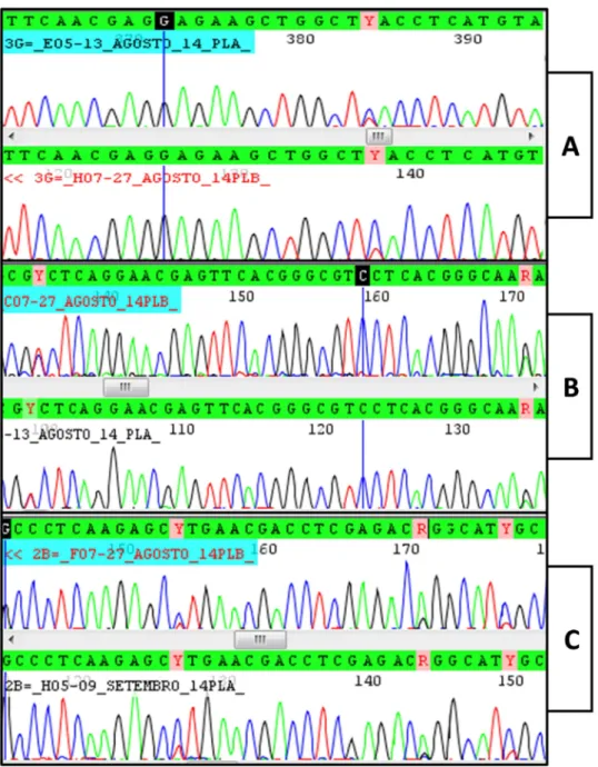
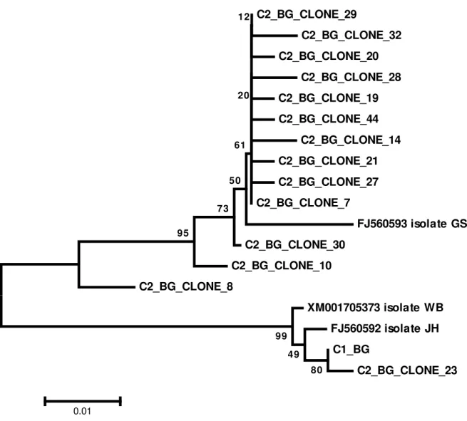
polymorphic site was found in those sequences. The bg chromatograms obtained from cyst C2 show several polymorphic sites compatible with overlap between sequences A and B (Figure 1 C). The nested PCR products from cyst C2 were cloned and 14 alleles were revealed. Twelve alleles were assemblage B, one allele was assemblage A and one allele had an assemblage A fragment interspersed with sequences of assemblage B, indicating that this allele may be a result of recombination between sequences A and B. Bg alignment between alleles found in the cyst 2 is shown in Figure 2. The phylogenetic relationships between the bg alleles from all the cysts investigated in this work and homologous sequences available in GenBank are shown in Figure 3.
31
220 230 240 250 260 270 280
. . . . | . . . . | . . . . | . . . . | . . . . | . . . . | . . . . | . . . . | . . . . | . . . . | . . . . | . . . . | . . . . | . . . . |
Isolate WB CAGAGGGCTT CGCCCGCATC TCCGCCGCGA TCGAGAAGGA GACGATCGCC CGCGAGAGGG CCGTTAGCGC
Isolate JH ... ... ... ... ... ... ...T..
C1_BG ... ... ... ... ... ... ...T..
C2_BG_CLONE_23 ... ... ... ... ... ... ...T..
Isolate GS ... ... ...T..C. ... ... ... ....C...
C2_BG_CLONE_8 ... ... ...C. ... ... ... ....C...
C2_BG_CLONE_10 ... ... ...C. ... ... ... ....C...
C2_BG_CLONE_30 ... ... ...C. ... ... ... ....C...
C2_BG_CLONE_14 ... ... ...C. ... ... ... ....C...
C2_BG_CLONE_29 ... ... ...C. ... ... ... ....C...
C2_BG_CLONE_21 ... ...C. ...C. ... ... ... ....C...
C2_BG_CLONE_28 ... ... ...C. ... ... ... ....C...
C2_BG_CLONE_7 ... ... ...C. ... ... ... ....C...
C2_BG_CLONE_19 ... ... ...C. ... ... ... ....C...
C2_BG_CLONE_20 ... ... ...C. ... ... ... ....C...
C2_BG_CLONE_27 ... ... ...C. ... ... ... ....C...
C2_BG_CLONE_32 ... ... ...C. ... ... ... ....C...
C2_BG_CLONE_44 ... ... ...C. ... ... ... ....C...
290 300 310 320 330 340
. . . . | . . . . | . . . . | . . . . | . . . . | . . . . | . . . . | . . . . | . . . . | . . . . | . . . . | . . . . | . . .
Isolate WB TGCCACGACA GAAGCGCTCA CAAACACGAA GCTCGTCGAG AAGTGCGTCA ACGAGCAGCT CGA
Isolate JH ... ... ... ... ... ... ...
C1_BG ... ... ... ... ... ... ...
C2_BG_CLONE_23 ... ... ... ... ... ... ...
Isolate GS C... ..G..C.... ... ... ... ... ...
C2_BG_CLONE_8 ... ..G..C.... ... ...T... ...T.... ... ...
C2_BG_CLONE_10 ... ..G..C.... ... ...T... ...T.... ... ...
C2_BG_CLONE_30 ... ..G..C.... ... ...T... ...T.... ... ...
C2_BG_CLONE_14 ... ..G..C.... ... ...T... ...C.T.... ... ...
C2_BG_CLONE_29 ... ..G..C.... ... ...T... ...T.... ... ...
C2_BG_CLONE_21 ... ..G..C.... ... ...T... ...T.... ... ...
C2_BG_CLONE_28 ...G.. ..G..C.... ... ...T... ...T.... ... ...
C2_BG_CLONE_7 ... ..G..C.... ... ...T... ...T.... ... ...
C2_BG_CLONE_19 ... ..G..C.... ... ...T... ...T.... ... ...
C2_BG_CLONE_20 ... ..G..C.... ... ...T... ...T.... ... ...
C2_BG_CLONE_27 ... ..G..C.... ... ...T... ...T.... ... ...
C2_BG_CLONE_32 ... ..G..C.... ... ...T... ...T.... ...T... ...
32
Figure 3. Evolutionary relationships of bg sequences. Sequences starting with C2 are alleles cloned from cyst 2. Sequence starting with C1 is the allele sequenced from cyst 1. The evolutionary history was inferred using the Neighbor-Joining method. The optimal tree with the sum of branch length = 0.90220161 is shown. The percentage of replicate trees in which the associated taxa clustered together in the bootstrap test (1000 replicates) are shown next to the branches. The tree is drawn to scale, with branch lengths in the same units as those of the evolutionary distances used to infer the phylogenetic tree. The evolutionary distances were computed using the Maximum Composite Likelihood method and are in the units of the number of base substitutions per site. The analysis involved 18 nucleotide sequences. Codon positions included were 1st+2nd+3rd+Noncoding. All positions containing gaps and missing data were eliminated. There were a total of 343 positions in the final dataset. Evolutionary analyses were conducted in MEGA6
C2_BG_CLONE_29 C2_BG_CLONE_32 C2_BG_CLONE_20 C2_BG_CLONE_28 C2_BG_CLONE_19 C2_BG_CLONE_44 C2_BG_CLONE_14 C2_BG_CLONE_21 C2_BG_CLONE_27 C2_BG_CLONE_7
FJ560593 isolate GS C2_BG_CLONE_30
C2_BG_CLONE_10
C2_BG_CLONE_8
XM001705373 isolate WB
33
3.4. Sequence analysis of orfc4
Genetic sequences from cysts C1, C3, C4, C6, C7, C8, C10, C11, C12, C+1, C+2 are all identical to each other. The closest to these sequences among those available in GenBank is the sequence of Giardia duodenalis isolate WB (XM_001704865) (sub-assemblage AI).
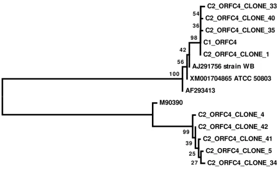
Polymorphic sites were not found in any of these sequences. The orfc4 chromatograms obtained from cyst C2 were difficult to interpret. The orfc4 chromatograms from cyst C2 are compatible with the occurrence of simultaneous amplification of two very different sequences to each other (data not shown). The nested PCR products of cyst C2 were cloned and nine alleles were revealed. Five alleles were assemblage B, and four allele was assemblage A. The phylogenetic relationships between the orfc4 alleles from all the cysts investigated in this work and homologous sequences available in GenBank are shown in Figure 4.
Figure 4. Evolutionary relationships of orfc4 sequences. Sequences starting with C2 are alleles cloned from cyst 2. Sequence starting with C1 is the allele sequenced from cyst 1. The evolutionary history was inferred using the Neighbor-Joining method. The optimal tree with the sum of branch length = 0.90220161 is shown. The percentage of replicate trees in which the associated taxa clustered together in the bootstrap test (1000 replicates) are shown next to the branches. The tree is drawn to scale, with branch lengths in the same units as those of the evolutionary distances used to infer the phylogenetic tree. The evolutionary distances were computed using the Maximum Composite Likelihood method and are in the units of the number of base substitutions per site. The analysis involved 14 nucleotide sequences. Codon positions included were 1st+2nd+3rd+Noncoding. All positions containing gaps and missing data were eliminated. There were a total of 393 positions in the final dataset. Evolutionary analyses were conducted in MEGA6.
C2_ORFC4_CLONE_33 C2_ORFC4_CLONE_40 C2_ORFC4_CLONE_35
C1_ORFC4
C2_ORFC4_CLONE_1 AJ291756 strain WB
34
4. DISCUSSION
Sequencing Giardia duodenalis genes directly from PCR amplified products reveals the
occurrence of polymorphic sites in cysts from stool samples of patients, cloned culture isolates and single cysts and / or trophozoites isolated by micromanipulation (Wielinga et al.
2011; Ankarklev et al. 2012). The detection of double peaks in the chromatograms from PCR
products amplified from single cysts is a plausible evidence that allelic sequence heterozigosity do occur in Giardia duodenalis (Ankarklev et al. 2012).
However, it is not possible to know how many alleles exist in a PCR mixture whose chromatograms present double peaks. In addition, considering amplicons containing two or more alleles it is possible that not all nucleotide differences between them are detected in the chromatogram because the different alleles may be present in different prevalence in the population of strands (Ankarklev et al. 2012). For this reason, we aimed in this study to clone
PCR products that had chromatograms with heterogeneous sites.
Among the analyzed cysts, only one of them (cyst 2) had sequences related to Giardia
duodenalis assemblage B. All other cysts were classified as sub-assemblage AII. Both
samples containing more than one cyst (C+1, C+2) showed no shared sites between asssemblage A and B, and both samples were assigned to sub-assemblage AII, which probably indicates that the frequency of assemblage B cysts in the original stool sample is very low. These results demonstrate that a specimen classified as sub-assemblage AII may actually contain other assemblages that are underrepresented.
Considering the single cysts classified as sub-assemblage AII, all chromatograms presented monomorphic sites, the only exception was the gdh sequence from cyst C3, in which only one polymorphic site was detected. This result contrasts with the results obtained with cyst C2. Although the chromatogram of tpi sequence of cyst C2 has no double peaks, allelic sequence heterozigosity was detected in all other loci.
Several heterogeneous sites was found in gdh sequences of cyst C2, but most of them were not differences between assemblages A and B. Therefore, the gdh chromatogram of cyst C2 is probably formed by the superposition of different alleles of assemblage B. Unfortunately it was not possible to clone the gdh products of cyst C2.
35
occurrence of an unexpected high number of alleles of both genes, besides supporting the simultaneous occurrence of assemblages A and B sequences in one single cyst.
Although axenic Assemblage B isolates was already demonstrated to possess A type allele for gdh, tpi and other genes, indicative of gene transfer events from Assemblage A–B individuals (Lasek-Nesselquist et al. 2009), the results presented here is unprecedented for
single cysts.
One can not completely exclude the possibility of this event is due to cross-contamination during the course of nucleic acid amplification. It is also possible that more than one cyst was aspirated together with cyst C2 though this possibility is remote because the cyst is aspirated from a drop containing only one cyst per microscopic field. After aspiration, the cyst is replaced on the plate, the droplet is re-examined, the cyst is aspirated again, and then transferred to a microtube. Secondly, it is very unlikely that multiple alleles of assemblage B are from more than one cyst, because the sampled population of cysts was largely formed by Assemblage AII cysts. Aspirate over one assemblage B cyst in a population where the ratio between assemblages A and B is at least 10: 1 would be a very unlikely event.
Assemblage B bg fragments flanking assemblage A bg fragment as observed in one of the clones of cyst 2 (C2_BG_CLONE8) is a strong evidence that alleles of different assemblages actually co-exist in the same cyst. No technical artifact could convincingly explain this event, which probably is the result of crossing-over recombination between strands of assemblage A and B. It is noteworthy mentioning that there are no similar sequences to C2_BG_CLONE8 sequence in Genbank.
The simultaneous occurrence of assemblage A and B alleles in one single cyst strongly suggests that genetic exchange occurs between these assemblages. This puts a check on the statement that different assemblages represent distinct species (at least as regards the distinction between AII and B) (Franzen et al. 2009), but the surprisingly high number of
alleles of bg and orfc4 in a single cyst is not supported by current knowledge on the biology of this agent. During the encystation, the DNA of Giardia duodenalis is replicated resulting a
cyst with a ploidy of 16N in four nuclei (Bernander et al. 2001). Such a high number of
alleles of bg and orfc4 (and probably gdh) detected in one single cyst may be the consequence of replication errors of DNA polymerase of Giardia duodenalis assemblage B or any other
36
From the results presented here and from those presented elsewhere (Lasek-Nesselquist et al. 2009; Ankarklev et al. 2012), an interesting hypothesis might emerge. Giardia duodenalis
does not infect only with clonally expanded individuals. Rather, a genetic heterogeneous population composes an infecting population with individuals highly specialized and adapted to the host and individuals that may act as genetic reservoir that are present in a much less number. The minority individuals may be responsible for the spillover capacity of the organism, providing the perpetuation of the genetic material by exchanging genes with the numerous host adapted individuals. For example, an animal challenged with cyst C1 would not be infected at all, but can, otherwise, be infected and transmit cyst C2 back to humans. Once entering human tissues again, cyst C2 would start a new infection by expanding cells containing AII genetic material. Thus, studies focusing on population of host-adapted cysts are welcome in order to confirm if only clonally expanded individuals form them or if they are actually formed with other genetic distinct individuals less represented but always present. The high numerous adapted individuals would be responsible for the infection itself while the less numerous individuals are responsible for the genetic exchange and perpetuation of the organism and its diversity, in a cooperative population.
ACKNOWLEDGEMENTS
Thanks are due to FAPESP (Research Support Foundation of the State of São Paulo) for the scholarship to JM (2011/13472-2). RMS is in receipt of grants from CNPq (Brazilian National Council for Scientific and Technological Development).
CONFLICT OF INTEREST STATEMENT
We confirm that the manuscript has been read and approved by all named authors and that there are no other persons who satisfied the criteria for authorship but are not listed. We further confirm that all of us have approved the order of authors listed in the manuscript.
ETHICS
37
REFERENCES
Almeida, A.A., Pozio, E. and Cacciò, S.M. (2010). Genotyping of Giardia duodenalis cysts
by new Real-Time PCR assays for detection of mixed infections in human samples. Applied
and Environmental Microbiology. 76, 1895–1201. doi: 10.1128/AEM.02305-09
Ankarklev, J.; Svärd, S. G. and Lebbad M. (2012). Allelic sequence heterozygosity in single Giardia parasites. BMC Microbiology 3, 12:65. doi: 10.1186/1471-2180-12-65.
Bernander, R., Palm, J.E. and Svard, S.G. (2001). Genome ploidy in different stages of the Giardia lamblia life cycle. Cellular Microbiol 3, 55–62.
Caccio, S.M., Beck, R., Lalle, M., Marinculic, A. and Pozio, E. (2008). Multilocus genotyping of Giardia duodenalis reveals striking differences between assemblages A and B.
International Journal for Parasitology 38, 1523–1531. doi: 10.1016/j.ijpara.2008.04.008.
Cacciò, S.M., de Giacomo, M. and Pozio, E. (2002). Sequence analysis of the b-giardin gene and development of a PCR-RFLP assay to genotype Giardia duodenalis cysts from
human faecal samples. International Journal for Parasitology 32, 1023–1030.
Cacciò, S.M. and Ryan, U. (2008). Molecular epidemiology of giardiasis. Molecular and
Biochemical Parasitology 160, 75–80. doi: 10.1016/j.molbiopara.2008.04.006.
Felsenstein, J. (1985). Confidence limits on phylogenies: An approach using the bootstrap. Evolution 39, 783-791.
Feng, Y. and Xiao, L. (2011). Zoonotic potential and molecular epidemiology of Giardia species and giardiasis. Clinical Microbiology Review 24, 110–140. doi:
10.1128/CMR.00033-10.
Franzen, O., Jerlström-Hultqvist, J., Castro, E., Sherwood, E., Ankarklev, J., Reiner, D.S., Palm, D., Andersson, J.O., Andersson, B. and Svärd, S.G. (2009). Draft genome sequencing of Giardia intestinalis assemblage B isolate GS: is human giardiasis caused by
two different species? PLoS Pathogens 5, e1000560. doi: 10.1371/journal.ppat.1000560.
Gelanew, T., Lalle, M., Hailu, A., Pozio, E. and Cacciò, S.M. (2007). Molecular characterization of human isolates of Giardia duodenalis from Ethiopia. Acta Tropica 102,
92–99.
38
Lalle, M., Pozio, E., Capelli, G., Bruschi, F., Crotti, D. and Caccio, S.M. (2005). Genetic heterogeneity at the beta-giardin locus among human and animal isolates of Giardia duodenalis and identification of potentially zoonotic sub-genotypes. International Journal for
Parasitology 35, 207–213.
Lasek-Nesselquist, E., Welch, D.M., Thompson, R.C.A., Steuart, R.F. and Sogin, M.L. (2009). Genetic Exchange Within and Between Assemblages of Giardia duodenalis Journal
of Eukaryotic Microbiology 56, 504–518. doi: 10.1111/j.1550-7408.2009.00443.x.
Lebbad, M., Ankarklev, J., Tellez, A., Leiva, B., Andersson, J.O. and Svärd, S., (2008). Dominance of Giardia assemblage B in León, Nicaragua. Acta Tropica 106, 44–53. doi:
10.1016/j.actatropica.2008.01.004.
Martins, J. (2015 in press) Multilocus amplification of genomic DNA from single cysts of Giardia duodenalis separated using micromanipulation technique.
Monis, P.T., Andrews, R.H., Mayrhofer, G. and Ey, P.L. (1999). Molecular systematics of the parasitic protozoan Giardia intestinalis. Molecular Biology and Evolution 16, 1135–1144.
Morrison, H.G., McArthur, A.G., Gillin, F.D., Aley, S.B., Adam, R.D., Olsen, G.J., Best, A.A., Cande, W.Z., Chen, F., Cipriano, M.J., Davids, B.J., Dawson, S.C., Elmendorf, H.G., Heh, A.B., Holder, M.E., Huse, S.M., Kim, U.U., Lasek-Nesselquist, E., Manning, G., Nigam, A., Nixon, J.E., Palm, D., Passamaneck, N.E., Prabhu, A., Reich, C.I., Reiner, D.S., Samuelson, J., Svard, S.G. and Sogin, M.L. (2007). Genomic minimalism in the early diverging intestinal parasite Giardia lamblia. Science 317, 1921–1926.
Ryan, U. and Cacciò, S.M. (2013). Zoonotic potential of Giardia. International Journal for
Parasitology 43, 943–956. doi: 10.1016/j.ijpara.2013.06.001.
Saitou, N. and Nei, M. (1987). The neighbor-joining method: A new method for reconstructing phylogenetic trees. Molecular Biology and Evolution 4, 406-425.
Sheather, A. L. (1923). The detection of intestinal protozoa and mange parasites by a flotation technique. Journal of Comparative Pathology 36, 266-275.
Sprong, H., Cacciò, S.M. and van der Giessen, J.W. (2009). ZOOPNET network and partners. Identification of zoonotic genotypes of Giardia duodenalis. PLoS Neglected
Tropical Diseases 3, e558. doi: 10.1371/journal.pntd.0000558.
39
potential zoonotic transmission of Giardia duodenalis. Emerging Infectious Diseases 9,
1444–1452.
Tamura K., Nei M. and Kumar S. (2004). Prospects for inferring very large phylogenies by using the neighbor-joining method. PNAS, Proceedings of the National Academy of Sciences
(USA) 101:11030-11035.
Tamura K., Stecher G., Peterson D., Filipski A. and Kumar S. (2013). MEGA6: Molecular Evolutionary Genetics Analysis version 6.0. Molecular Biology and Evolution 30,
2725-2729. doi: 10.1093/molbev/mst197.
Wielinga, C., Ryan, U., Andrew Thompson, R.C. and Monis, P. (2011). Multi-locus analysis of Giardia duodenalis intra-Assemblage B substitution patterns in cloned culture
isolates suggests sub-Assemblage B analyses will require multi- locus genotyping with conserved and variable genes. International Journal for Parasitology 41, 495 – 503. doi:
40
5 General Conclusions
The molecular characterization of G. duodenalis has been widely used in clinical and
environmental samples to assist in epidemiological research of infection. Several genotyping studies are based on analysis of a single genetic marker, however, when the multilocus analysis is employed, researchers are faced with the lack of agreement between the results, occurring often the detection of different assemblages of G. duodenalis in a given sample.
As shown in this study G. duodenalis is a complex organism due its high genetic
variability, thus genetically distinct cysts may be present in the same population (mixed infection). In a multilocus analysis, if there amplification by distinct assemblages for one or more genes, is not possible to know, in fact, whether that sample has genetically distinct cysts or exists ASH or even genetic recombination. The only way to confirm the occurrence of such events is studying a single individual of G. duodenalis.
In this work was developed a micromanipulation technique of a single Giardia cyst