Braz Dent J 13(3) 2002
212Braz Dent J (2002) 13(3): 212-214 V.A. Pereira Filho et al.
Nasolabial Cyst: Case Report
Valfrido Antonio PEREIRA FILHO1
Alessandro Costa da SILVA2
Márcio de MORAES2
Roger William F. MOREIRA2
Halbert VILLALBA3
1Department of Oral and Maxillofacial Surgery, Faculty of Dentistry of Araraquara,
UNESP, Araraquara, SP, Brazil
2Department of Oral and Maxillofacial Surgery, and 3Department of Oral Pathology, Faculty of Dentistry of Piracicaba,
UNICAMP, Piracicaba, SP, Brazil
The nasolabial cyst is classified as a fissural cyst, found outside the bone, and on the region corresponding to the nasolabial furrow and alar nose. This cyst is frequently asymptomatic with the most usual sign being alar nose elevation. In spite of the low occurrence of nasolabial cysts, it is important to recognize the clinical characteristics of this lesion. The purpose of this paper is to review the literature and discuss the histomorphology and etiology of this condition, showing treatment by surgical excision.
Key Words: nasolabial cyst, nasoalveolar cyst.
Correspondence: Prof. Valfrido Antonio Pereira Filho, Faculdade de Odontologia de Araraquara, UNESP, Rua Humaitá, 1680, 14801–903 Araraquara, SP, Brasil. Tel: +55-16-201-6359. e-mail: dinho@foar.unesp.br
ISSN 0103-6440
INTRODUCTION
The nasolabial cyst is a rare non-odontogenic cyst originating in maxillofacial soft tissues. According to Allard (1), this lesion was first described in 1882 by Zuckerkandl, and since then, two main etiological theo-ries have been proposed. One holds that the lesion arises from trapped nasolacrimal duct tissue (2), and the other affirms that it is an embryonic fissural cyst (3). Klestadt (4) first postulated an embryologic origin for these cysts and considered that these lesions must origi-nate from embryonic epithelium, entrapped in the de-velopmental fissures between the lateral nasal and max-illary processes. Since then, many authors have classi-fied this entity based on Klestadt’s embryologic theory as a fissural cyst (5).
This lesion presents an extraosseous location in the region of the nasolabial fold and can cause swelling in the furrow, alar nose elevation and upper lip projec-tion (3). Despite the uncommon occurrence of nasola-bial cysts, it is important to recognize the characteris-tics of this lesion.
The purpose of this paper was to review the
literature and discuss the histomorphology and etiology of this condition, showing the treatment of this pathol-ogy by surgical excision.
Report of case
A 42-year-old, Caucasian woman sought treat-ment in the Oral and Maxillofacial Surgery Departtreat-ment at the University of Campinas (UNICAMP), Piracicaba Dental School. The patient’s main complaint was swell-ing and elevation of the right nasolabial region. Clini-cally, there was a flattened upper lip, particularly on the region of the right alar base and the sulcus nasolabialis appeared indistinct, suggesting the possible diagnosis of nasolabial cyst. On palpation, a soft, fluctuant mass was observed which extended from the buccal area up to the floor of the nose.
Braz Dent J 13(3) 2002
Nasolabial cyst: case report 213
medical history did not reveal any pathological condi-tion. Therefore, surgery was proposed.
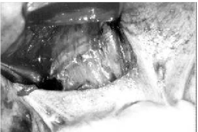
The cyst was excised under local anesthesia by infraorbital block injection and infiltration with 2% lidocaine containing 1:100,000 epinephrine. Incision was made in the labial mucosa 5 mm above the mucogingival junction, and the lesion was carefully separated with sharp dissection from the mucous mem-brane of the floor of the nose (Figure 1), leaving a 2 mm oronasal communication which was repaired with 3-0 chromic catgut. The insertion of the alar base was re-established by suturing it in a perforation made in the anterior nasal spine, with 3-0 nylon suture.
The lesion measured 10x10x8 mm, presenting a flat surface. Microscopically, the cyst was lined by thin, nonpapillated cuboidal to columnar epithelium with occasional goblet cells. The cyst wall consisted of condensed fibrous connective tissue containing only scattered chronic inflammatory cells. The histologic
diagnosis was nasolabial cyst (Figure 2).
The histologic, clinical and radiologic findings were compatible with nasolabial cyst. The postopera-tive course was uncomplicated and there was no lesion recurrence up to one year of follow-up and the patho-logic signs and symptoms of the lesion had disap-peared.
Discussion
Many names have been suggested for nasolabial cysts, with the nasoalveolar cyst and nasolabial cyst being the two most widely used. Nasolabial cysts repre-sent about 0.7% of all cysts in the maxillofacial region, and 2.5% of non-odontogenic cysts. Many authors believe that its prevalence is actually higher than pre-sented in the literature; however, due to misdiagnosis, indexes remain low (6).
Nasolabial cysts are usually unilateral, with no prevalence of side occurrence but bilateral cases have been also reported (2). It has been estimated that ap-proximately 10% of the cases are bilateral (6,7). Other significant findings include a greater incidence in fe-males (4:1) and possibly greater prevalence among Blacks (8). It frequently occurs during middle age (2,5,9). Although we report only one case, this patient revealed the same characteristics that the literature presents: middle-aged, female and asymptomatic. This lesion is usually asymptomatic unless secondarily in-fected.
The clinical findings of the nasolabial cyst are fairly typical. Patients usually complain of a swelling adjacent to the nose, and sometimes the cyst may be observed on routine examination (10,11). The develop-ment of swelling in the maxillary buccal sulcus may reach great dimensions, causing discomfort with the use of dentures, breathing obstruction and facial asym-metry. In this report, the patient mainly complained about alar nose flaring, upper lip swelling, diminished nasolabial sulcus, nasal floor elevation and greater volume in the maxillary labial sulcus. The latter was felt to be soft and fluctuant during intraoral palpation.
Due to similar signs and symptoms, this lesion may be misdiagnosed as a dental or periodontal ab-scess, odontogenic cyst, tumor and choanal polyp (12). In the present case, the patient reported endodontic treatment in the left canine region. This could cause a false diagnosis.
Figure 1. Transoperatory view.
Braz Dent J 13(3) 2002
214 V.A. Pereira Filho et al.
Although it is a soft-tissue cyst, the nasolabial cyst can sometimes cause erosion of the underlying maxillary bone which may be observed in radiographic examination (13). Schroff (14) pointed out that these are not bone lesions and thus detailed radiographic examination must be obtained to distinguish them from odontogenic or other non-odontogenic etiologies (15). In the literature, rare cases of radicular absorption were observed due to these cysts. However, an alteration in the shape of the maxillary bone in the periapical region of the tooth involved was noted in the present case. This bone resorption was also observed by other authors (16,17).
Injection of esclerotic substances, marsupializa-tion and surgical removal may be considered for treat-ment. However, unlike some of the large intraosseous cysts, this soft-tissue lesion does not respond to marsu-pialization (18) and surgical excision is the treatment of choice. Because this cyst is usually closely related to the floor of the nose (1,19), perforation of the nasal mucosa may be expected during its removal. When very small perforations are caused, they can be left untreated, however, larger ones must be sutured (18).
In the present case, the nasal and buccal struc-tures healed well without any recurrence of the lesion after one year. Despite the suture, there was an enlarge-ment of the alar nose. However, this did not compro-mise facial esthetics.
RESUMO
Pereira Filho VA, da SilvaAC, de MoraesM, Moreira RWF, Villalba H. Cisto Nasolabial: Relato de Caso. Braz Dent J 2002:13(3):212-214.
O cisto nasolabial é classificado como um cisto fissural, localizado externamente ao tecido ósseo, na região correspondente ao sulco nasolabial e asa do nariz. Estes cistos são freqüentemente assintomáticos e geralmente promovem a elevação da asa do nariz. Apesar da sua difícil ocorrência, é importante
reconhecermos as características desta lesão. O objetivo deste artigo é o de revisar a literatura e de discutir aspectos histológicos e etiológicos desta condição, bem como o tratamento por meio da excisão cirúrgica.
Unitermos: cisto nasolabial, cisto nasoalveolar.
REFERENCES
1. Allard RHB. Nasolabial cyst. A review of the literature and report of cases. Int J Oral Surg 1982;11:351-359.
2. Precious DS. Chronic nasolabial cyst. J Canad Dent Assoc 1987;53:307-308.
3. Wesley RK, Scannel T, Nathan LE. Nasolabial cyst: Presentation of a case with a review of the literature. J Oral Maxillofacial Surg 1984;42:188-192.
4. Klestadt WD. Nasal cyst and facial cleft cyst theory. Ann Otol Rhinl Laryngol 1953;62:84-89.
5. Egervary G, Csiba A. Bilateral nasolabial cyst. Dental Digest 1969;75:504-507.
6. Smith RA, Katibah RN, Merrell P. Nasolabial cyst: report of a case. J Canad Dent Assoc 1982;11:727-729.
7. Roed-Petersen B. Nasolabial cysts: a presentation of five patients with a review of the literature. Br J Oral Surg 1969;7:84-95. 8. Cohen MA, Hertzanu Y. Huge growth potential of the nasolabial
cyst. Oral Surg 1985;59:441-445.
9. Santora E, Ballantyne AJ, Hinds EC. Nasoalveolar cyst: report of case. J Oral Surg 1970;28:117-120.
10. Campbell RL, Burkes Jr EF. Nasolabial cyst: report of case. J Am Dent Assoc 1975;91:1210-1213.
11. Rao RV. Naso-labial cyst. J Laryngol Otol 1955;69:352-355. 12. Werner PE, Lehman RH, Collentine ME, Darling RJ. Intraoral
presentation of a nasal (choanal) polyp: report of case. J Oral Surg 1968;26:589-592.
13. Adams A. Roentgeno-Oddities. Oral Surg 1985;60:118-119. 14. Schroff J. Unusual cysts in the maxilla. Cysts of nasopalatine
duct and fissural cysts. Dent Items Interest 1929;51:109-113. 15. Chinellato LEM, Damante JH. Contribution of radiographs to the
diagnosis of the naso-alveolar cyst. Oral Surg 1984;58:729-735. 16. Balfour RS. Nasoalveolar cyst. J MD State Dent Assoc
1977;20:92-94.
17. Seward GR. Nasolabial cysts and their radiology. Dent Pract 1962;12:154-161.
18. Crowford W, Korchin L, Greskovich FJ. Nasolabial cysts: report of two cases. J Oral Surg 1968;26:582-588.
19. Brandão GS, Ebling H, Souza IF. Bilateral nasolabial cyst. Oral Surg 1974;37:480-484.